Human Biology - Body Systems
1/63
Earn XP
Description and Tags
Respiratory system, Circulatory system, DIgestive system
Name | Mastery | Learn | Test | Matching | Spaced |
|---|
No study sessions yet.
64 Terms
Label a diagram of the respiratory system
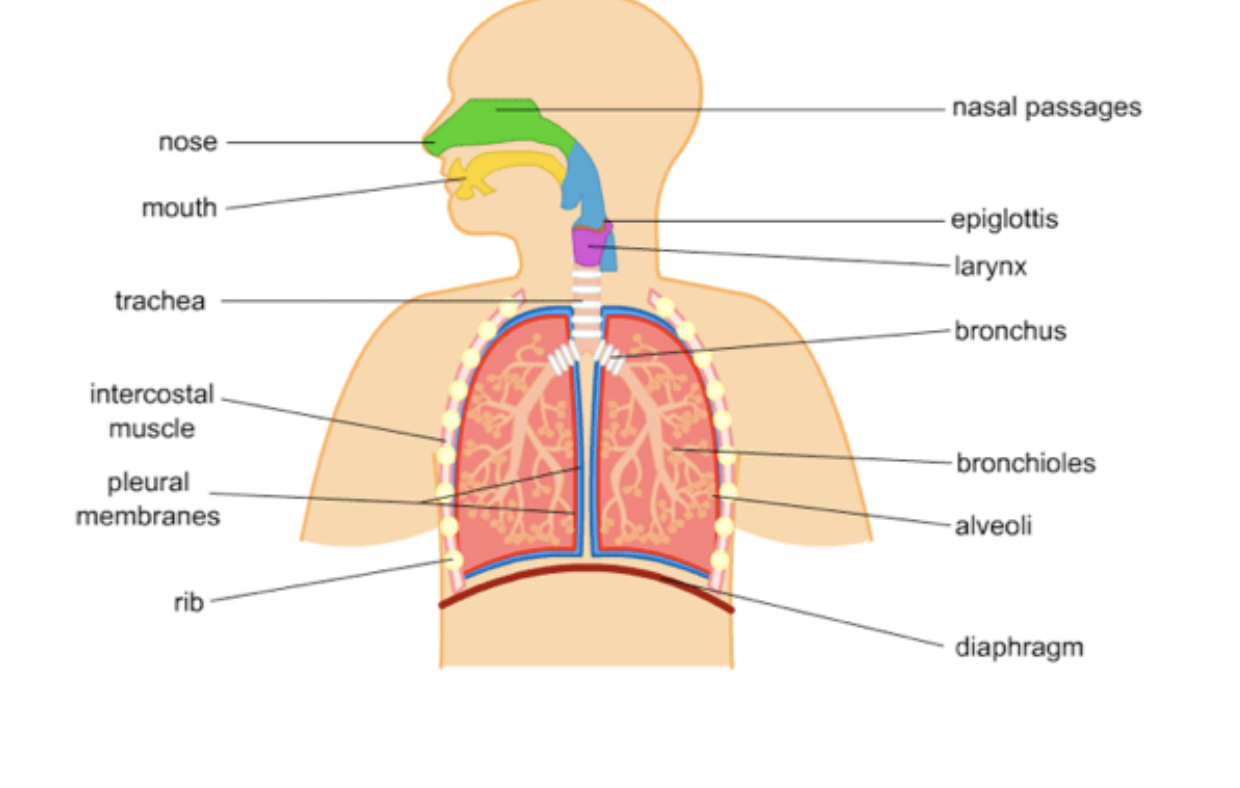
describe the function of the Nose
- Filters/warms/moistens air before it enters the lungs
- Hairs and mucus trap dust
- Acts as a resonating chamber for speech sounds
Smell receptors
describe the function of the Pharynx
- Throat
- Air from the nasal cavity passes through
describe the function of the Larynx
(vocal cords)
- Air passes through the larynx (to and from the lungs)
- Contains vocal cords which vibrate to make sound
describe the function of the Trachea
- Windpipe
- Carries air to and from the lungs
- Lined with mucous and cilia (moved trapped particles upward)
describe the function of the Lungs
- Covered with a pleural membrane (pleural fluid holds the lungs against the inside of the chest)
- Site for gas exchange
describe the function of the Bronchi
- Two primary bronchi branch from the trachea, then divide into secondary and tertiary branches
- Carry air to and from the lungs
describe the function of the Bronchioles
- Very fine tubes with walls of smooth muscle
- Finest branch finishes at the alveoli (air sacs)
- Carry air to and from the lungs
describe the function of the Alveoli
- Tiny air sacs that make up the majority of the lungs
- Increased surface area
- Have thin walls and are well supplied with blood capillaries
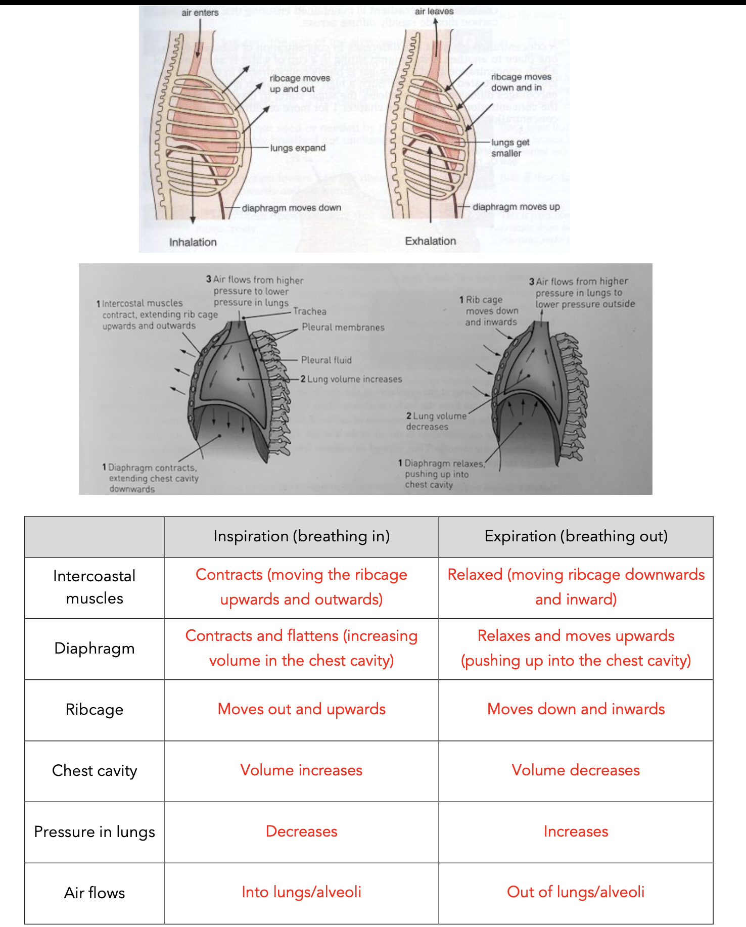
describe the function of the Diaphragm
- Muscle that separates the chest from the abdomen
- Contracts and flattens to increase the volume of the chest and lungs = air moves in
describe the function of the Intercostal muscles
- Muscle between the ribs
- Contracts and move rib cage upwards and outwards to
increase the volume of the chest and lungs = air moves in
describe the function of the Ribs
Ribs - form the framework for the
chest. and protect it
describe the function of the Pleural membranes
- Pleura
- Covers the lungs surface and inside of the chest
- Works with the pleural fluid to allow breathing to occur
describe the function of the Epiglottis
§Epiglottis is a flap of elastic cartilage which covers the oesophagus to guide air into the trachea. It does the reverse when swallowing food.
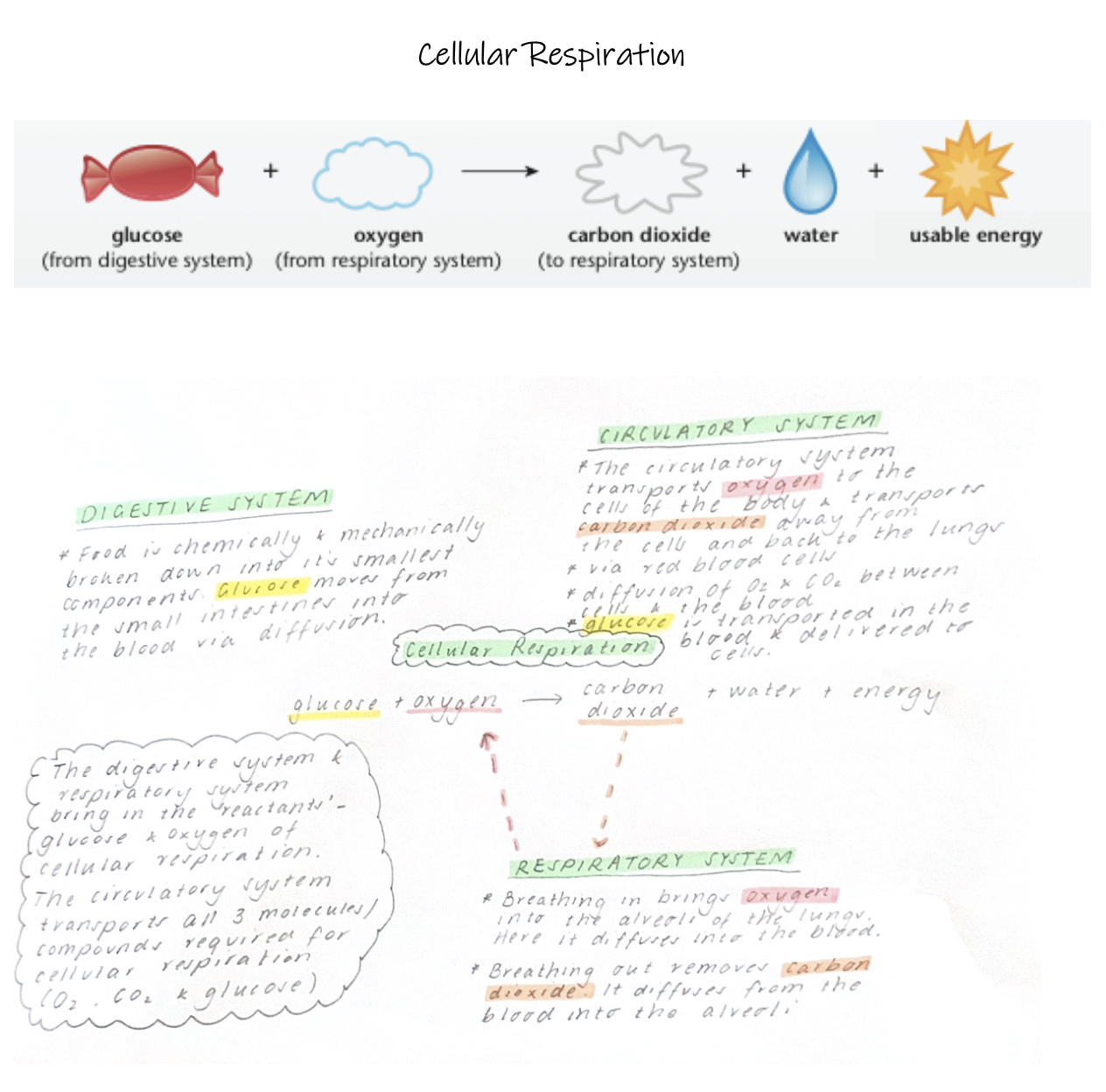
Distinguish between breathing and respiration
Breathing is the movement of air into and out of the lungs, while respiration is the process occurring in cells to produce ATP energy.
Outline and explain the mechanics of breathing
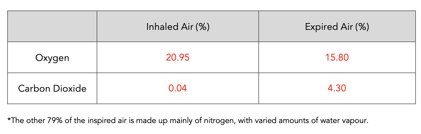
Compare the composition of of inhaled and exhaled air.
Describe where gas exchange occurs and how it occurs.
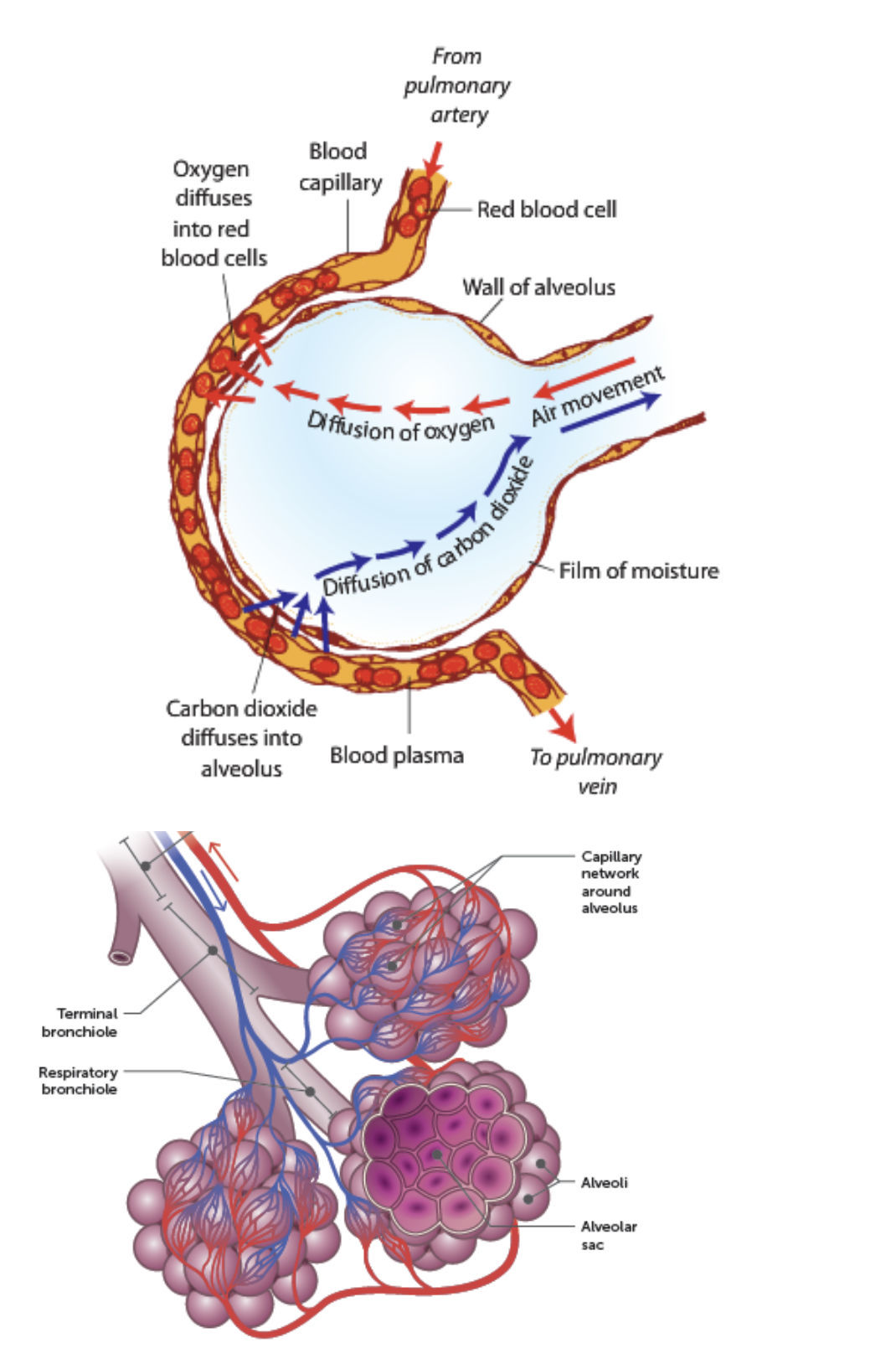
Describe the characteristics of the respiratory surfaces that enable efficient gas exchange.
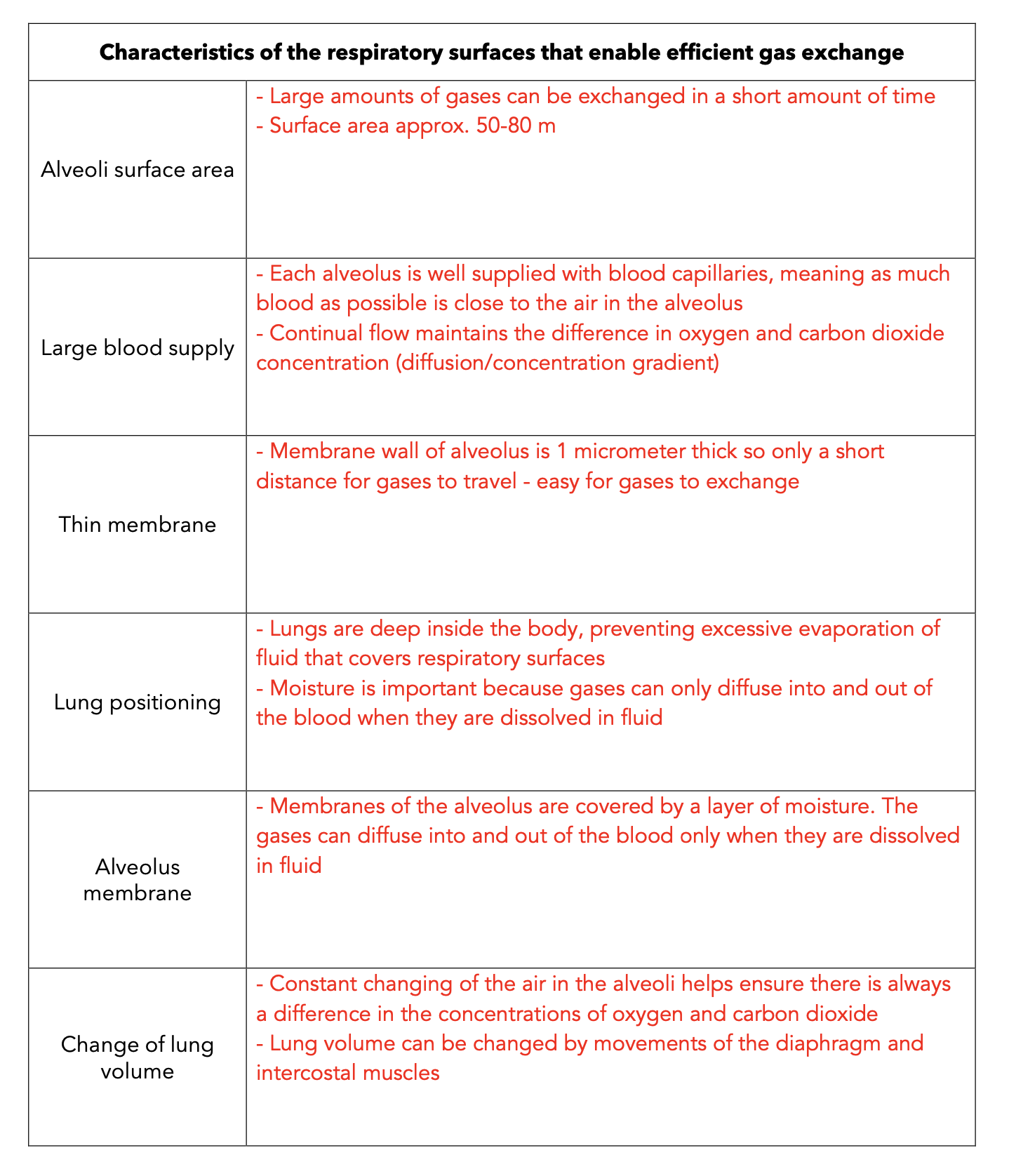
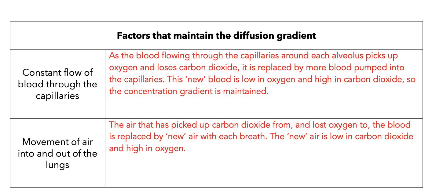
Describe how the following factors that maintain a diffusion gradient between the gases in the air, in the alveoli and in the blood
· Constant blood supply
Constant air flow (breathing)
Describe the functions of blood.
§ Transport of:
- O2 and nutrients to cells of body
- CO2 and waste away from cells of body
- Chemical messengers (hormones) to cells
§ Maintain pH of body fluids
§ Distribute heat/maintain body temp
§ Maintain water content & ion concentration
§ Protect against disease causing micro-organisms
§ Prevention of blood loss (clotting when vessels damaged)
§ 55% Plasma
- 91% water
- carries dissolved substances e.g. nutrients (glucose, amino acids, lipids), ions (Na+, K+, Ca2+, Cl-, HCO3-), dissolved gases (O2, CO2), hormones, plasma proteins and waste (urea)
§ 45% Formed Elements (cells/cell fragments) - Erythrocytes (Red Blood Cells)
- Leucocytes (White Blood Cells)
- Thrombocytes (Platelets)
Plasma, erythrocytes (red blood cells), leucocytes (white blood cells) and thrombocytes (platelets).
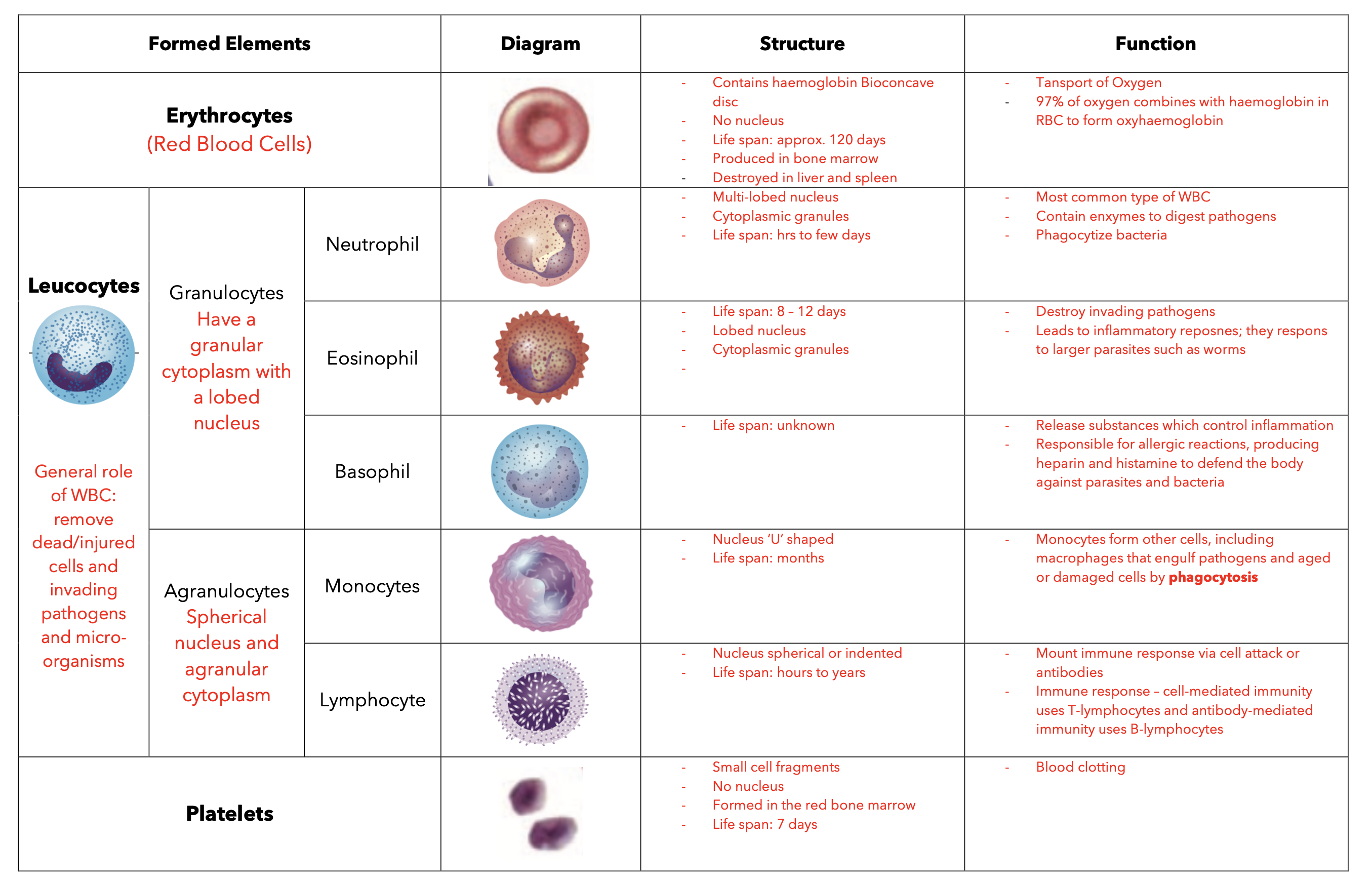
Red blood cells contain haemoglobin
Describe the structure of an erythrocyte.
- Contains haemoglobin
- Bioconcave disc
- No nucleus
- Life span: approx. 120 days
Where are erythrocytes forms?
Bone Marrow
Why do red blood cells lack a nuclei?
Increased volume for haemoglobin and therefore oxygen transporations
What is haemoglobin and its role?
- Oxygen transportation - 97% combines with haemoglobin in RBC to form oxyhaemoglobin
- Carbon dioxide transporation - 22% combines with haemoglobin in RBC to form carbaminohaemoglobin
9. What is the normal lifespan of an erythrocyte? 120 days
10.How does the bioconcave shape of the erythrocyte facilitate oxygen transport?
- Increase SA:Vol ratio – increasing the amount of space available for haemoglobin and therefore ozygen and carbon dioxide transport
11.Leucocytes are white blood cells. How do they compare to erythrocytes?
- Larger in size
- Have a nucleus
- Function: remove dead/injured cells and invading pathogens and micro-organisms
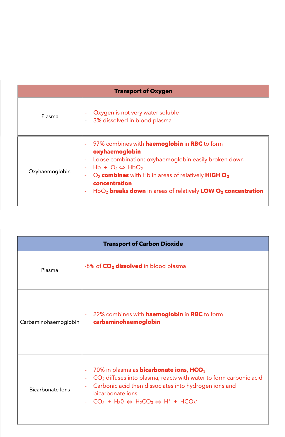
Plasma facilitates the transport of carbon dioxide by dissolving it in the liquid component of blood and by carrying bicarbonate ions formed from carbon dioxide through reactions with water, aiding in maintaining blood pH balance.
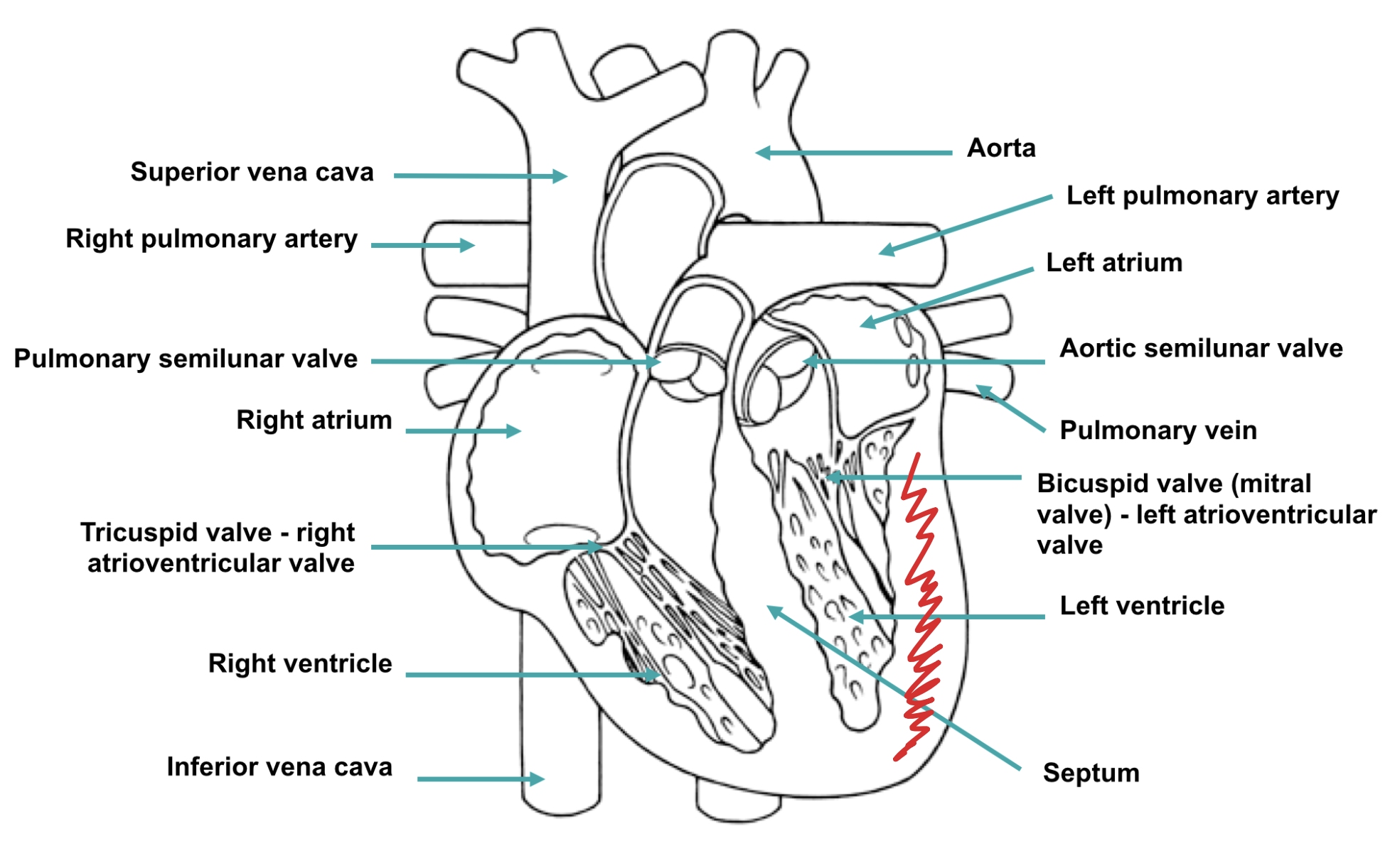
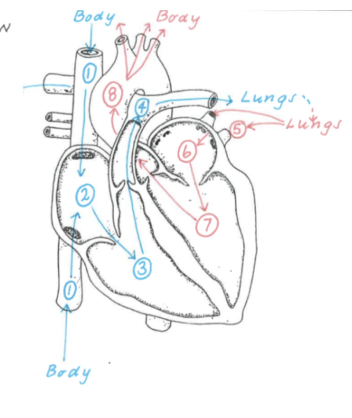
Pulmonary Circulation
Passage of blood from the heart to the lungs, to remove excess carbon dioxide and pick up oxygen, and back to the heart
Systemic Circulation
Passage of blood from the heart to the organs and tissues of the body, to deliver oxygen and collect carbon dioxide, and back to the heart
- Double pump- left and right sides pump ‘separately’
- Right side receives deoxygenated blood and pumps blood to the lungs
- Left side receives oxygenated blood and pumps blood to the whole body - Blood from the right side never mixes with blood from the left side
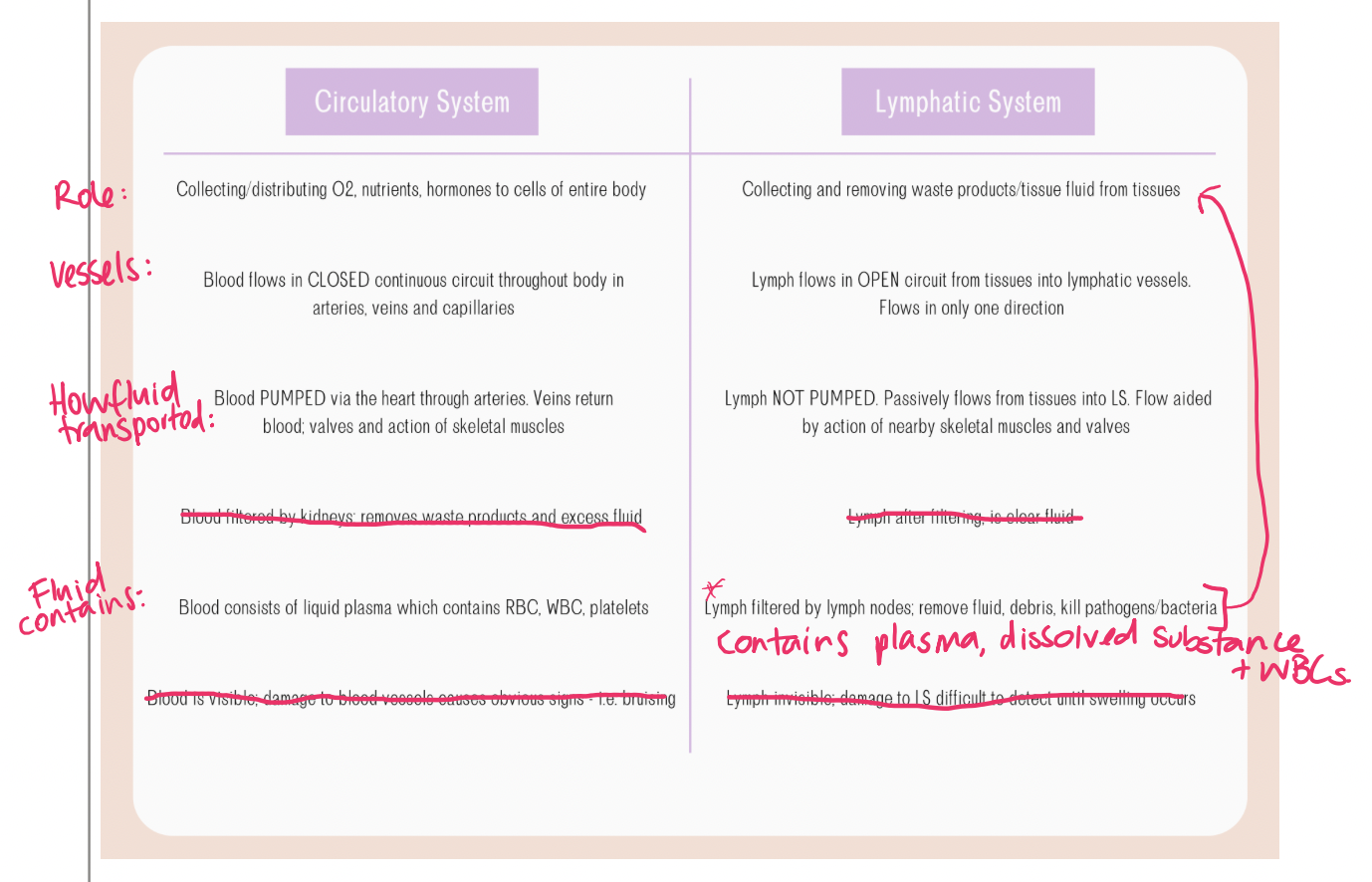
Arteries, arterioles, veins, venules and capillaries.

The Lymphatic System functions to
1) Return tissue fluid to the circulatory
system.
2) Immune surveillance- to assist in
protecting the body from disease.
3) Helps large molecules to enter the
blood (e.g., lipids and hormones).
4) Absorbs fats via lacteals (specialised
lymph vessel) in the digestive system
and transport them back to the blood.
Lymph vessels transport lymph fluid back to the bloodstream and include lymph capillaries, which absorb interstitial fluid, and lymph veins, which carry lymph to lymph nodes. Lymph nodes filter lymph, trapping pathogens and facilitating immune responses.
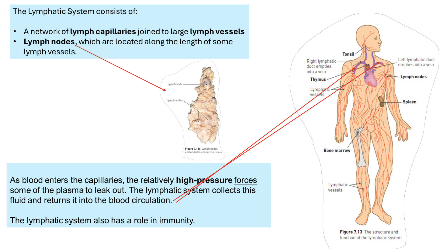
Compare the structure of the blood circulatory system and the lymphatic system.
The blood circulatory system consists of a closed network of arteries, veins, and capillaries that transport blood, while the lymphatic system is an open system made up of lymph vessels and nodes that helps return interstitial fluid to the bloodstream and plays a vital role in immune function.
Describe the following as reasons why cells require nutrients:
· To supply energy for metabolism
· Matter for synthesis To regulate cell processes.
All cells require nutrients in order to provide energy for the cell’s activities and materials for cell growth, cell reproduction, secretion and other metabolic processes. The digestive system extracts nutrients from the food we eat and absorbs them into the body for use by the cells.
For carbohydrates, proteins and lipids describe the following:
· biological function
· energy value
dietary sources
Carbohydrates
Carbohydrates are the main source of energy for cells.
Simple sugars (monosaccharides), particularly glucose, are used in cellular respiration to release energy.
Complex carbohydrates, such as starch, are broken down to simple sugars.
All carbohydrates contain atoms of carbon, hydrogen and oxygen.
Simple sugars can join together to form larger molecules (disaccharides).
Polysaccharides are larger carbohydrate molecules formed when many simple sugars join together.
Lipids
Lipids include fats and oil and are another important energy source. They are broken down to fatty acids and glycerol.
Glycerol is broken down to release energy in a similar way to glucose.
Each lipid molecule consists of one molecule of glycerol and one, two or three fatty acid molecules.
Proteins
Proteins are organic compounds that are made up of many amino acids. The most import
An amino acid is a molecule that contains an amino group and a carboxylic acid group.
When two amino acids bond together, these two groups react to form a peptide bond, releasing a water molecule. There are 20 different amino acids found in proteins, proteins, each one differing in the structure of the side chain.
Proteins consist of 100 or more amino acids; their type and order are determined by the DNA that codes for the protein’s production. Each protein has a characteristic shape due to the folding of the chain.
Shorter lengths of amino acids include dipeptides and polypeptides.
Water
Water is important in metabolism because it is the fluid in which other substances are dissolved. Some of the cell’s chemical reactions occur in water, and in others water molecules actually take part in the reaction.
Minerals
Minerals are important for metabolism because they may be a part of enzymes, function as cofactors for enzymes, or be a part of substances such as adenosine triphosphate (ATP) that are involved in metabolism.
Vitamins
Vitamins act as coenzymes for many of the chemical reactions of metabolism.
The alimentary canal refers specifically to the continuous tube through which food passes, including the mouth, esophagus, stomach, intestines, and anus, while the digestive system encompasses the entire system involved in processing food, including accessory organs like the liver and pancreas.
²ingestion of food and water
²mechanical digestion of food
²chemical digestion of food
²movement of food along the alimentary canal
²absorption of digested food and water into the blood and lymph
²elimination of material that is not absorbed.
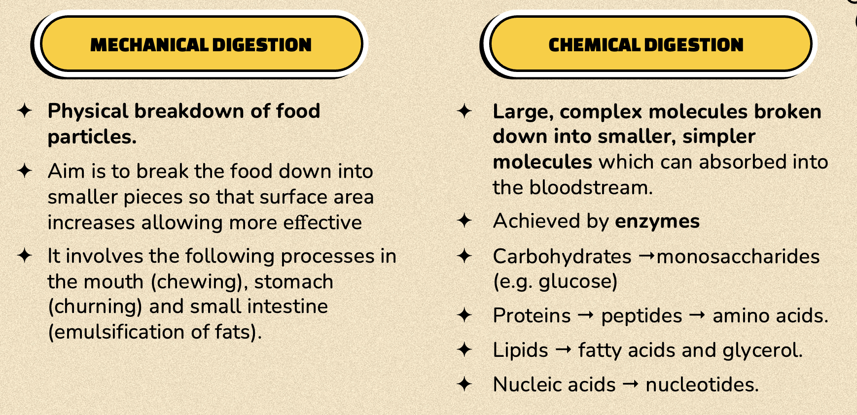
öAim is to break the food down into smaller pieces so that surface area increases allowing more effective
öIt involves the following processes in the mouth (chewing), stomach (churning) and small intestine (emulsification of fats).
· where it takes place
· the enzymes involved and where they are produced.
the substrates and end products.
öAchieved by enzymes
öCarbohydrates "monosaccharides (e.g. glucose)
öProteins " peptides " amino acids.
öLipids " fatty acids and glycerol.
öNucleic acids " nucleotides.
The ileum has a large surface area due to villi and microvilli, thin walls for easy transfer, a rich blood supply for efficient nutrient transport, and specialized transport mechanisms for various nutrients.
Describe the process of absorption of nutrients in terms of the following:
· when they are absorbed
· where the nutrients are absorbed
· how they are absorbed (simple diffusion or active transport).
When chemical digestion is complete, the complex carbohydrates will have been broken down into simplesugars, the proteins into amino acids, and the fats into fatty acids and glycerol. These products of digestion, along with substances such as vitamins, minerals and water, are then absorbed through the wall of the small
intestine into the blood. Nutrients are absorbed through the internal surface of the small intestine, so efficient
absorption requires a large surface area. A large internal surface area is achieved in a number of ways:
• The small intestine is very long – about 6–7 m.
• The inner lining, known as the mucosa, has folds that extend into the interior of the small intestine.
• The mucosa has small, finger-like projections called villi that extend from the folded surface.
• The cells covering the outside of the villi have tiny microscopic projections from their external
surfaces. These are the microvilli.
Circulatory System
After digestion, glucose, amino acids, water-soluble vitamins, and minerals are absorbed into blood capillaries in the villi of the small intestine.
These nutrients are carried via the hepatic portal vein directly to the liver.
In the liver, nutrients are processed:
Glucose may be stored as glycogen.
Toxins are detoxified.
Amino acids are modified or stored.
After processing, nutrients enter the general circulation through hepatic veins and are transported to tissues for energy, repair, or storage.
Lymphatic System
Fats (as fatty acids and glycerol) and fat-soluble vitamins (A, D, E, K) are absorbed into lacteals (lymph vessels in villi).
These travel through the lymphatic system and bypass the liver at first.
They enter the bloodstream at the subclavian vein, near the heart.
From there, fats are delivered to body tissues for storage or energy use.
Eventually, the liver may also process some fats after they circulate in the blood.
Excretion is the removal of metabolic waste – waste that has been produced by chemical activity of the body cells. Except for the bile pigments, the contents of faeces are not metabolic waste, so defecation is better referred to as elimination, rather than excretion.
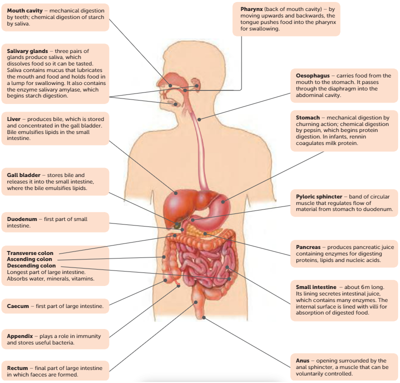
mechanical digestion by teeth; chemical digestion of starch by saliva.
The action of the jaws and teeth begins mechanical digestion. There are four types of teeth, each with
a different function in mechanical digestion. A full adult set of teeth in the lower jaw consists of:
• four incisors – chisel-shaped teeth used for biting or cutting, as when taking a bite of an apple
• two canines, one on each side of the incisors. These are conical teeth used for tearing. Human
canines are the same length as the other teeth
• four premolars, two on each side of the jaw
• six molars, three on each side of the jaw. The premolars and molars have broad crowns with
rounded cusps. The cusps of the teeth of one jaw fit into depressions on the surface of teeth on
the other jaw, making the premolars and molars ideal for crushing and grinding food.
The same number and type of teeth occur in the upper jaw.
After chewing, the tongue shapes the food into a rounded lump called a bolus. To swallow, the tongue moves upwards and backwards, pushing the bolus into the back of the mouth, the pharynx, which leads to the oesophagus.
(back of mouth cavity) – by moving upwards and backwards, the tongue pushes food into the pharynx for swallowing.
carries food from the mouth to the stomach. It passes through the diaphragm into the abdominal cavity.
The cardiac sphincter, or lower esophageal sphincter, controls the passage of food from the esophagus into the stomach. It prevents the backflow of stomach contents into the esophagus.
mechanical digestion by churning action; chemical digestion by pepsin, which begins protein digestion. In infants, rennin coagulates milk protein.
band of circular muscle that regulates flow of material from stomach to duodenum.
about 6m long. Its lining secretes intestinal juice, which contains many enzymes. The internal surface is lined with villi for absorption of digested food.
✦1.5m long, large in diameter than the small intestine.
✦Made up of the caecum, colon, rectum and anus.
✦No villi or digestive juices secreted here and no digestion occurs but large amount of mucus secreted by mucosa.
✦Movement is slow, taking ~18-24hrs for material to pass through.
✦Main functions:
²Most of the remaining water is absorbed during this time making contents more solid. ²Bacteria in large intestine break down remaining organic compounds - some also produce vitamins which are absorbed through walls into blood.
²Mineral nutrients absorbed.
final part of large intestine in which faeces are formed.
opening surrounded by the anal sphincter, a muscle that can be voluntarily controlled.
three pairs of glands produce saliva, which dissolves food so it can be tasted. Saliva contains mucus that lubricates the mouth and food and holds food in a lump for swallowing. It also contains the enzyme salivary amylase, which begins starch digestion.
produces bile, which is stored and concentrated in the gall bladder. Bile emulsifies lipids in the small intestine.
stores bile and releases it into the small intestine, where the bile emulsifies lipids.
produces pancreatic juice containing enzymes for digesting proteins, lipids and nucleic acids.
prevents food from entering the trachea during swallowing. a flap of cartilage behind the root of the tongue,
What is Peristalsis
Peristalsis is the wave-like muscle contractions that move food through a tube such as the oesophagus.