BIO121 Unit 2 (CH19 & 22)
 Call Kai
 Learn
Learn Practice Test
 Spaced Repetition
 Match
Match Flashcards
 Knowt Play
1/87

There's no tags or description
Looks like no tags are added yet.
Name | Mastery | Learn | Test | Matching | Spaced | Call with Kai | Chat |
|---|
No analytics yet
Send a link to your students to track their progress
88 Terms
Blood
• Liquid connective tissue that circulates through the cardiovascular system
• pH = 7.35 - 7.45 (very closely regulated)
• Composed of:
• Non-living plasma = liquid matrix
• Formed elements = cells and cell fragments
• Contains many proteins and dissolved substances
• More viscous and has a higher specific gravity than water
• Makes up about 8% of total body weight
Function of blood (transport)
• Gasses (i.e. oxygenand carbon dioxide), nutrients, waste products
• Processed molecules (i.e. Vitamin D from the skin → liver → kidneys)
• Regulatory molecules (i.e. hormones)
function of blood (regulation & maintenance)
• pH (buffers)
• Osmosis (water movement)
• Body temperature
function of blood (protection)
• Immune cells, antibodies and inflammatory chemicals
• Clot formation
plasma composition
• Plasma is approximately 91% water
• plasma composition remains relatively stable due to homeostatic regulation
• Plasma Proteins = 7% by weight

albumins (58% of plasma protein)
• Viscosity
• Osmotic pressure
• Buffers
• Transports hormones, certain drugs, and fatty acids
• Bilirubin
• Thyroid hormones
globullins (38% of plasma protein)
• Transport many substances
• Involved in immunity (alpha globulin, beta globulin, gamma globulin)
fibrinogen (4% of plasma protein)
• Key protein in blood clotting
• Converted into fibrin during coagulation
erythrocytes (RBCs)
• 95% of the volume of formed elements
• Overall function = transport of respiratory gasses

leukocytes (WBCs)
• Includes: neutrophils, eosinophils, basophils, lymphocytes and monocytes
• Overall function = immunity

thrombocytes (platelets)
• Platelets are not true cells - they are fragments of megakaryocytes
• Overall function = hemostasis (bloodclotting)

Hemoglobin (Hb) Structure
red iron-containing pigment protein
globin chains
• 4 polypeptide chains
• Carries CO2
heme groups
• 4 iron-containing groups (1 per globin)
• 1 molecule of O2 can bind to each heme group
• Max O2 per Hb = 4 molecules of O2

Hemoglobin (Hb) O2 transport
oxyhemoglobin = Hb when it is bound to O2
• Unsaturated = 1-3 oxygen bound
• Saturated = 4 oxygen bound
• Appears bright red in color
deoxyhemoglobin = Hb when it is NOT bound to O2
• Appears dark red in color
carbaminohemoglobin = Hb when it is bound to carbon dioxide
Hematocrit
• Percentage of total blood volume occupied by RBCs
• Average for males is about 47%
• Average for females is about 42%
Hematopoiesis
• Hematopoiesis = production of blood cells
• occurs in red bone marrow, with lymphoid cells also developing in lymphatic tissue
• Red bone marrow is most abundant in the ribs, sternum, vertebrae, pelvis, proximal femur and proximal humerus of adults
Erythropoiesis
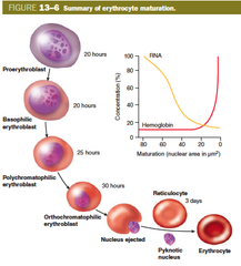
• Production of RBCs
• Regulated primarily by the hormone erythropoietin
• Reticulocytes are immature RBCs that enter the bloodstream (take 1-2 days to mature into functional RBCs)
• Reticulocyte "retic" count indicates the rate of RBC production (about 0.5- 1.5% of RBCs)
Erythropoietin
• Low blood oxygen levels stimulate the kidneys to release erythropoietin
• Promotes RBC production in the bone marrow
• also released slowly by the liver under normal conditions

anemia
• Condition in which the ability of the blood to carry oxygen is reduced • Caused by decrease in the amount of RBCs or Hb in the blood
Polycythemia
abnormal increase in red blood cells, causing thickened, slow-flowing blood that increases the risk of clots, stroke, and heart attack
Type A
RBCs with type A surface antigens and plasm with anit-B antibodies
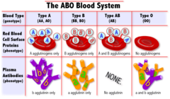
Type B
RBCs with type B surface antigens and plasm with anti-A antibodies
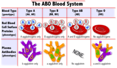
Type AB
RBCs with both type A and B surface antigens and neither anti-A nor anti-B antibodies

Type O
RBCs with neither type A nor B surface antigens but both anti-A and anti-B antibodies

Rh factor
• Most Americans are Rh+ = means they carry the Rh antigen
• Rh- individuals do not have the Rh antigen
• A person who is Rh+ can receive blood from Rh+ and Rh- individuals
• A person who is Rh- can only receive blood from Rh- individuals
• Anti-Rh antibodies are not automatically formed in the blood of Rh-negative individuals (unlike the antibodies of the A B O system)
• If an Rh+ person receives Rh- blood, there is usuallyno transfusion reaction
• If an Rh- person receives Rh+ blood:
• First transfusion - immune system will becomesensitized and produces antibodies - reaction does notoccur
• Subsequent transfusions - antibodies made during thefirst transfusion will attack the Rh factor - reactionoccurs
• If a Rh- person has a Rh+ baby:
• First pregnancy - immune system builds up antibodies- usually no issues
• Subsequent pregnancies - immune system attacks theRh+ fetus• RhoGAM is an injection used to prevent the build up ofRh+ antibodies

hemolytic disease of the newborn
a blood disorder occurring when maternal antibodies cross the placenta and destroy fetal red blood cells due to ABO or Rh incompatibility
Blood transfusion compatibility

Leukocytes (WBCs)
• Leukopoiesis = formation of whiteblood cells in the red bone marrow
• Functions
• Protect the body from invadingmicroorganisms
• Remove dead cells and debris fromthe body
• Each WBC type has specific functionsrelated to immunity

granulocytes
have large granules and lobed nuclei
arganulocytes
have granules that are not clearly visible
neutrophils (granulocytes)
% of WBCs
• 55-70%
Location
• Produced in red bone marrow & then enter blood circulation
• Don't stay in circulation long - usually enter the tissues after about 10-12 hours
Functions
• Phagocytize bacteria, antigen-antibody complexes and other foreign matter
• Secrete lysozyme - enzyme that acts as antimicrobial agent
Average Lifespan (in Tissues)
• 1-2 days

eosinophils (granulocytes)
% of WBCs
• 1-4%
Location
• Produced in red bone marrow & then enter blood circulation
• Usually only leave circulation & enter tissues during inflammatory response
Functions
• Prevalent in allergic reactions
• Destroy inflammatory chemicals (i.e. histamine)
• Release chemicals that destroy parasitic worm infections
Average Lifespan (in Tissues)
• 2-5 days

basophils (granulocytes)
% of WBCs
• 0.5-1% = rarest WBC
Location
• Produced in red bone marrow & then enter blood circulation
• Usually only leave circulation & enter tissues during inflammatory response
Functions
• Prevalent in allergic reactions
• Produce histamine (chemical that enhances inflammation)
• Produce heparin (anticoagulant)
Average Lifespan (in Tissues)
• 1-2 days

lymphocytes (arganulocyte)
% of WBCs
• 20-40%
Location
• Produced in red bone marrow
• B lymphocytes (B cells) proliferate in the bone marrow
• T lymphocytes (T cells) leave the bone marrow and proliferate in the thymus
Functions
• B cells produce antibodies (immune protein)
• There are a variety of T cells that function to destroy virus-infected and cancer cells
• Both B and T cells produce memory cells - important forfuture exposure to recognized pathogens
Average Lifespan (in Tissues)
• Depends on the cell, some live for only a few days and some live for many years
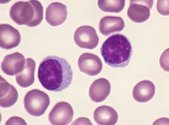
monocytes (arganulocyte)
% of WBCs
• 2-8%
• Note: These are the largest WBCs (size)
Location
• Remain in blood circulation for about 3 days
• Leave circulation and become macrophages
Functions
• Phagocytes
• Break down antigens and present them to lymphocytes for recognition
Average Lifespan (in Tissues)
• Once they become macrophages, they can live months to years

diapedesis
• Cells leave the blood stream
• Cells become thin, elongated, and stick to the vessel walls (pavementing)
• The cells then squeeze out between the endothelial cells of the capillaries (margination)
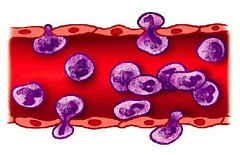
chemotaxis
• Attraction to and movement toward foreign materials and damaged cells
• Ability to travel to the site of infection
phagocytosis
• engulf anything at the site of infection that doesn't belong (damaged cell debris, pathogens, etc.)
• happens in neutrophils and monocytes

Differential White Blood Cell Count
• A differential WBC count is a standard blood test that identifies the percentage of each type of WBC present in the patient's blood
• This is important to help identify abnormal levels of specific blood cell populations - helpful for making diagnoses
• Normal = 5,000 to 10,000 per mictoliter of blood
• leukocytosis - elevated WBC count
• leukopenia - low WBC count
Thrombopoiesis
• formation of platelets in the red bone marrow
• Platelets are fragments of a larger cell called a megakaryocyte

Thrombocytes
• Have surface glycoproteins and proteins that allow for adhesion to other molecules like collagen
• Important in preventing blood loss:
• Produce platelet plugs
• Promotes the formation and contraction of blood clots
• Lifespan = 5-9 days

hemostasis
prevent excessive blood loss

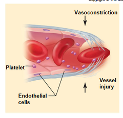
vascular spasm
• Vasoconstriction of the injured blood vessel immediately after injury - reduces blood flow to the area
• Triggered by:
• Injury to vascular smooth muscle
• Chemicals released by platelets
• Reflex actions triggered by pain receptors
• Blood vessel compression caused by build up of escaped blood insurrounding tissues
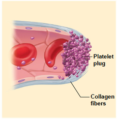
platelet plug formation
• Formation of a temporary seal shortly after injury occurs
• This is not a clot - that comes later
• Three major steps:
• Platelet adhesion
• Platelet release reaction
• Platelet aggregation - platelets change shape & express receptors that can bind to fibrinogen (protein in clotting)
Coagulation
• Coagulation is the last stage of hemostasis
• Involves several clotting factors:
• Proteins found in the blood plasma
• Circulate in an inactive state until tissue injury occurs
• Damaged tissues & platelets produce chemicals that will begin the activation of the clotting factors
• Result is a blood clot - a network of threadlike fibrin fibers, trapped blood cells, platelets & fluid
Clotting Factors
• Factor X is the clotting factor that initiates the common pathway
• Vitamin K is required for the formation of most of the clotting factors

fibrin
an insoluble protein formed from fibrinogen during the clotting of blood
Clot Dissolution (Fibrinolysis)
Process in which clots are removed after repair is completed
Substances affecting clotting
heparin and anticoagulants
functions of the lymphatic system
Fluid balance
• Excess interstitial fluid enters lymphatic capillaries & becomes lymph
Lipid absorption
• Absorption of fat and other substances from thedigestive tract via lacteals
• Fatty lymph is called chyle
Defense
• Microorganisms & other foreign substances are filtered from the lymph by lymph nodes
• Spleen filters the blood
Lymph compostition
large amount of water, proteins, lipids, and other materials
Lymph circulation
• All lymphatic vessels function to carry fluid away from the tissues to ultimately return it to the blood (similar to veins)
• There is NO pump to move lymph through the vessels so other mechanisms are needed to move lymph against gravity:
• One-way valves prevent backflow
• Contraction of smooth muscle in vessel walls
• Skeletal muscle contractions during movement
• Pressure changes during breathing
Lymph flow
• lymp capillaries to lymph vessels to lymph trunks to lymph ducts
Lymphatic capillaries
• More permeable than blood capillaries
• Flap-like endothelial cells function as one-way valves for fluid to enter the capillaries
• Blind-ended = they just end,they don't create a closed circuit like blood vessels

Lymphatic vessels
• Lymphatic capillaries join together to form larger lymphatic vessels
• Very structurally similar to veins
• Along the length of the lymphatic vessels there are lymph nodes that filter the lymph

Lymphatic trunks
• Lymphatic vessels join to form even larger vessels called lymphatic trunks
• Drain specific large areas of the body
• Some carry lymph directly to the veins, others join to form lymphatic ducts

Lymphatic ducts
• Lymphatic ducts return lymph to the bloodstream at the junction of the internal jugular and subclavian veins

Right lymphatic duct
• Right lymphatic duct drains the:
• Right side of the head
• Right upper limb
• Right side of the thorax

Thoracic duct
Thoracic duct drains the rest of the body

lymph nodes
• Small, round/bean-shaped structures 1 to 25 mm long
• Responsible for filtering lymph
• Found both deep and superficially
• Superficial lymph nodes area bundant in the axillary, cervical and inguinal regions
• Substances removed by phagocytosis or stimulate lymphocytes to proliferate in germinal centers
spleen
• Located in left upper quadrant of the abdomen - tucked behind the stomach
• Filters BLOOD (not lymph)
• Functions:
• Contains macrophages - destroys old & defective RBCs & removes other debris from the blood
• Stores breakdown products RBCs to be recycled
• Stores platelets
• Site of RBC production in the fetus

thymus
• Bilobed gland located in the superior part of the mediastinum (just anterior to the heart)
• Grows rapidly in first year of life, and then gradually atrophies as we age
• Establishes its part of the immune system in first year of life (works the hardest)
• Maintains its part of the immune systemfor the rest of life (less use = atrophy)
• Site of the maturation of T cells
• Secretes thymosin which is important in T cell development

tonsils
• Groups of lymphatic nodules associated with the digestive & respiratory mucosa (part of the MALT)
• Provide protection against pathogens that enter through the nose and mouth
• Form a ring around the boarder between the oral cavity and pharynx
• One large pharyngeal tonsil (adenoids)
• Two palatine tonsils (sides of throat)
• One large lingual tonsil (base of tongue)

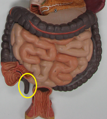
appendix
• Worm-like structure that hangs off the cecum of the large intestine (proximal end of the large intestine)
• Contains a large number of lymphoid follicles
• Have the same function has Peyer's patches
• Because of its structure, the appendixis prone to trapping pathogens and becoming infected
Peyer's patches
• Groups of lymphoid nodules located in the distal end of the small intestine
• Structurally similar to tonsils
• Destroy pathogens within the intestine to prevent them from passing through the intestinal wall into the lymph, blood or tissue spaces

lacteals
in the small intestine and absorbs lipids (forming chyle)
innate immunity
• Natural defenses presentat birth
• Provides a broad, nonspecific defense againsta variety of pathogens
mechanical (physical) barriers (innate)
• Skin
• Mucous membranes
• Mucus traps microorganisms
• Hairs and cilia lining respiratory tract, nose hairs
• Coughing and sneezing
• Mechanical flushing includes urination, vomiting, defecation, salivation, and tear production
chemical barriers (innate)
• Also called chemical mediators - includes certain chemicals that destroy or prevent the growth of bacteria and viruses:
lysozyme
interferons
histamines & kinins
acids
complement
pyrogens
cytokines
general responses (innate)
acting within minutes to hours via inflammation, phagocytosis, and complement system activation without long-term memory
Inflammation functions
• Triggered in response to tissue injury
• Attempt to repair damage, prevent spread of infection, and neutralize toxins
local inflammation
confined to a specific area of the body
• Cardinal signs of inflammation: redness, heat, swelling, pain & loss of function
systemic inflammation
occurs in many parts of the body
• Local symptoms at inflammation sites
• Increased neutrophil numbers released by red bone marrow
• Fever improves performance of the immune system
Signs of Inflammation
heat, redness, swelling, pain, loss of function
Inflammation mediators
histamine - vasodilation & increased vascular permeability during inflammation
prostaglandins - promote inflammation
cytokines - proteins secreted by cells that bind to receptors on cell surfaces
Adaptive immunity
• This is the part of the immune system that develops over our lifetime as we are exposed tovarious antigens
• Provides defense against specific substances
• Has memory - the ability to remember previous encounters with a specific substance to allow for a more rapid response
• Adaptive immunity develops as a result of exposure to antigens
Antibody Mediated Immunity
• Also referred to as humoral immunity
• Involves B Lymphocytes
• Most effective against bacteria in the body fluids
• Specialized B cells produce antibodies
plasma cells
produced antibodies (activated by B cells) in response to antigen exposure
Cell Mediated Immunity
• Involves T Lymphocytes
• Most effective against parasites, viruses & cancer cells
• Bind to an antigen and then divide to form several types of T cells
cytoxic T cell
destroy cells by lysis or by producing cytokines
helper T cell
activates B cels and cytotoxic T cells
regulatory T cell
inhibits the actions of B cells, helper T cells, and cytoxic T cells
memory T cell
• Remembers a previously encountered antigen
• Allows for more rapid response if the antigen is encountered again
• Memory cells are what provide adaptive immunity
dendritic cell
• Process (identify) antigens
• Involved in the activation of B cells and T cells
antibodies
• A protein molecule produced during specific immunity
• Binds to a specific antigen and renders it harmless = antigen-antibody complexes
antigens
• Molecules that trigger specific immune responses from the body
• Pathogen = disease causing antigen
• Normally the body can recognize self-antigens and non-self antigens
natural immunity
arises from direct, active infection with a pathogen, often providing strong, long-lasting, but risk-prone protection
artificial immunity
acquired safely through vaccinations or antibody transfers, providing targeted protection without the danger of contracting the disease
hypersensitivity reactions (allergy)
• An overreaction to an antigen that causes injury to body tissues