review Ch 5 Erythrocyte Production and Destruction
1/52
There's no tags or description
Looks like no tags are added yet.
Name | Mastery | Learn | Test | Matching | Spaced |
|---|
No study sessions yet.
53 Terms
RBCs
are formally called erythrocytes
erythroblasts or normoblasts
Nucleated RBC precursors, normally restricted to the bone marrow
normoblast
which refers to developing nucleated RBC precursors (i.e., blasts) with normal appearance.
1. Normoblastic
2.Rubriblast (parallels the nomenclature used for granulocyte)
3.Erythroblast
Three nomenclatures are used for naming erythroid precursors.
Burst Forming Unit - Erythroid (BFU-E)
The earliest committed progenitor gives rise to large colonies because they are capable of multisubunit colonies (called bursts)
colony-forming unit-erythroid (CFU-E)
gives rise to smaller colonies
pronormoblast
is the first morphologically identifiable RBC precursor.
three to five divisions before maturing further
at the CFU-E stage, the cell completes approximately:
18 to 21 days
how many days are required to produce a mature RBC from the BFU-E?
8 to 32 mature RBCs usually result
How many Erythrocytes may result from a single pronormoblast?
1. peripheral blood film or
2. bone marrow smear
Morphologic identification of blood cells can be on :
Wright or Wright-Giemsa
stain for identification of blood cells are:
1. nuclear chromatin pattern (texture, density, homogeneity)
2. nuclear diameter
3.nucleus-to-cytoplasm (N:C) ratio
4.presence or absence of nucleoli
5.cytoplasmic color
The most important features in the identification of RBCs are:
1. Overall diameter of the cell decreases
2. Diameter of the nucleus decreases more rapidly than does the diameter of the cell. As a result, the N:C ratio also decreases.
3. 3. Nuclear chromatin pattern becomes coarser, clumped, and condensed.
It becomes even coarser and more clumped as the cell matures, developing a raspberry-like appearance, in which the dark staining of the chromatin is distinct from the almost white appearance of the parachromatin.
This chromatin/ parachromatin distinction is more dramatic than in other cell lines. Ultimately the nucleus becomes quite condensed, with no parachromatin evident at all, and the nucleus is said to be pyknotic. CFU
4. Nucleoli disappear. Nucleoli represent areas where the ribosomes are formed and are seen early in cell development as cells begin actively synthesizing proteins.
As erythroid precursors mature, nucleoli disappear, which precedes the ultimate cessation of protein synthesis.
5. Cytoplasm changes from blue to gray-blue to salmon pink.
-Blueness or basophilia
-eosinophilia or acidophilia
As erythroid precursors mature, several general trends affect their appearance:
pyknotic.
The term used to describe a nucleus that has died. The chromatin has become densely compacted, so no pattern is visible. the nucleus becomes quite condensed, with no parachromatin evident at all.
Blueness or basophilia
is due to acidic components that attract basic stains, such as methylene blue. the degree of it correlates with the amount of ribosomal RNA. When ribosomes and other organelles decline over the life of the developing
erythroid precursor, and the _____________fades.
eosinophilia or acidophilia
Pinkness of erythrocyte due to accumulation of more basic components that attract acid stains, such as eosin
Eosinophilia of erythrocyte cytoplasm
correlates with the accumulation of hemoglobin as the cell matures
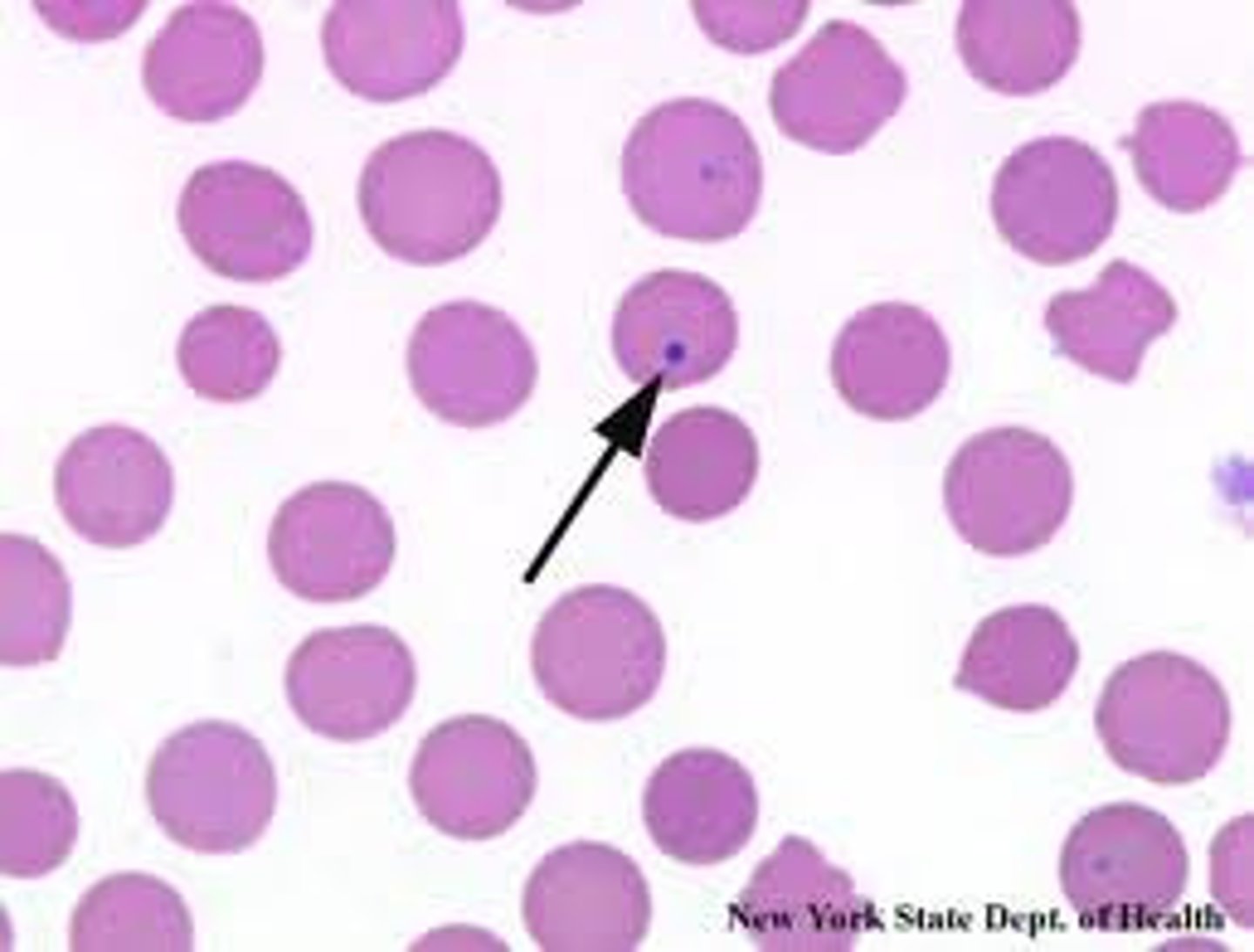
reticulocyte
immature erythrocyte
size:12 to 20 μm
Nucleus
- The nucleus takes up much of the cell
(N:C ratio of 8:1)
- The nucleus is round to oval, containing one or two nucleoli. The purple red chromatin is open and contains few, if any, fine clumps.
Bone
cytoplasm
- The cytoplasm is dark blue because of the concentration of ribosomes and RNA.
- The Golgi complex may be visible next to the nucleus as a pale, unstained area.
- Pronormoblasts may show small tufts of irregular cytoplasm along the periphery of the membrane.
Pronormoblast characteristics
size: 10 to 15 μm
Nucleus
- The chromatin begins to condense
-N:C ratio decreases to about 6:1
Cytoplasm
- When stained, the cytoplasm may be a deeper, richer blue than in the pronormoblast
location
- bone marrow
cellular activity
- Detectable hemoglobin synthesis occurs, but completely mask by many cytoplasmic organelles
lenght
- slightly more than 24h
basophilic normoblast characteristics:
size: 10 to 12 μm
Nucleus
- The condensation of chromatin
reduces the diameter of the nucleus considerably, so the N:C ratio decreases from 4:1 to about 1:1 by the end of the stage.
- Notably, no nucleoli are present.
cytoplasm
- mixture of pink and blue, resulting in a mixture of pink and blue, resulting in a murky gray-blue
division
- the last stage in which the cell is capable of
undergoing mitosis
location
-is present only in the bone marrow
cellular activity
- Hemoglobin synthesis increases, and
the accumulation begins to be visible as a pinkish color in the cytoplasm.
- Cellular RNA and organelles are still present,
particularly ribosomes, which contribute a blue color to the cytoplasm.
-The progressive condensation of the nucleus and disappearance of nucleoli are evidence of progressive decline in transcription of deoxyribonucleic acid (DNA).
lenght
- 30h
characteristics of Polychoromatic Normoblast
size: 8 to 10 μm
Nucleus
- The nucleus is completely condensed (i.e., pyknotic)
- As a result, the N:C ratio is low or approximately 1:2.
cytoplasm
- The increase in the salmon pink color of the
cytoplasm reflects nearly complete hemoglobin production.
division
- is not capable of division because of the condensation of the chromatin.
location
-bone marrow
cellular activity
- Hemoglobin production continues on the
remaining ribosomes using messenger RNA produced earlier. Late in this stage, the nucleus is ejected from the cell.
-The nucleus moves to the cell membrane and into a pseudopod-like projection
-loss of vimentina protein responsible for holding organelles in proper location in the cytoplasm, is probably important in the movement of the
nucleus to the cell periphery
-Ultimately the nucleus-containing
projection separates from the cell by having the membrane seal and pinch off the projection with the nucleus enveloped by cell
membrane
- The enveloped extruded nucleus,
called a pyrenocyte is then engulfed by bone marrow macrophages.
lenght
- 48 hours
Orthochromic Normoblast characteristics:
phosphatidylserine
The macrophages recognize ______________on the pyrenocyte surface as an "eat me" flag. Other organelles are
extruded and ingested in similar fashion.
Howell-Jolly bodies
small fragments of nucleus are left behind if the projection is pinched off before the entire nucleus is enveloped
size: 8 to 8.5 μm
Nucleus
- no nucleus
- when a cell loses its nucleus,
regardless of cytoplasmic appearance, it is a polychromatic erythrocyte.
Cytoplasm
-the cell is the same color as a mature RBC, salmon pink.
-It remains larger than a mature cell, however. - - The shape of the cell is not the mature biconcave disc but is irregular in electron micrographs
division
- no division
location
-Bone marrow (2 days)
-peripheral blood or sequestered by spleen (1 day)
cellular activity
- completesproduction of hemoglobin from a small amount of residual messenger RNA using the remaining ribosomes
- small amount of residual ribosomal RNA is present
-The residual
ribosomes appear as a mesh of small blue strands, a reticulum,
or, when more fully digested, merely blue dots
lenght
-3 days
Polychromatic Erythrocyte
Endoribonucleas
digests the ribosomes in Polychromatic Erythrocyte
reticulocyte
When so stained, the polychromatic erythrocyte is called_____________________
However, the name reticulocyte is often used to refer
to the stage immediately preceding the mature erythrocyte.
the spleen
RBC after 120 are removal by:
Erythrokinetics
is the term describing the dynamics of RBC production and destruction.
erythron
is the name given to the collection of all stages of erythrocytes throughout the body. It conveys the concept of a unified functional tissue.
- is the entirety of erythroid cells in the body,
RBC mass
cells in circulation.
- refers only to the cells in circulation
hypoxia
- it is too little tissue oxygen
- is detected by the peritubular fibroblasts of the kidney
erythropoietin (EPO)
-the major stimulatory cytokine for RBCs.
-1% of RBCs that normally die each day
hypoxiainducible factors (HIFs)
a family of transcription factor proteins that increase the EPO production
binding to kidney hypoxia responsive elements located at the 5' flanking region of the EPO gene
This results in
- increased EPO gene transcription,-
- EPO production, and
ultimately increased RBC production
HIFs respond to hypoxia by:
- is a thermostable, nondialyzable, glycoprotein hormone with a molecular weight of 34 kD.
-It consists of a carbohydrate unit and a terminal sialic acid unit, both of which
play a role in the biologic activity of the hormone.
Structure of EPO
--is a true hormone, being produced at one location (kidney) and acting at a distant location (bone marrow).
- It is a growth factor (or cytokine) that initiates an intracellular message to the developing erythroid cells; this process is called signal transduction
EPO in action:
EPOR
EPO (the ligand) must bind to its receptor____________on the surface of EPO-responsive immature erythroid cells to initiate the signal or message
sensitivity to EPO
EPOresponsive cells vary in their ______________________as some are able
to respond to low levels of EPO, whereas others require higher
levels
The EPO receptor
is a transmembrane protein homodimer with extracellular and cytoplasmic domains
in a change in the conformation of the receptor. This activates Janusactivated
tyrosine kinase 2 (JAK2) signal transducers that are associated with the cytoplasmic domains of the EPO receptor. JAK2 then activates downstream signal transduction pathways
(such as the signal transduction and activator of transcription 5 or STAT5 pathway) that ultimately promotes transcription of
specific genes in the RBC nucleus
The binding of EPO to the extracellular domain of the EPO receptor (on erythrocyte progenitors and early precursors) results in:
(1) allowing early release of reticulocytes from
the bone marrow,
(2) preventing apoptotic cell death, and
(3) reducing the time needed for cells to mature in the bone marrow
three major effects of EPO:
1. EPO induces changes in the adventitial cell
layer of the bone marrow/sinus barrier that increase the width of the spaces for RBC egress into the sinus
RBCs are held in the marrow because they express surface membrane receptors for adhesive molecules located on the
bone marrow stroma, such as fibronectin
2. EPO downregulates the expression of these receptors so that cells can
exit the marrow earlier than they normally would.
Early release of reticulocytes its caused by two mechanisms
shift reticulocytes
Reticulocytes that are released from the marrow prematurely.
-are still very basophilic because they have not spent as much
time degrading their ribosomes and RNA or making hemoglobin
as they normally would before entering the bloodstream.(polychromasia)
- nucleated RBCs (i.e., erythroblasts or normoblasts) can be released early in cases of extreme anemia when the demand for RBCs in the peripheral circulation is great
-it is limited in effectiveness because the available precursors
in the marrow are depleted within several days and still may
not be enough to meet the need in the peripheral blood for more
cells..
Inhibition of apoptosis
A second, and probably more important, mechanism by which EPO increases the number of
circulating RBCs is by increasing the number of cells that will
be able to mature into circulating erythrocytes. It does this by
_________________, the programmed death of RBC progenitors.
Apoptosis: programmed cell death
-the degradation of chromatin
into fragments of varying size that are multiples of 180 to 185 base pairs long
- protein clustering
- activation of transglutamase
-is not associated with inflammation
-condensation of the nucleus
-causing increased basophilic staining of the chromatin
-nucleolar disintegration
-shrinkage of cell volume with concomitant
increase in cell density and compaction of cytoplasmic organelles, whereas mitochondria remain normal
-This is followed by
a partition of cytoplasm and nucleus into membrane-bound apoptotic bodies that contain varying amounts of ribosomes, organelles, and nuclear material.
-The last stage of degradation
produces nuclear DNA fragments consisting of multimers of 180 to 185 base pair segments.
morphologic changes in apoptosis:
macrophages
The apoptotic cell contents remain membrane
bound and are ingested by____________ which prevents
an inflammatory reaction.
One effect of EPO is an indirect avoidance of apoptosis by removing an apoptosis
induction signal inside or outside of the cell.
Evasion of apoptosis by erythroid progenitors and precursors.
external messaging apoptosis: the death receptor Fas on the membrane of the earliest erythroid precursors, whereas its ligand, FasL, is expressed by more mature erythroid precursors
the younger Fas-positive precursors are allowed to develop, which increases the overall output of RBCs from the marrow.
FasL-bearing cells are depleted
direct EPO rescue from apoptosis
the major way in which EPO is able to increase RBC production
-When EPO binds to its receptor on the CFU-E, one of the
effects is to reduce production of Fas ligand.32 Thus the younger
cells avoid the apoptotic signal from the older cells