2. Cellular Adaptations to Stress

0.0(0)
learnLearn
examPractice Test
spaced repetitionSpaced Repetition
heart puzzleMatch
flashcardsFlashcards
Card Sorting

1/20

Study Analytics
Name
Mastery
Learn
Test
Matching
Spaced

No study sessions yet.

21 Terms

1
New cards

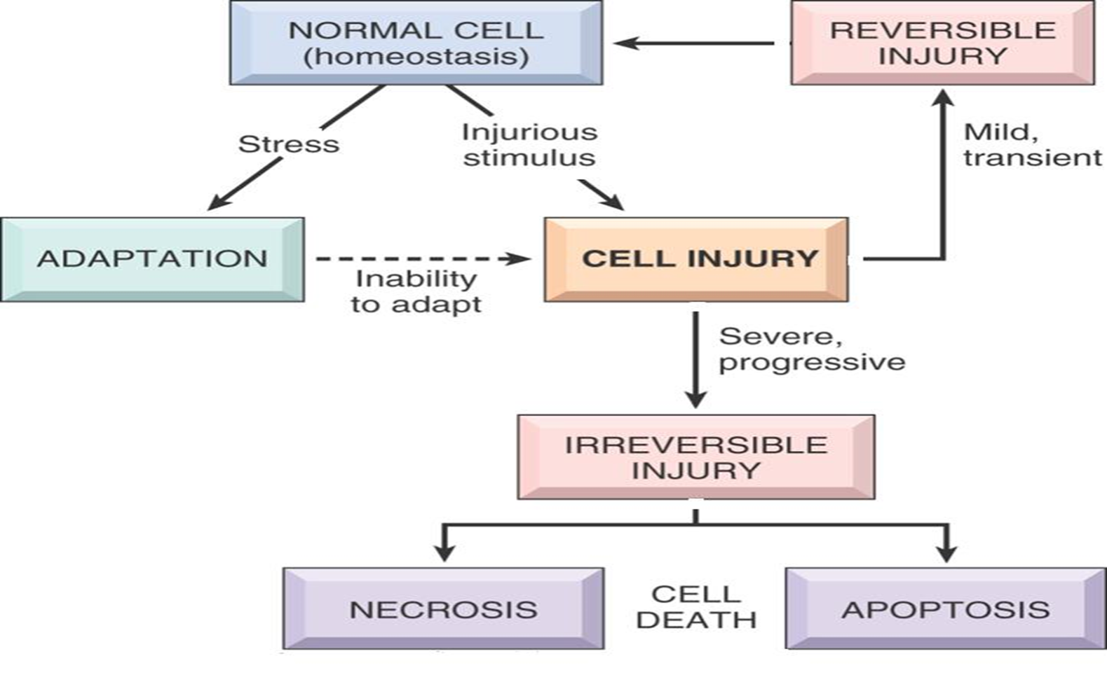
Describe the stages in the cellular response to stress and injurious stimuli.

  • Normal cell undergoes stress, adapts to the stress

  • If the stress goes on for long, it will no longer be able to adapt and cell injury occurs

  • If a normal cell encounters an injurious stimulus, cell injury occurs

  • In both cases, if cell injury is mild/transient, it is reversible and the cell returns back to normal (homeostasis)

  • If the cell injury is severe and progressive, it becomes an irreversible injury

  • This leads to cell death by either necrosis or apoptosis
<ul><li><p><strong>Normal </strong>cell undergoes <strong>stress</strong>, <strong><mark data-color="blue" style="background-color: blue; color: inherit">adapts</mark></strong> to the stress</p></li><li><p>If the stress goes on for <strong>long</strong>, it will <strong>no longer be able to adapt</strong> and <strong><mark data-color="blue" style="background-color: blue; color: inherit">cell injury occurs</mark></strong></p></li><li><p>If a normal cell encounters an <strong>injurious stimulus,</strong> cell injury occurs</p></li><li><p>In both cases, if cell injury is <strong><mark data-color="blue" style="background-color: blue; color: inherit">mild/transient</mark></strong>, it is <strong><mark data-color="blue" style="background-color: blue; color: inherit">reversible</mark></strong> and the cell <strong>returns back to normal (homeostasis)</strong></p></li><li><p>If the cell injury is <strong><mark data-color="blue" style="background-color: blue; color: inherit">severe</mark></strong> and <strong><mark data-color="blue" style="background-color: blue; color: inherit">progressive,</mark></strong> it becomes an <strong><mark data-color="blue" style="background-color: blue; color: inherit">irreversible injury</mark></strong></p></li><li><p>This leads to <strong>cell death</strong> by either <strong><mark data-color="blue" style="background-color: blue; color: inherit">necrosis</mark></strong> or <strong><mark data-color="blue" style="background-color: blue; color: inherit">apoptosis</mark></strong></p></li></ul><p></p>
2
New cards

Define and describe cellular adaptation.

  • A state lies between normal cell and injured/overstressed cell

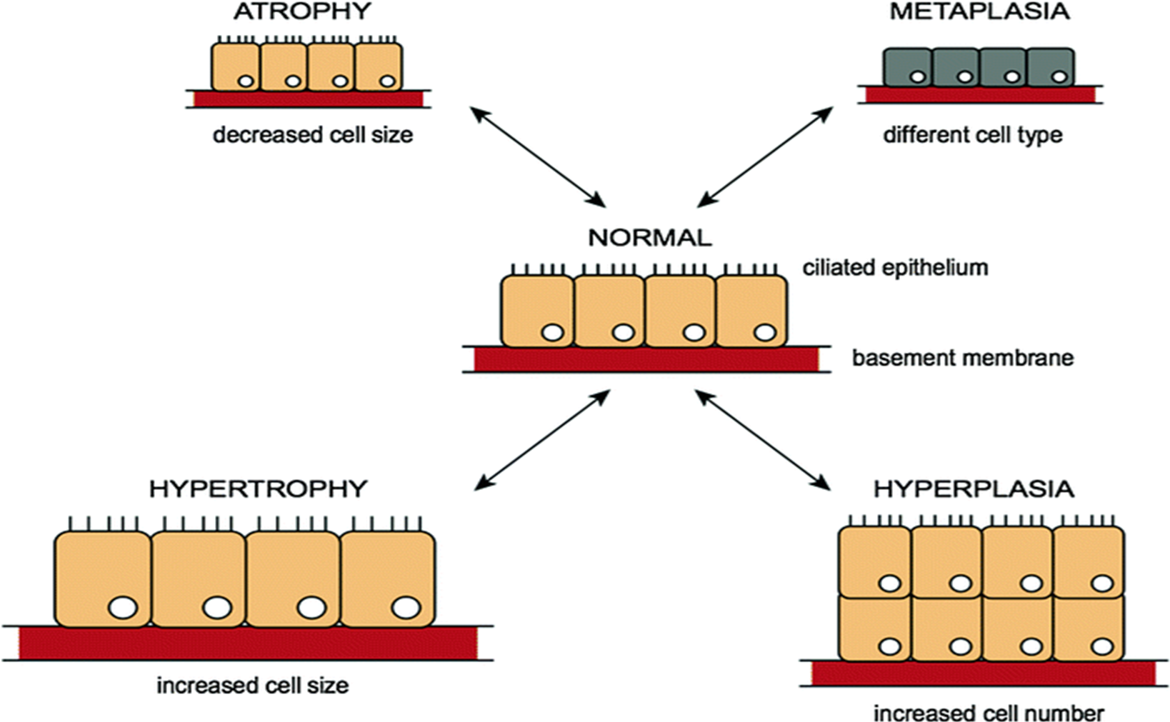
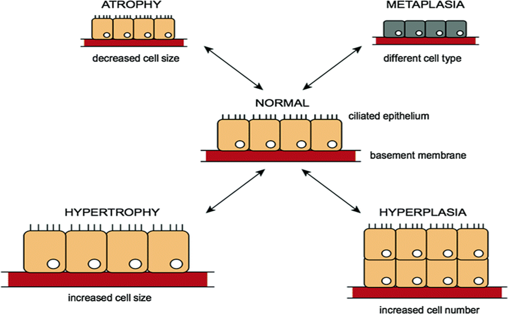
  • Adaptations are reversible changes in the size, number, phenotype, metabolic activity, or functions of cells in response to changes in their environment

3
New cards

What are the responses of cells to stress/injury?

  1. Adaptive responses

    • Atrophy

    • Hypertrophy

    • Hyperplasia

    • Metaplasia

  2. Cell injury

    • Reversible

    • Irreversible (cell death; necrosis, apoptosis)

4
New cards

Define atrophy.

  • Decrease in size of an organ due to decrease in size and no. of its cells

  • Decreased cell size

<ul><li><p>Decrease in size of an organ due to decrease in size and no. of its cells</p></li><li><p><strong><mark data-color="blue" style="background-color: blue; color: inherit">Decreased cell size</mark></strong></p></li></ul><p></p>
5
New cards

What are the two categories that atrophy is split into?

Physiologic atrophy

Pathologic atrophy

6
New cards

Physiological atrophy is normal. Give examples of physiologic atrophy.

  • Some embryonic structures undergo atrophy during development; e.g. thyroglossal duct

  • Uterus decreases in size shortly after menopause

7
New cards

Pathologic atrophy depends on underlying cause and can be local or generalized. What are the common causes/types of atrophy?

  1. Decreased workload

  2. Loss of nerve supply/innervation

  3. Diminished blood supply

  4. Inadequate nutrition

  5. Loss of endocrine stimulation

  6. Pressure

8
New cards

Describe decreased workload (atrophy of disuse).

  • When broken limb is immobilized in plaster cast/when patient is on bed rest, skeletal muscle atrophy appears

  • Is reversible once activity is resumed

9
New cards

Describe loss of innervation (denervation atrophy).

  • Normal function of skeletal muscle is dependent on its nerve supply

  • Damage to nerves leads to rapid atrophy of muscle fibers supplied by those nerves

10
New cards

Describe diminished blood supply.

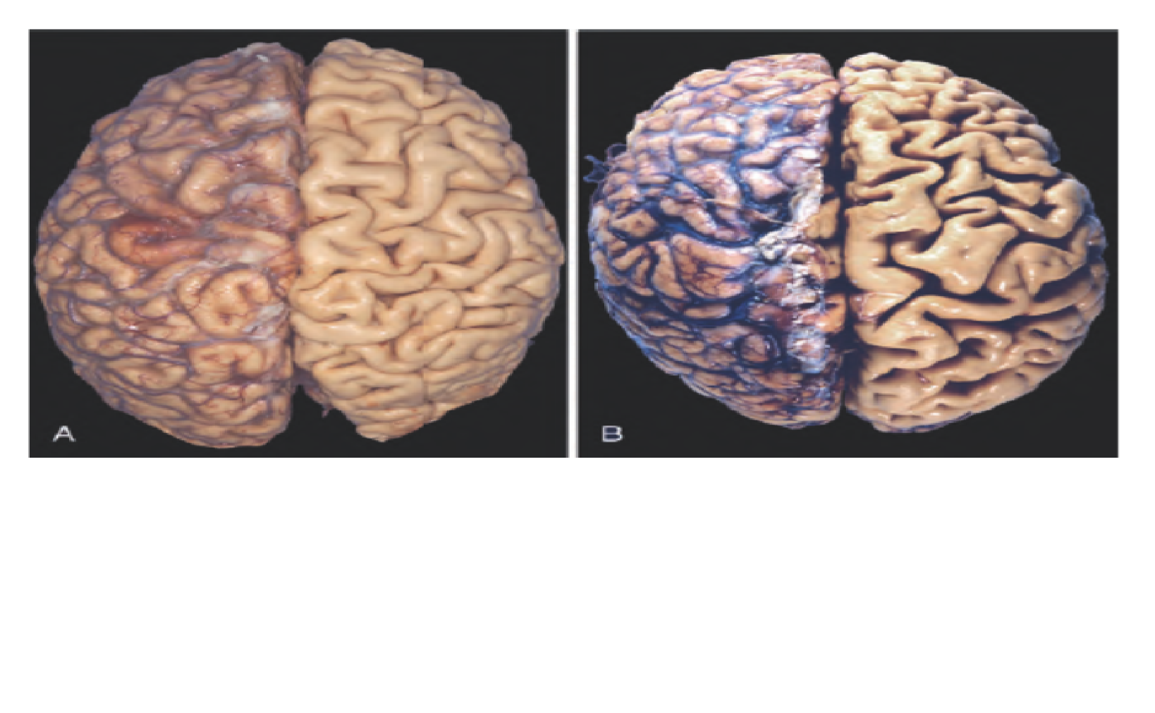
  • Decrease in blood supply to tissue because of arterial occlusive disease results in atrophy of tissue because of progressive cell loss

  • In late adult life, brain undergoes progressive atrophy as atherosclerosis narrows its blood supply
<ul><li><p>Decrease in blood supply to tissue because of <strong><mark data-color="blue" style="background-color: blue; color: inherit">arterial occlusive disease</mark></strong> results in atrophy of tissue because of <strong><mark data-color="blue" style="background-color: blue; color: inherit">progressive cell loss</mark></strong></p></li><li><p>In late adult life, <strong>brain </strong>undergoes <strong><mark data-color="blue" style="background-color: blue; color: inherit">progressive atrophy</mark></strong> as <strong><mark data-color="blue" style="background-color: blue; color: inherit">atherosclerosis</mark> </strong>narrows its blood supply</p></li></ul><p></p>
11
New cards

Describe inadequate nutrition.

  • Severe protein-calorie malnutrition (marasmus)

  • Causes cachexia in chronic inflammatory diseases and cancer

12
New cards

Describe loss of endocrine stimulation.

  • Loss of estrogen stimulation after menopause results in physiologic atrophy of endometrium, vaginal epithelium, and breast

  • It is pathologic atrophy in YOUNG women

13
New cards

Describe pressure atrophy.

  • Tissue compression for any length of time can cause atrophy

  • An enlarging benign tumor can cause atrophy in surrounding compressed tissues

14
New cards

Describe hyperplasia.

  • Increase in size of an organ due to increase in number of cells

  • Cells capable of dividing undergo hyperplasia

  • Hyperplasia may be physiologic or pathologic
<ul><li><p><strong><mark data-color="blue" style="background-color: blue; color: inherit">Increase in size</mark></strong> of an organ due to <strong><mark data-color="blue" style="background-color: blue; color: inherit">increase in number of cells</mark></strong></p></li><li><p>Cells <strong>capable of dividing</strong> undergo hyperplasia</p></li><li><p>Hyperplasia may be <strong><mark data-color="blue" style="background-color: blue; color: inherit">physiologic or pathologic</mark></strong></p></li></ul><p></p>
15
New cards

Give examples of hyperplasia being physiologic.

  1. Enlargement of breast during puberty and pregnancy

  2. Enlargement of pregnant uterus

  3. Compensatory hyperplasia

    • Liver regeneration after donation of a lobe for transplantation

    • Rapid bone marrow hyperplasia in response to blood donation

16
New cards

Give examples of hyperplasia being pathologic.

  1. Enlargement of prostate in benign nodular hyperplasia in elderly males

  2. Endometrial hyperplasia due to high levels of circulating estrogen

  3. Certain viral infections, e.g. papillomaviruses, which cause skin warts (hyperplastic epithelium)
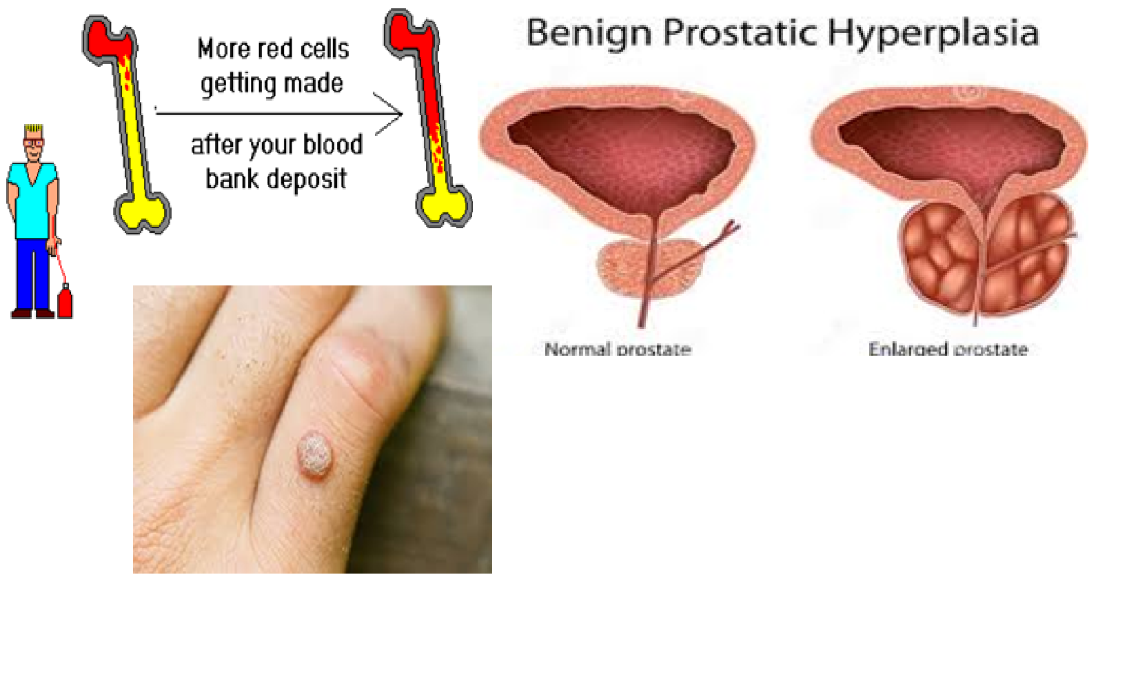
<ol><li><p><strong><mark data-color="blue" style="background-color: blue; color: inherit">Enlargement of prostate</mark></strong> in <strong><mark data-color="blue" style="background-color: blue; color: inherit">benign nodular hyperplasia</mark></strong> in <strong>elderly males</strong></p></li><li><p><strong><mark data-color="blue" style="background-color: blue; color: inherit">Endometrial hyperplasia</mark></strong> due to <strong>high levels of circulating estrogen</strong></p></li><li><p>Certain <strong>viral infections</strong>, e.g. <strong><mark data-color="blue" style="background-color: blue; color: inherit">papillomaviruses</mark></strong>, which cause <strong><mark data-color="blue" style="background-color: blue; color: inherit">skin warts (hyperplastic epithelium)</mark></strong></p></li></ol><p></p>
17
New cards

Describe hypertrophy.

  • Increase in size of an organ due to increase in size of the cells

  • Cells that are not capable of dividing cannot undergo hyperplasia, so they undergo hypertrophy and become larger in size

  • There are no new cells; they just grow in size

  • Hyperplasia and hypertrophy may occur together

  • Hypertrophy may be physiologic or pathologic

18
New cards

Give examples of hypertrophy being physiologic.

  1. Enlargement of skeletal muscles after exercise in bodybuilders

  2. Breast and uterus also undergo hypertrophy along with hyperplasia in pregnancy

19
New cards

Give an example of hypertrophy being pathologic.

Enlargement of wall of left ventricle of the heart in longstanding hypertension
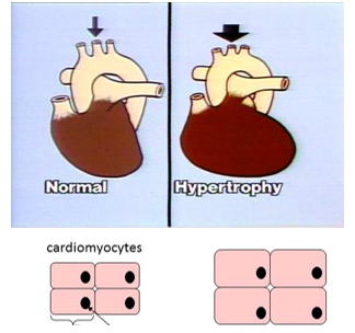
<p><strong><mark data-color="blue" style="background-color: blue; color: inherit">Enlargement of wall of left ventricle of the heart in longstanding hypertension</mark></strong></p>
20
New cards

Define metaplasia.

A reversible change in which one adult cell is replaced by another cell type (function and shape)

21
New cards

Name and describe the 2 types of metaplasia.

  1. Epithelial metaplasia from columnar to squamous epithelium: in habitual cigarette smoker, normal ciliated columnar epithelial cells of trachea and bronchi are often replaced focally or widely by stratified squamous epithelial cells

  2. Epithelial metaplasia from squamous to columnar: occurs in Barrett esophagus, in which esophageal squamous epithelium is replaced by intestinal-like columnar cells, under the influence of refluxed gastric acid
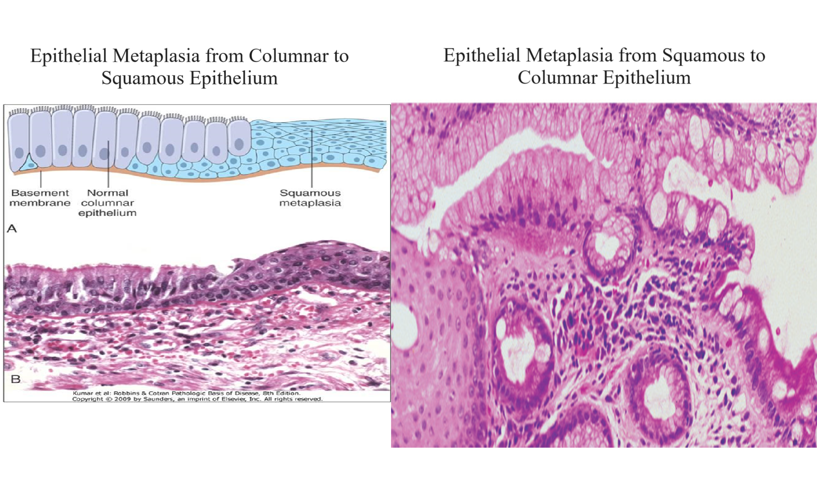
<ol><li><p><strong><mark data-color="blue" style="background-color: blue; color: inherit"><u>Epithelial metaplasia from columnar to squamous epithelium:</u></mark></strong> in habitual cigarette smoker, <strong>normal ciliated columnar epithelial cells</strong> of <strong>trachea </strong>and <strong>bronchi </strong>are often <strong>replaced </strong>focally or widely by <strong>stratified squamous epithelial cells</strong></p></li><li><p><strong><mark data-color="blue" style="background-color: blue; color: inherit"><u>Epithelial metaplasia from squamous to columnar:</u></mark></strong> occurs in <strong>Barrett esophagus</strong>, in which esophageal <strong>squamous epithelium</strong> is <strong>replaced </strong>by<strong> intestinal-like columnar cells</strong>, under the influence of <strong>refluxed gastric acid</strong></p></li></ol><p></p>