biochemistry
 Call Kai
 Learn
Learn Practice Test
 Spaced Repetition
 Match
Match Flashcards
 Knowt Play
1/63

There's no tags or description
Looks like no tags are added yet.
Name | Mastery | Learn | Test | Matching | Spaced | Call with Kai | Chat |
|---|
No analytics yet
Send a link to your students to track their progress
64 Terms
cell death
2 pathways: apoptosis and necrosis, representing programmed and accidental death.
caspase definition
proteolytic enzymes destroy cell components, it contains cysteine and are able to cleave aspartic acid residues (cysteine-aspartic-acid-proteases).
result of cell death
both pathways activation caspases
Disorders of phenylalanine metabolism 3 diseases
PKU
Albinism
Alkaptonuria

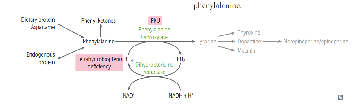
PKU
deficiency of phenylalanine hydroxylase
signs and symptoms: musty smell in urine, CNS, blue eyes, fair hair, pale skin
treatment: phenylalanine restriction, tyrosine supplement essential

Deficiency of BH4 enzyme in PKU
• Often due to defective dihydropteridine reductase
• Also impaired BH4 synthesis → decreased the synthesis of:
• Epinephrine, Norepinephrine, Serotonin
• Dopamine (↑prolactin)
→ Treatment:
• Dietary restriction of phenylalanine
• Supplementation of BH4
• L-dopa, carbidopa → dopamine
• 5-hydroxytryptophan → serotonin
different between deficiency of phenyalanine hydroxylase and BH4 enzyme in PKU
BH4: progressive neurologic injury (eg, axial hypotonia, dystonia, autonomic dysfunction). Dopamine inhibits prolactin → decrease dopa makes prolactin increase
phenylalanine hydroxylase: neurologic symptoms would not progress, no effect on dopamine.

Reverse transcription polymerase chain reaction (RT-PCR)
to detect and quantify levels of messenger RNA (mRNA) in a sample, not identify protein
In RT-PCR, this template is generated by the action of reverse transcriptase on the mRNA sample, producing a complementary DNA (cDNA) strand that can then be amplified by PCR. Because cDNA is complementary to the mRNA sequence, it contains the exons of a gene along with the 5' and 3' untranslated regions
example: identify mRNA transcribed from the BCR-ABL fusion gene → diagnose CML

Disorders of fructose metabolism
essential fructosuria
hereditary fructose intolerance
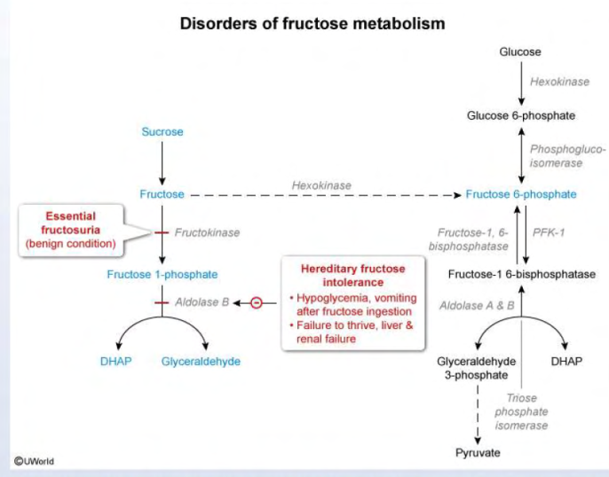
essential fructosuria
deficiency of fuctokinase
benign condition but fructose appears in urine (urine dipstick not identify)
not symptoms because hexokinase becomes first pathway for converting fructose to F6P
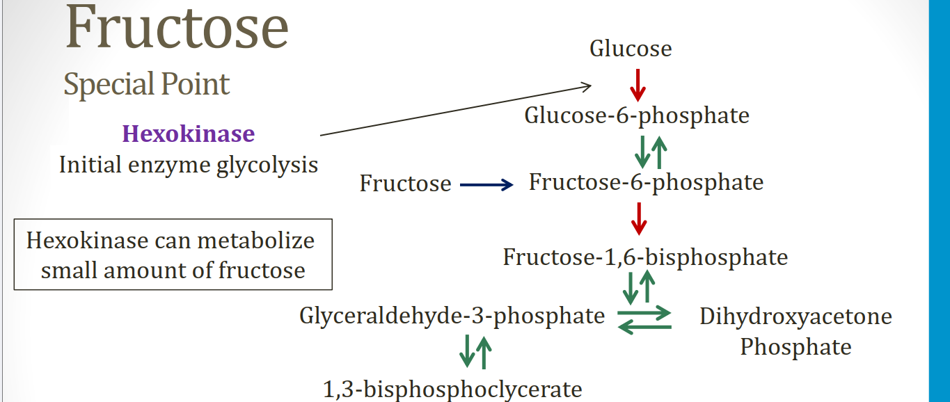
hereditary fructose intolerance
deficiency of aldolase B
→ lead toxic accumulation Fructose 1-phosphate in the liver → depletion ATP
Hypoglycemia, jaundice, cirrhosis, and vomiting.
Symptoms only present following consumption of fruit, juice, or honey.
treatment: decrease intake of fructose, sucrose (glucose + fructose), and sorbitol (metabolized to fructose).

polyol pathway
An alternative method of trapping glucose in the cell is to convert it to its alcohol counterpart, sorbitol, via aldose reductase.
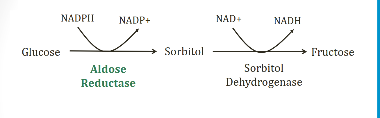

insufficient amount/activity of sorbitol dehydrogenase are at risk of
intracellular sorbitol accumulation, causing osmotic damage (eg, cataracts, retinopathy, and peripheral neuropathy seen with chronic hyperglycemia in diabetes).

deficiency of aldose reductase in polyol pathway
High blood levels of galactose also result in conversion to the osmotically active galactitol via aldose reductase
Lens has primarily Aldose reductase. Retina, Kidneys, and Schwann cells have only aldose reductase (LARKS).
in polyol pathway, which organs have sorbitol dehydrogenase and aldose reductase?
Liver, ovaries, and seminal vesicles

galactose metabolism
classic galactosemia
galactokinase deficiency
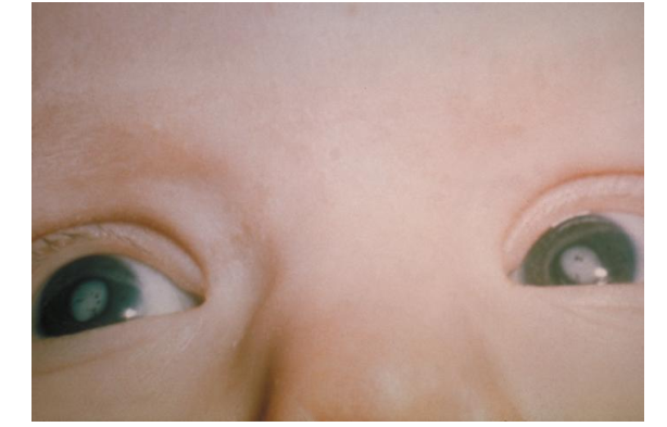
classic galactosemia
Deficiency of galactose 1-phosphate uridyltransferase
Autosomal recessive disorder
Galactose-1-phosphate accumulates in cells
Leads to accumulation of galactitol in cells
Presents in infancy
Liver accumulation galactose/galactitol
Cataracts
Treatment: avoid galactose
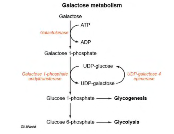

Galactokinase Deficiency
Milder form of galactosemia
Galactose not taken up by cells
Accumulates in blood and urine
Main problem: cataracts as child/young adult
In the liver, alanine is transaminated by alanine aminotransferase to pyruvate with
amino group being transferred to a-ketoglutarate to form glutamate.
Transport of ammonia by alanine
In the liver, before alanine is transaminated by ALT to pyruvate, its amino groups are transferred to a-ketoglutarate to form glutamate.
Glutamate is metabolized by the enzyme glutamate dehydrogenase → liberates free ammonia + regenerates aketoglutarate.
Ammonia then enters the urea cycle to form urea → excretion in urine
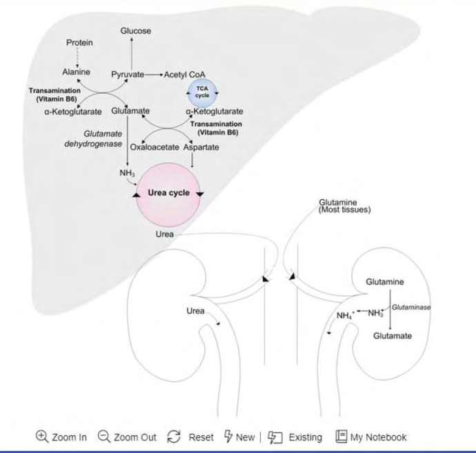

Ammonia metabolism
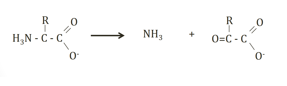
amino acid breakdown → No storage form of amino acids, Unused amino acids broken down →
Amino group removed → NH3 + α-keto acid
Converted by liver to urea (non toxic) for excretion
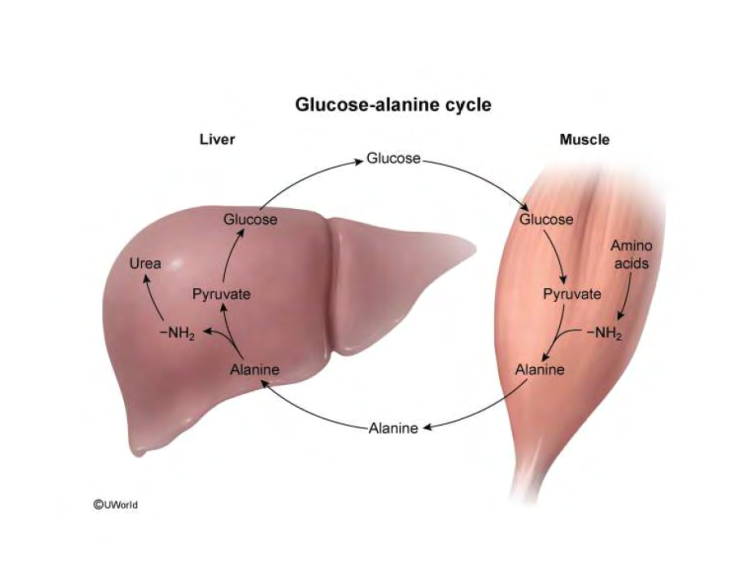
glucose - alanine cycle
Muscle tissue lacks a functional urea cycle to dispose of toxic free ammonia NH4. When amino acids are broken down for fuel, excess amino groups → alpha-ketoglutarate → form glutamate. Alanine aminotransferase (ALT) then transfers the amino group from glutamate onto pyruvate (from glycolysis), → alanine. Alanine serves as a neutral, non-toxic carrier to transport nitrogen safely through the bloodstream
Alanine enters hepatocytes, hepatic ALT reverses the reaction, transferring the amino group from alanine back onto alpha-ketoglutarate → glutamate and pyruvate.
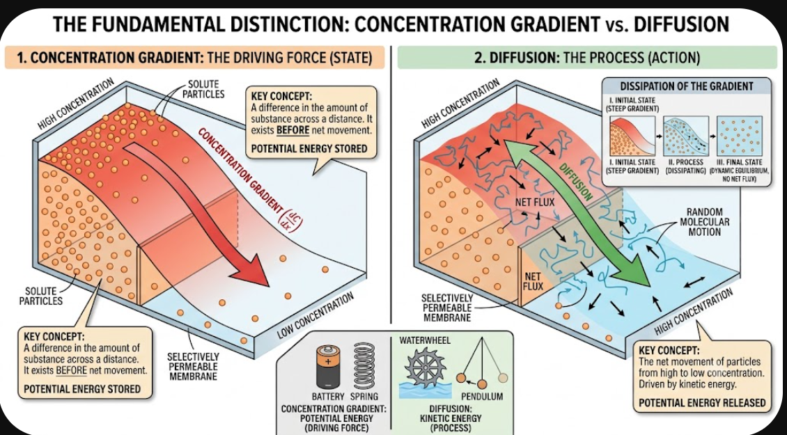
diffusion gradient and concentration gradient
Moving down the gradient: Particles naturally move from high to low concentration through diffusion without using energy.
Moving against the gradient: Moving from low to high concentration requires cell energy (ATP) via active transport.
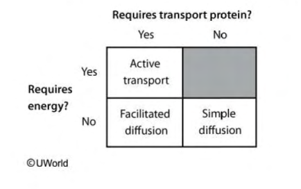
Classification of transport across cell membranes
three main pathways:
active transport
simple diffusion
faciliated diffusion: Transport that is facilitated by transmembrane proteins without the expenditure of energy

carrier-mediated transport.
Transport across the cell membrane by carrier proteins (which undergo conformational changes as the substrate is transported, unlike channel proteins)
These proteins are stereoselective and preferentially bind a specific molecule: for example, GLUT only recognizes glucose; if different molecules like amino acids, the carrier protein does not work
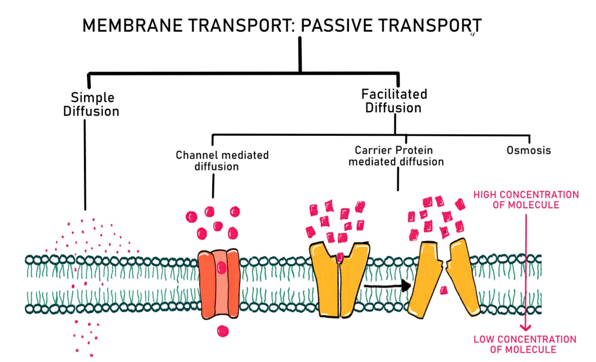
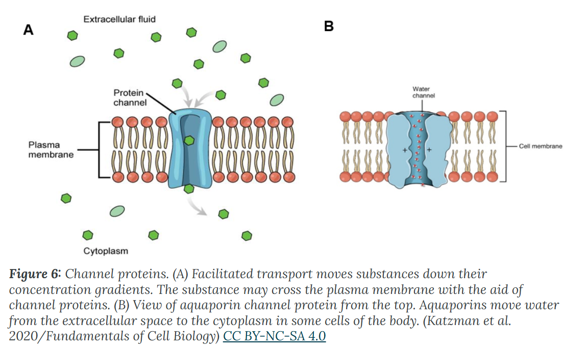
Facilitated Transport - channel protein
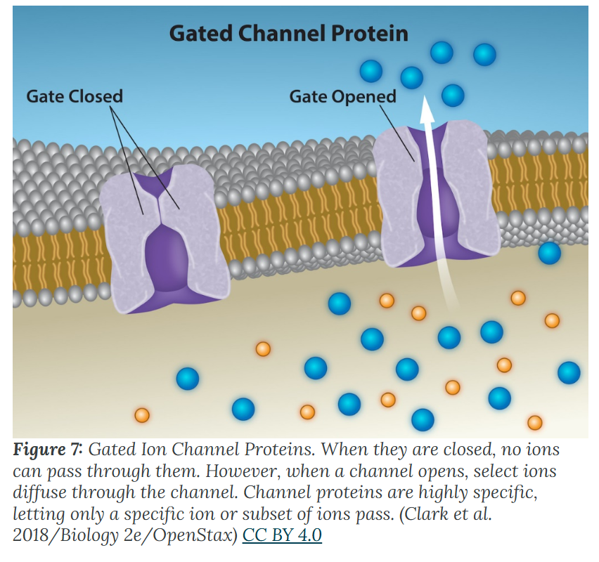
three types of gated channels in more detail:
Voltage-gated
Ligand-gated
Mechanically gated
classification of receptors
cell surface receptor: peptide hormone, neurotransmitters
intracellular receptor: steroid hormone, thyroid hormone

type of enzyme inhibition
two groups:
irreversible: permanently covalently bond to and destroy the enzyme, like aspirin or penicillin
reversible: temporarily bind and detach. Include 3 types: competitive, non competitive, uncompetitive
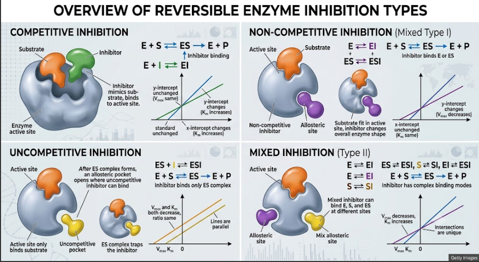
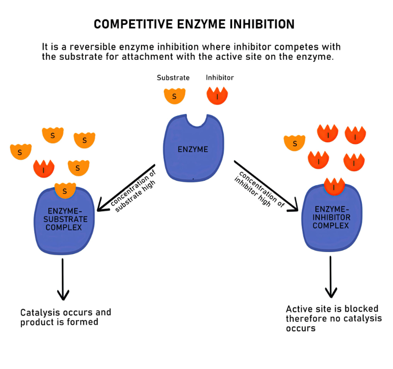
In competitive inhibition
The inhibitor physically blocks the active site. The enzyme can bind the substrate or the inhibitor, but never both at the same time.
Vmax Unchanged, Km: Increases
Elevated substrate outcompetes the inhibitor.
Example: statins (enz HMG - CoA reductase), ACE inhibitors, …
In noncompetitive inhibition
The inhibitor binds to a secondary (allosteric) site on the enzyme → changes the enzyme's 3D shape → non-functional
Km unchanged, Vmax decreased
Example: oxamic acid, monoamine oxidase

In uncompetitive inhibition
The inhibitor only binds to the Enzyme-Substrate (ES) complex after the substrate has already attached → locks the substrate in place, preventing the reaction from finishing.
Vmax: Decreases, Km: Decreases
Adding more substrate actually creates more targets for the inhibitor.
Example: lithium

Factors That Affect Diffusion
Higher concentration gradients across the membrane
molecular weight
surface area
membrane thickness
Temperature
Solvent Density
Distance Travelled
explain:
Heavy or large molecules move more slowly than lighter ones. It takes more energy in the medium to move them along.
Increasing or decreasing temperature increases or decreases the energy in the medium, affecting molecular movement.
The denser a solution is, the harder it is for molecules to move through it, causing diffusion to slow down due to friction.
Living cells require a steady supply of nutrients and a steady rate of waste removal. If the distance these substances need to travel is too great, diffusion cannot move nutrients and waste materials efficiently to sustain life.


collagen synthesis
intracellular (synthesis, hydroxylation, glycosylation) → Extracellular (exocytosis, proteolytic processing, assembly and alignment, cross-linking)

required for collagen synthesis
vitamin C
copper

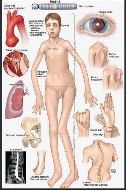
marfan syndrome
FBN1 mutation on chromosome 15 → defect fibrillin 1 (Fibrillin-1 is a major component of the microfibrils that form a sheath around elastin. Microfibrils are abundantly present in blood vessels and in the suspensory ligaments of the lens)
Findings: very tall and long, pigeon chest, pectus excavatum, hypermobile joints, long and tapering fingers and toes, cystic medial necrosis of the aorta; aortic root aneurysm rupture or dissection (the most common cause of death); mitral valve prolapse; increased risk of spontaneous pneumothorax. normal intellectual disability

homocystinuria
cystathionine beta synthase deficiency
Presentation similar to Marfan syndrome with pectus deformity, tall stature, increase arm:height ratio, decrease upper:lower body segment ratio, arachnodactyly, joint hyperlaxity, skin hyperelasticity, scoliosis, fair complexion (vs Marfan syndrome), intellectual disability and development delay.
pulmonary embolism + DVT → early atherosclerosis
treatment: avoid methionine, increase cysteine, B6 supplementation

osteogenesis imperfecta
brittle bone disease
autosomal dominant with decrease production of collagen type 1 → altered triple helix formation
gene COL1A1, COL1A2
Symptoms: BITE
Bones: multiple fractures
I (eye): blue sclerae
Teeth: dental imperfections
Ear: hearing loss


Ehlers Danlos syndrome
Faulty collagen synthesis causes skin to be hyperextensible and often thin or transparent, joints to be hypermobile , and tendency to bleed (easy bruising).
Multiple types.
Inheritance and severity vary.
Can be autosomal dominant or recessive.
May be associated with joint dislocation, berry and aortic aneurysms, organ rupture.
Hypermobility type (joint instability): most common type.
Classical type (joint and skin symptoms): mutation in type V collagen (eg, COL5A1, COL5A2).
Vascular type (fragile tissues including vessels [eg, aorta], muscles, and organs that are prone to rupture [eg, gravid uterus]): mutations in type III procollagen (eg, COL3A1).
Can be caused by procollagen peptidase deficiency.
![<p>Faulty collagen synthesis causes skin to be hyperextensible and often thin or transparent, joints to be hypermobile , and tendency to bleed (easy bruising).</p><p>Multiple types. </p><p>Inheritance and severity vary. </p><p>Can be autosomal dominant or recessive. </p><p>May be associated with joint dislocation, berry and aortic aneurysms, organ rupture. </p><p>Hypermobility type (joint instability): <strong>most common type. </strong></p><p>Classical type (joint and skin symptoms):<strong> mutation in type V collagen (eg, COL5A1, COL5A2).</strong> </p><p>Vascular type (fragile tissues including vessels [eg, aorta], muscles, and organs that are prone to rupture [eg, gravid uterus]): <strong>mutations in type III procollagen (eg, COL3A1).</strong> </p><p>Can be caused by procollagen peptidase deficiency. <br></p>](https://assets.knowt.com/user-attachments/6a4c220c-87b5-4e1e-a538-aac3eff227df.png)

menkes disease
X-linked recessive connective tissue disease caused by impaired copper absorption and transport due to defective Menkes protein ATP7A (Absent copper), vs ATP7B in Wilson disease (copper Buildup).
Leads to decreased activity of lysyl oxidase (copper is a necessary cofactor), resulting in defective collagen cross-linking.
→ brittle, “kinky” hair, failure to thrive, and severe neurological deterioration, hypotonia, increase risk of cerebral aneurysms.

Elastin, a fibrous protein in the connective tissue such as
skin, blood vessels, lung alveoli
De novo pyrimidine synthesis
enzyme: carbamoyl synthase II → UMP synthase → ribonucleotide reductase →thymidylate synthase, dihydrofolate reductase

orotic aciduria
Autosomal recessive → Defect in UMP synthase → Buildup of orotic acid
→ Loss of pyrimidines
findings
• Orotic acid in urine
• Megaloblastic anemia
• No B12/folate response
• Growth retardation
Treatment:
• Uridine
• Bypasses UMP synthase

Ornithine transcarbamylase
Key urea cycle enzyme
• Combines carbamoyl phosphate with ornithine → Makes citrulline
• OTC deficiency → increased carbamoyl phosphate
• ↑ carbamoyl phosphate → ↑ orotic acid
• Don’t confuse with orotic aciduria (defect in UMP synthase)
• Both have orotic aciduria
• OTC only: ↑ ammonia levels (urea cycle dysfunction)
• Ammonia → encephalopathy (baby with lethargy, coma)


Megaloblastic anemia caused by defective DNA production
• Folate deficiency → loss dTMP production
• B12 deficiency (neuro symptoms, MMA)
• Orotic aciduria
• Drugs (MTX (block dihydrofolate reductase), 5-FU (block thymidylate synthase), hydroxyurea (block ribonucleotide reductase))
• Zidovudine (HIV NRTIs)

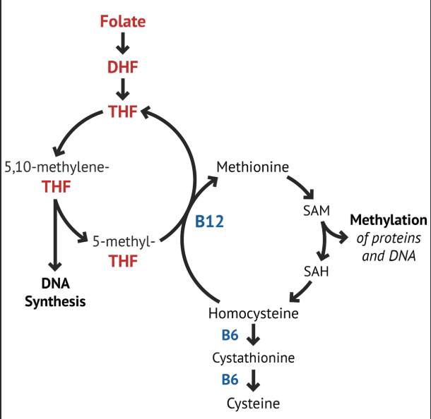
B12 and folate
Both folate and B12 are required to convert to methionine
deficiencies → elevated homocysteine, macroblastic anemia
B12 deficiency = ↑ methylmalonic acid (MMA) level
Folate deficiency = normal MMA level

Methionine cycle defect → homocystinuria
Cystathionine synthase deficiency (treatment: decrease methionine, increase cysteine + B6, B12, and folate in diet)
Decrease affinity of cystathionine synthase for pyridoxal phosphate (treatment: increase B6 + cysteine in diet)
Methionine synthase (homocysteine methyltransferase) deficiency (treatment: increase methionine in diet)
Methylenetetrahydrofolate reductase (MTHFR) deficiency (treatment: increase folate in diet)
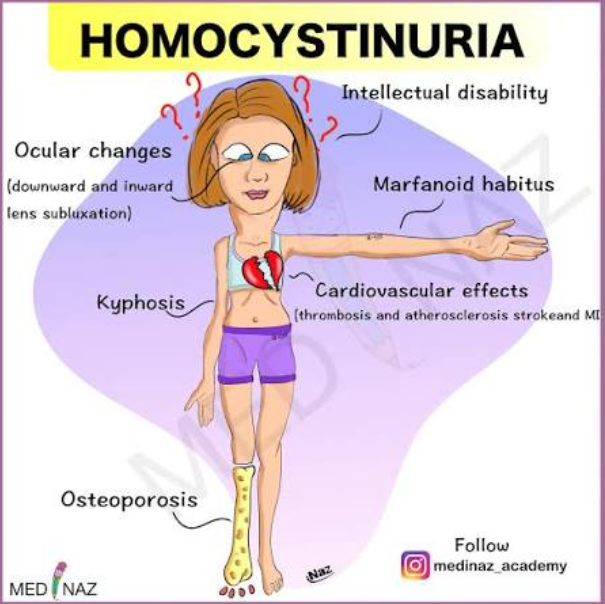
Symptoms: HOMOCYstinuria: elevated Homocysteine in urine, Osteoporosis, Marfanoid habitus, Ocular changes (downward and inward lens subluxation), Cardiovascular effects (thrombosis and atherosclerosis → stroke, MI), kYphosis

level of consciousness

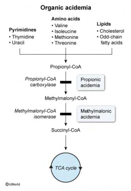
VOMIT: Valine, Odd chain fatty acids, Methionine, Isoleucine, Threonine, cholesterol
organic acidemia
Symptoms in infancy: poor feeding, vomiting, hypotonia, high anion gap metabolic acidosis, hepatomegaly, seizures.
Organic acid accumulation:
Inhibits gluconeogenesis → hypoglycemia, hyper-ketoacidosis → high anion gap metabolic acidosis
Inhibits the urea cycle → hyperammonemia
Treatment: low-protein diet limited in substances that metabolize into propionylCoA (VOMIT)
Cofactor ??? in propionic acidemia and methylmalonic acidemia
enzymes in beta-oxidation of fatty acids, the TCA cycle, and the carboxylation of pyruvate (gluconeogenesis) all occur
mitochondria
enzymes responsible for glycolysis, fatty acid synthesis, and the pentose phosphate pathway reside in
cytosol

TCA cycle, Krebs cycle
Citrate is the Krebs starting substrate for making oxaloacetate
Purpose of TCA cycle: Cellular Energy Production, like NADH, FADH2, Biosynthetic Precursors (succinyl CoA, a-KG, citrate, and Metabolic Integration
Production: 3NADH, 1 FADH2, 2CO2, 1GTP = 1 ATP
Occurs in mitochondria

α-ketoglutarate dehydrogenase complex requires
same cofactors as the pyruvate dehydrogenase complex (vitamins B1, B2, B3, B5, lipoic acid)


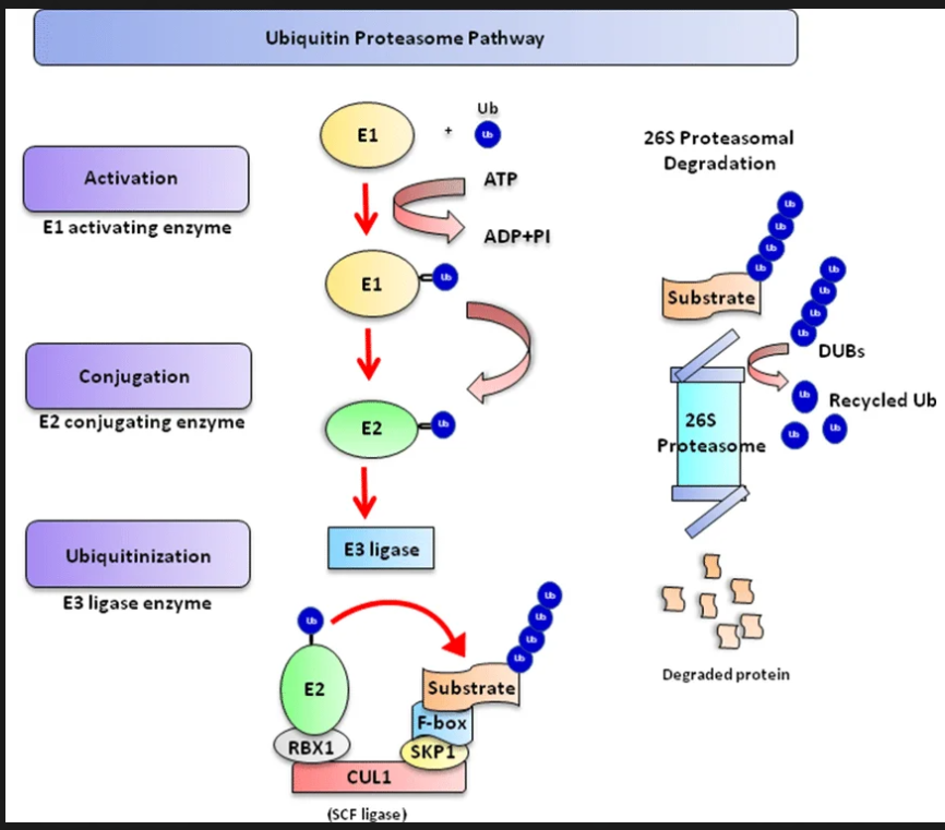
ubiquitin proteasome pathway
E1: activator
E2: delivery truck
E3 ligase: enzyme recognizes target cells and bring them to E2
Proteasome: barrel shaped to shred. E2 bind polyubiquitin (at least 4 ub) and move to proteasome. Enzyme DUBs cuts poly ub and bings target cells into the barrel shaped which will destroy the cell
Phosphorylation
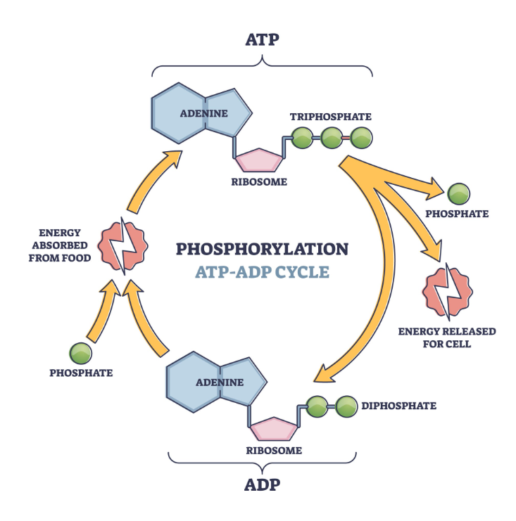
attachment of a phosphate group to a molecule, most commonly a protein. It serves as the primary "on/off" switch for cellular activity. kinases: add the phosphate group (typically extracting it from an ATP molecule), while phosphatases: remove it.

Glucuronidation
addition of a glucuronic acid molecule (a sugar derivative) to a substrate.
This is a major "Phase II" detoxification reaction carried out primarily by the liver. One step in the hepatic processing of bilirubin includes bilirubin conjugation with glucuronic acid in the endoplasmic reticulum.


Gamma-carboxylation
adds a carboxyl group to specific amino acids (glutamates) within a protein. requires Vitamin K to occur. critical for blood clotting—without gamma-carboxylation, coagulation proteins like prothrombin cannot function.
warfarin inhibits vitamin K synthesis → prevents carboxylation


heterochromatin - euchromatin
Heterochromatin refers to condensed DNA that has a low level of transcriptional activity
Euchromatin (loosely arranged) has high levels of transcriptional activity.
Histone acetylation promotes the formation of euchromatin; this process is impaired in Huntington disease,