intrinsic muscles, attachments & actions
 Call Kai
 Learn
Learn Practice Test
 Spaced Repetition
 Match
Match Flashcards
 Knowt Play
1/90

There's no tags or description
Looks like no tags are added yet.
Name | Mastery | Learn | Test | Matching | Spaced | Call with Kai | Chat |
|---|
No analytics yet
Send a link to your students to track their progress
91 Terms
Deltoideus m. scapular part
What muscle is highlighted in the picture

Deltoideus m. acromion part
What muscle in highlighted in the picture

spine of the scapula, acromion, deltoid tuberosity of the humerus
What are the attachments of the deltoideus muscles
flex the shoulder joint
What are the action of the deltoideus muscles
Supraspinatus m.
What muscle is highlighted in the picture?
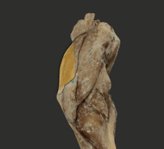
supraspinatous fossa, spine of the scapula, cranial part of greater tubercle (LA lesser tubercle)
What are the attachments of the supraspinatus m?
stablize shoulder joint, extend shoulder joint
What are the actions of the supraspinatus m?
Infraspinatus m.
What muscle is highlighted in the picture?
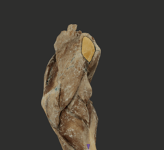
infraspinous fossa, spine of scapula, lateral surface of the greater tubercle of humerus
What are the attachments of the infraspinatus m?
stablize the shoulder joint, extend or flex shoulder joint
What are actions of the infraspinatus m?
teres minor m.
Not pictured, what muscle is deep to the deltoideus m. on the lateral side?
distal, caudal border of the scapula, teres minor tuberosity of humerus
What are the attachments of the teres minor m?
flexes the shoulder joint
What is the function of the teres minor muscle?
subscapularis m.
What muscle is highlighted in the picture?
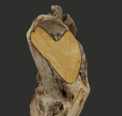
subscapular fossa and lesser tubercle of the humerus
What are the attachments of the subscapularis m?
stablize shoulder joint, extend shoulder joint
What are the actions of the subscapularis m?
teres major m.
What muscle is highlighted in the picture?
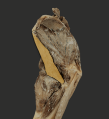
caudal angle and border of scapula, teres major tuberosity of humerus
What are the attachments of the teres major m?
flexes shoulder joint, adduction of the brachium
What are the actions of the teres major m?
coracobrachialis m
What muscle is being pointed to in the picture?

coracoid process of the scapula, proximal or teres major tuberosity of humerus
What are the attachments of the coracobrachialis m?
outward rotion of shoulder, adduction of brachium, extend shoulder joint (ca), flex shoulder joint (eq)
What are the actions of the coracobrachialis m?
tensor fasciae antebrachii m
Not pictured, what is the thin strap muscle that covers the medial surface of the long head of the triceps brachii m?
fascia of the latissimus dorsi m, olecranon and fascia of antebrachium
What are the attachments of the tensor fasciae antebrachii m?
tenses the fascia of the antebrachium, extends elbow joint
What are the actions of the tensor fasciae antebrachii m?
long head, lateral head, medial head, accessory head
What are the four heads of the triceps brachii m?
medial and accessory, accessory
In cats, which heads of triceps are fused? What head is not present in equine?
long head of the triceps brachii m.
What muscle is highlighted in the picture?

medial head of the triceps brachii m.
What muscle is highlighted in the picture?
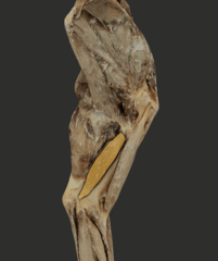
lateral head of the triceps brachii m.
What muscle is highlighted in the picture?

accessory head of triceps brachii m.
Not pictured, what head of the triceps brachii m. is visible after removing the lateral head of triceps brachii and deltoideus m.?
olecranon tuber
What is the common attachment of the triceps brachii m.?
caudal border of scapula
What is the attachment for the long head of triceps?
tricipital line of humerus
What is the attachment for the lateral head of triceps?
medial shaft and lesser tubercle of humerus
What is the attachment for the medial head of triceps?
caudal part of neck of humerus
What is the attachment for accessory head of triceps?
extend elbow joint, flex shoulder joint
What is the action of triceps brachii m? What action is specific to long head?
anconeus m.
What muscle is highlighted in the picture?
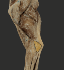
lateral supracondylar crest of humerus, olecranon tuber
What are the attachments of the anconeus m?
extend elbow joint
What are the actions of the anconeus m?
biceps brachii m.
What is the muscle highlighted in the picture?

supraglenoid tuberosity of scapula, radial and ulnar tuberosities
What are the attachments of biceps brachii m?
extend shoulder joint, flex elbow joint, stablize shoulder and carpal joints in LA
What are the actions of biceps brachii m?
lacertus fibrosus
Not pictured, what is the fibrous structure that links the central tendon of biceps brachii muscle to extensor carpi radialis tendon, playing role in passive stay apparatus of equids?
brachialis m.
What muscle is highlighted in the picture?
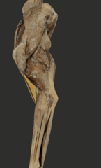
proximal third of the caudolateral surface near neck of humerus, radial and ulnar tuberosities (ca), radial tuberosity (la)
What are the attachments of brachialis m?
flex elbow joint
What is the action of the brachialis m?
extensor carpi radialis m
What muscle is highlighted in the picture?

lateral supracondylar crest and radial fossa, dorsal tuberosity at base of metacarpal bones
What are the attachments of the extensor carpi radialis m?
extend the carpal joints
What are the actions of the extensor carpi radialis m?
common digital extensor m.
what muscle is highlighted in the picture?

lateral humeral epicondyle and lateral collateral ligament of elbow, extensor processes of distal phalanges
what are the attachments of the common digital extensor m.?
extend carpal joints and digit joints
what are the actions of common digital extensor m?
lateral digital extensor m.
what muscle is highlighted in the picture?

lateral humeral epicondyle, lateral collateral ligament of elbow, and lateral tuberosity of radius
what are the attachments of the lateral digital extensor m?
extend carpal and digit joints
what are the actions of the lateral digital extensor m.?
ulnaris lateralis m
what muscle is highlighted in the picture?

lateral humeral epicondyle and collateral ligament of elbow, accessory carpal bone and proximal end of lateral metacarpal bone
what are the attachments of the ulnaris lateralis m?
abductor digiti 1 longus m.
not pictured- what m. is only seen in carnivores and attaches to craniolateral surface of radius and ulna, and proximal end of metacarpal bone 1 (think dew)
extensor carpi obliquus m.
not pictured- what is the large animal muscle that is equivalent to the abductor digiti 1 longus m?
craniolateral surface of radius and ulna, medial surface on proximal end of metacarpal 2 and 3
what are the attachments of the extensor carpi obliquus m.?
extensor retinaculum
not pictured- what is the fibrous band that holds the tendons of extensor muscles in place over the dorsal surface of the carpus?
flexor carpi radialis m.
what muscle is highlighted in the picture?

medial humeral epicondyle, palmar surface on proximal end of metacarpal bones
what are the attachments for the flexor carpi radialis m?
flex the carpal joint
what is the action for the flexor carpi radialis m.?
flexor carpi ulnaris m.
what muscle is highlighted in the picture?
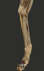
medial humeral epicondyle, olecranon, accessory carpal bone
what are the attachments of the flexor carpi ulnaris m?
flex the carpal joints
what are the actions of the flexor ulnaris m?
superficial digital flexor m.
what muscle is highlighted in the picture?

medial humeral epicondyle, palmar surface of middle phalanges
what are the attachments of the superficial digital flexor m.
flex carpal, metacarpophalangeal, and proximal interphalangeal joint
what are the actions of the superficial digital flexor m.?
deep digital flexor m.
not pictured- what is deep to the superficial digital extensor m?
3 heads- humeral, radial, and ulnar
what is distinct about the deep digital flexor m.?
medial humeral epicondyle
what is the attachment for the humeral head of the deep digital flexor m?
caudal surface of proximal radius
what is the attachment for the radial head of the deep digital flexor m?
caudal surface of proximal ulna
what is the attachment for the ulnar head of the deep digital flexor m?
flex carpal joints and digital joints
what are the actions of the deep digital flexor m?
flexor retinaculum
not pictured- what is the fibrous band that holds the digital flexor tendons in the carpal canal and has superficial and deep parts
manica flexoria
what is the sleeve formed by the superficial digital flexor tendon called?
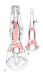
allows passage of deep digital flexor tendon to insert in the distal phalanges
what is the function of the manica flexoria
1 per weight bearing digit (4 ca, 2 rum, 1 equid)
how many of the manica flexoria's do each species have?
palmar, distal (p2), and proximal (p3)
what are the three annular ligaments
supinator m and pronator teres m
not pictured- what are the two rotator muscles?
lateral humeral epicondyle and caranial surface of radius
what are the attachments of the supinator m?
supination of arm, flex elbow
what are the actions of the supinator m?
pronator teres m
what muscle is highlighted in picture?

medial humeral epicondyle and cranial surface of radius
what are the attachments of the pronator teres m
pronation of forearm and flex elbow joint
what are the actions of the pronator teres m
pronator quadratus m
not pictured- what fills the space between the radius and ulna, pronates the forearm and is another rotator muscle?
interosseus m
what is the suspensory ligament called?
caudal surface of metacarpal bones, proximal seasmoid bones
what are the attachments of the interosseus m?