Ch 7 - The Nervous System Part 1
 Call Kai
 Learn
Learn Practice Test
 Spaced Repetition
 Match
Match Flashcards
 Knowt Play
1/80

There's no tags or description
Looks like no tags are added yet.
Name | Mastery | Learn | Test | Matching | Spaced | Call with Kai | Chat |
|---|
No analytics yet
Send a link to your students to track their progress
81 Terms
central nervous system and peripheral nervous system
what 2 parts is the nervous system divided into
brain and spinal cord
what makes up the central nervous system
cranial and spinal nerves
what makes up the peripheral nervous system
neurons and neuroglia
what 2 types of cells is the nervous tissue composed of
1) neurons can conduct impulses but generally cannot divide
2) neuroglia support the neurons and can’t conduct impulses, but can divide
main differences between neurons and neuroglia in terms of impulses and dividing
1) respond to chemical and physical stimuli
2) conduct electrochemical impulses
3) release chemical regulators
4) enable perception of sensory stimuli, learning, memory, and control of muscles and glands
what are the 4 general functions of the neurons
1) cell body → contains the nucleus, Nissl bodies, and other organelles (most proteins are translated here)
2) dendrites → receive impulses and conducts a graded impulse toward the cell body (conduct chemical stimuli into electrical stimuli)
3) axon → conducts action potentials away from the cell body
3 parts of the structure of a neuron and its function
the secondary metabolic center (some proteins translated here)
what are the axon terminals considered
CNS → nuclei
PNS → ganglia
what is a cluster of cell bodies in the CNS vs in the PNS called
collateral axon → allows single neuron to have contact with many cells
what is a branched axon called and what does it allow
some need to make shorter or longer connections
why do axons vary in length
the axon hillock where action potentials are generated at the initial segment of the axon (also where synthesis from dendrites occurs)
what is the axon connected to the cell body by AND what occurs here
myelin sheath → open spots are nodes of Ranvier
what are axons covered in and what are the open, uncovered spots called
an active process needed to move organelles and proteins from the cell body to axon terminals
what is axonal transport
1) fast components move membranous vesicles
2) slow components move microfilaments, microtubules, and proteins
with axonal transport, what do fast vs slow components move
1) from cell body to dendrites and axon
2) uses kinesin molecular motors
3) carries neurotransmitters and ions
which way is anterograde transport, what does it use to transport, and what does it carry
1) from dendrites and axon to the cell body
2) uses dynein molecular motos with its activator dynactin
3) carries lysosomes, autophagosomes, endosomes, and other molecules AND certain viruses and tetanus toxin
which way is retrograde transport, what does it use to transport, and what does it carry
conduct impulses exclusively in the CNS and integrate functions of the NS
where do association/interneurons conduct impulses and what does it do
from sensory receptors to the CNS (afferent)
which way do sensory neurons conduct impulses
from the CNS to target organs (muscles or glands; efferent)
which way do motor neurons conduct impulses
for reflexes and voluntary control of skeletal muscles
what are somatic motor neurons responsible for
they innervate involuntary targets such as smooth muscle, cardiac muscle, and glands
what are autonomic motor neurons responsible for
1) parasympathetic → normal functions; “rest and digest”
2) sympathetic → emergency situations; “fight or flight”
both involve autonomic motor neurons
parasympathetic vs sympathetic nervous system AND what neurons involved
pseudounipolar, bipolar neurons, and multipolar neurons → based on the number of processes that extend from the cell body
what are the 3 structural classifications of neurons and what are they based on
1) pseudounipolar → single short process that branches like a T to form 2 longer processes (sensory neurons)
2) bipolar neuron → have 2 processes, one on either end (found in retina of eye)
3) multipolar neuron → several dendrites and one axon; most common (motor neurons)
pseudounipolar vs bipolar vs multipolar
pseudounipolar
what type of neuron is this
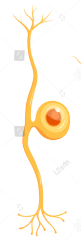
bipolar neuron
what type of neuron is this

multipolar neuron
what type of neuron is this

bundles of axons located outside the CNS
what are nerves
both sensory and motor neurons (mixed nerves)
what are most nerves composed of
sensory fibers
what do some of the cranial nerves only have
a tract
what is a bundle of axons in the CNS called
Schwann cells and satellite cells
what are the 2 types of neuroglia (glial cells) found in the PNS
they form myelin sheaths around peripheral axons
what do Schwann cells in the PNS do
they support cell bodies within the ganglia of the PNS (can migrate short distances also)
what do satellite cells do in the PNS
1) oligodendrocytes
2) microglia
3) astrocytes
4) ependymal cells
what are the 4 types of neuroglial cells found in the CNS
they form myelin sheaths around the axons of the CNS neurons
what do oligodendrocytes in the CNS do
they migrate around CNS tissue and phagocytize foreign and degenerated material
what do microglia in the CNS do
they regulate the external environment of the neurons
what do astrocytes in the CNS do
they line the ventricles central canal of the spinal cord and secrete cerebrospinal fluid
what do ependymal cells of the CNS do
they were produced in the embryonic yolk sac and migrated to the developing neural tube
what is unique about the microglia compared to other cells in the CNS
related to macrophages however when they die, they are replaced by cell division of existing microglia instead of monocytes of the blood like macrophages
what are microglial embryonically related to, and how do they differ from them also
they have a small cell body with many fine processes (helps maintain healthy neuronal synaptic function)
brief description of structure of microglia
infection or trauma → they become phagocytotic, mobile cells and sense ATP released from damaged cells and migrate towards them
what can lead to microglial activation and what does it lead to
they kill endogenous pathogens and remove damaged dendrites, myelin, and other debris
what do microglial cells kill and what do they remove in the CNS
pruning synapses
what can microglial cells do to axons to eliminate inappropriate synaptic connections
neurilemma (extends its plasma membrane and wraps it around axon)
sheath of Schwann cells that surrounds all axons in the PNS
myelin sheath
all axons in the PNS are surrounded by Schwann cells that have neurilemma, but not all of those cells have what
layers upon layers of plasma membrane (lipids)
what is the myelin sheath composed of
usually unmyelinated
in the PNS are small axons usually myelinated or unmyelinated
conduct impulses
what can myelinated axons do quicker than unmyelinated axons
1 Schwann cell : 1 axon
1 oligodendrocyte : multiple axons
ratio of Schwann cell to axon vs oligodendrocyte to axon
it sends extensions to several axons and each wraps around a section of an axon giving myelin sheath
how do oligodendrocytes produce myelin sheath for axons in CNS
myelin sheaths give white color while those without myelin sheath are gray
what color are tissues with myelination vs without it
those in which the immune system attacks the myelin sheaths that protect nerve fibers
what are demyelinating diseases
the T cells of the immune system attack the myelin sheaths of the PNS which produces rapid onset of symptoms that includes muscle weakness
what occurs in Guillain-Barre syndrome
autoimmune attack by T lymphocytes causing lymphocytes and monocyte-derived macrophages to enter the brain (CNS) and target the myelin sheaths leading to range of neurological symptoms
what occurs in multiple sclerosis (MS)
1) portion of neve fiber is cut, the severed part degenerates and Schwann cells form a regeneration tube
2) growth factors are released, stimulating the growth of axon spurts within this tube
3) new axon connects to the undamaged axon or the effector
(no-go inhibitor protein produced by Schwann cells are removed by phagocytosis to allow this to happen)
explain process of PNS regeneration
CNS axons have a limited ability to regenerate, unlike PNS axons
how does the ability of CNS axons to regenerate compare to PNS axons?
axon collaterals, but central axons have a much more limited ability to regenerate than peripheral axons
what does injury in the CNS stimulate growth of
1) oligodendrocyte apoptosis due to death receptors forming
2) inhibitory proteins in myelin like Nogo produce by oligodendrocytes
3) glial scar formation by astrocytes
what are the 3 major factors that make CNS regeneration difficult
1) promote neuronal growth in the fetal brain (nerve growth factor, etc.)
2) in adults, they aid in the maintenance of sympathetic ganglia and the regeneration of sensory neurons
what are the 2 main roles of neurotrophins through life
astrocytes
what is the most abundant glial cell in the brain
astrocyte end-feet
what is considered the 3rd metabolic center of the neuron
end-feet; they cover the entire surface of blood capillaries and associate with axon terminals
what are astrocytes processes known as and what do they cover
interactions between neurons and also between neurons and blood; helps regulate blood flow, maintain the blood-brain barrier, and support neuronal function
what do astrocytes influence interactions between and what do those interactions help regulate
1) K+ ions from extracellular environment, maintaining the ionic balance necessary for neurons
2) glutamate from CNS axon terminal (excitatory neurotransmitter) which can be converted to glutamine and then GABA (inhibitory neurotransmitter)
what 2 things can the astrocytes (not around the capillaries) take up and what does it do
glucose from blood and convert it to lactic acid for neurons to use in ATP production OR store it as glycogen and produce lactate for neurons when needed
what can astrocytes’ end-feet around capillaries absorb and do with it
the hippocampus and subventricular zone
where do astrocytes regulate neurogenesis in the adult brain
synapses
astrocytes create crucial environment for what to form in the CNS
glial-derives neurotrophic factor (GDNF)
what do astrocytes produce that is needed for survival of spinal cord neurons and dopamine-releasing neurons
the blood-brain barrier
what do astrocytes form that protects the brain from harmful substances
gliotransmitters, such as glutamate, ATP, adenosine, and D-serine
what do astrocytes release that can stimulate or inhibit neurons by responding to calcium signals
changes in intracellular Ca2+ concentration
astrocytes don’t produce action potentials, but they are excited by what
1) when some neurons are active, they release ATP; which increases Ca2+ of adjacent astrocytes; creates a Ca2+ wave
2) a rise in Ca2+ can also cause the astrocyte release prostaglandin E2 from the end-feet on a blood capillary, increasing blood flow
explain intracellular Ca+ concentration changes exciting astrocytes
it’s a critical structure in the brain’s capillaries, where cells are joined by tight junctions, preventing pores between adjacent cells
where is the blood-brain barrier and how are cells joined
that substances can only move through very selective processes such as diffusion endothelial cells, ion channels, transport proteins, and active transport
what does the blood-brain barrier ensure
transcellular
what type of movement is it across the blood-brain barrier
they influence the production of ion channels, carrier proteins, and enzymes that can destroy toxin substances through the secretion of glial-derives neurotrophic factor
how do astrocytes play a significant role in the BBB
they surround the endothelium of CNS capillaries and produce transporters that facilitate two-way communication between endothelial cells and astrocytes
where are pericytes in the BBB and what do they produce
challenges in treating brain cancers, diseases, and infections because many drugs cannot penetrate this barrier
what challenges does the BBB create