Case 11: I Think I Have to Pee!
 Call Kai
 Learn
Learn Practice Test
 Spaced Repetition
 Match
Match Flashcards
 Knowt Play
1/30

There's no tags or description
Looks like no tags are added yet.
Name | Mastery | Learn | Test | Matching | Spaced | Call with Kai | Chat |
|---|
No analytics yet
Send a link to your students to track their progress
31 Terms
b. The differential absorption of x-rays.
Why can we visualize abdominal organs within the abdominal cavity in a radiograph?
a. The fact that x-rays are electromagnetic waves.
b. The differential absorption of x-rays.
c. The fact that each structure within the abdomen has a different atomic number.
d. None Of The Above
False
It is always really easy to palpate the urinary bladder in dogs.
TRUE OR FALSE
True
The descending colon's position in relationship to the urinary bladder is within normal limits in this image.
TRUE OR FALSE
Liver
What is A?
Stomach
What is B?
Spleen
What is C?
Small Intestines
What is D?
Descending colon
What is E?
True
You should be worried that the patient in this VD abdominal projection has urinary calculi.
TRUE OR FALSE
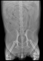
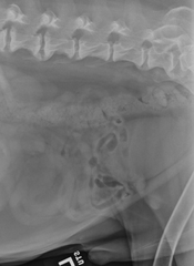
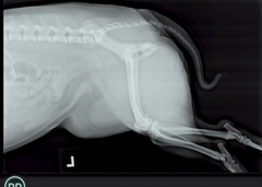
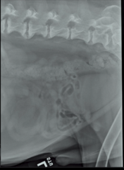
Two lateral abdominal projections are available for review.
Describe the study
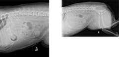
Patient positioning is not adequate for interpretation in the first image as the hindlimbs are not extended caudally. Also, in the second image, the caudal extent of the abdomen is not included.
Study technique is adequate for interpretation.
Describe the patient position and study technique
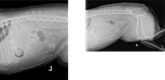
Abdominal serosal detail is within normal limits. In the first image, the stomach contains a mild amount of gas and possibly irregularly shaped mineral objects (need orthogonal view to confirm in stomach or if this mineralization is in the colon). The liver and spleen are within normal limits. The kidneys are not well visualized. The small intestines are uniform in size and shape. The descending colon is displaced ventrally and contains amorphous granular material consistent with feces. The caudal aspect of the ventral abdominal wall is thickened with the impression of irregular margins at its caudal most extent. The urinary bladder is not visualized. The extra-abdominal structures are within normal limits. No evidence of any fractures is seen. In the second image, the cranial aspect of the bladder is seen ventral to the caudal abdominal wall in the inguinal region.
Describe your findings
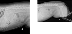
Urinary bladder herniation into the ventral and caudal soft tissues.
Describe your conclusions
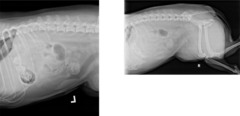
Surgery.
Describe your next steps
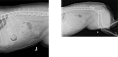
The legs are not pulled all the way back = superimposition of soft tissue
What is wrong with this patient positioning?
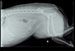
The pelvic limbs and caudal abdomen are not included
What is wrong with this patient positioning?
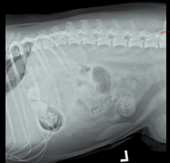
Normally, it runs straight thru the abdomen, but here is dips ventrally as it heads more cranially due to the bladder being displaced
What is the normal position like of the colon?
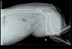
Herniation (displacement) of the bladder backed up by the ventral location of the descending colon
There is a soft tissue, round opacity in the center of this image. What do you think is happening?
Normal
Is this NORMAL OR ABNORMAL colon, bladder abdominal relationship?
e. All Of The Above
If you do not readily visualize the urinary bladder in the caudal abdominal cavity, what are some possible reasons to explain this?
a. The bladder could be empty (or not very full).
b. It could be outside the abdomen.
c. There could be poor serosal detail in the abdomen due to the patient being young, ascites, or the patient being emaciated.
d. The bladder could be ruptured.
e. All Of The Above
EXTERNAL: external abdominal oblique
internal abdominal oblique
rectus abdominus
INTERNAL: transversus abdominus
The linea alba includes aponeuroses from these four muscles, the muscles that make up the abdominal wall: transverse abdominus, external abdominal oblique, rectus abdominis, internal abdominal oblique. Put them in order, from exterior (1) to interior (4).
Sort elements:
internal abdominal oblique
external abdominal oblique
rectus abdominis
transversus abdominis
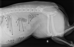
urinary bladder
What structure is outlined by the red arrows?
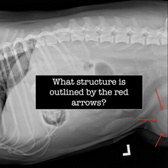
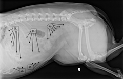
c. Superimposition of the soft tissues of the legs with the urinary bladder.
This is one of the images from this case. Why is it very difficult to see the herniated bladder where we now know it is located?
a. The bladder is empty.
b. The mAs is not high enough to penetrate the patient appropriately.
c. Superimposition of the soft tissues of the legs with the urinary bladder.
d. All of the above.
Liver
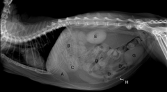
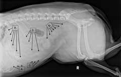
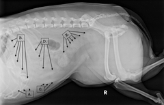
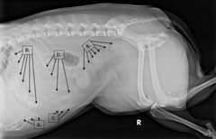
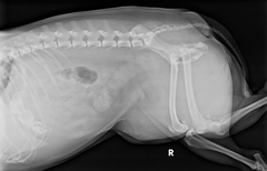
This case involved a dog, but it is important to get a feel for the differences between canine and feline abdominal anatomy. Identify the labeled structures in this feline patient.
What is A?
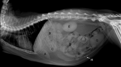
Fundus of the stomach
This case involved a dog, but it is important to get a feel for the differences between canine and feline abdominal anatomy. Identify the labeled structures in this feline patient.
What is B?
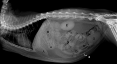
Pylorus
This case involved a dog, but it is important to get a feel for the differences between canine and feline abdominal anatomy. Identify the labeled structures in this feline patient.
What is C?
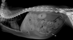
Small intestines
This case involved a dog, but it is important to get a feel for the differences between canine and feline abdominal anatomy. Identify the labeled structures in this feline patient.
What is D?
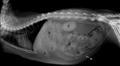
Superimposed right and left kidney
This case involved a dog, but it is important to get a feel for the differences between canine and feline abdominal anatomy. Identify the labeled structures in this feline patient.
What is E?
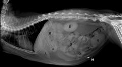
Descending colon
This case involved a dog, but it is important to get a feel for the differences between canine and feline abdominal anatomy. Identify the labeled structures in this feline patient.
What is F?
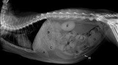
Urinary bladder
This case involved a dog, but it is important to get a feel for the differences between canine and feline abdominal anatomy. Identify the labeled structures in this feline patient.
What is G?
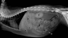
Metallic sutures in ventral body wall
This case involved a dog, but it is important to get a feel for the differences between canine and feline abdominal anatomy. Identify the labeled structures in this feline patient.
What is H?