Maya Cards - PowerPoint 3 (28 SLide one)
 Call Kai
 Learn
Learn Practice Test
 Spaced Repetition
 Match
Match Flashcards
 Knowt Play
1/27

Earn XP
Description and Tags
Facial Prominences and Embryologic Development of the Face
Name | Mastery | Learn | Test | Matching | Spaced | Call with Kai | Chat |
|---|
No analytics yet
Send a link to your students to track their progress
28 Terms
From how many prominences does the face develop, and what tissue are they made of?
5 facial prominences, made predominantly of neural crest-derived mesenchyme from the first pharyngeal arches.
What are the 5 facial prominences?
Frontonasal prominence (1, unpaired),
paired maxillary prominences (2),
paired mandibular prominences (2).
At week 4, how are the 5 prominences positioned around the stomodeum?
Maxillary = lateral;
Mandibular = caudal;
Frontonasal = rostral.
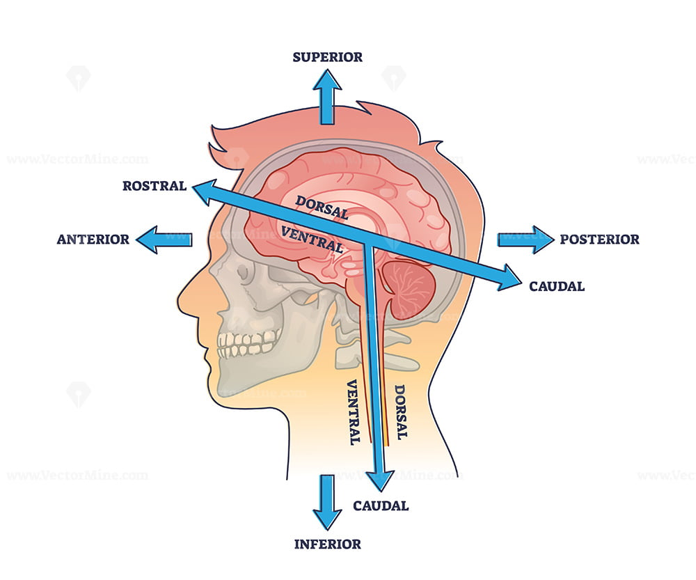
What is the stomodeum, and what membrane must rupture near it?
The stomodeum is the precursor to the mouth;
the buccopharyngeal membrane ruptures to connect the oral and pharyngeal cavities.
During week 5, what do the nasal placodes form, and what two prominences arise beside them?
Placodes invaginate to form :
Nasal pits; ridges on each side form the medial nasal and lateral nasal prominences.
During weeks 6-10, in which direction do the maxillary prominences grow and what do they compress?
They grow medially,
compressing the medial nasal prominences toward the midline.
The upper lip is formed by fusion of which prominences?
The 2 medial nasal prominences
The 2 maxillary prominences.
The lower lip and mandible are formed by which prominences?
The mandibular prominences, merging across the midline.
What forms the nasolacrimal groove, and what develops at its base?
The groove separates the maxillary and lateral nasal prominences
ectoderm at its base forms the nasolacrimal duct.
What do the maxillary prominences enlarge to form?
The cheeks and the maxillae.
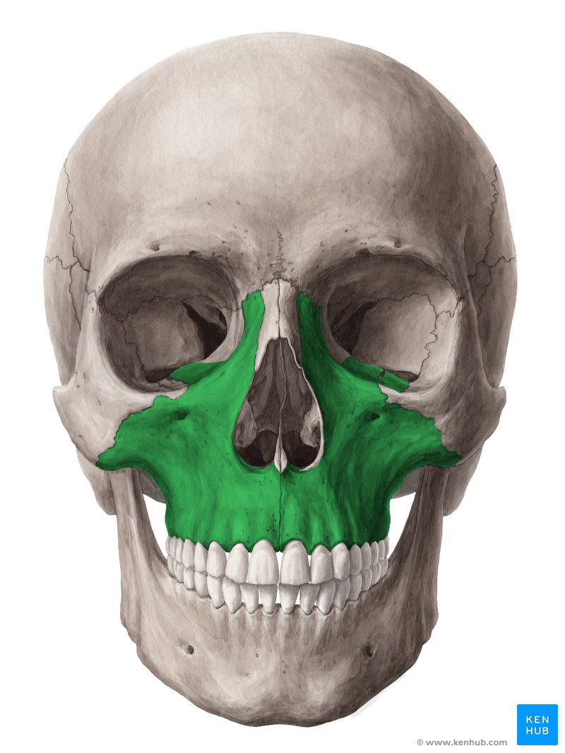
Which prominences form the bridge, tip/crest, and alae of the nose?
Bridge = frontonasal prominence;
tip/crest = merged medial nasal prominences;
alae = lateral nasal prominences.
By what week is the intermaxillary segment formed, and from what?
By week 7, from merging of the maxillary and medial nasal prominences.
What are the 3 components of the intermaxillary segment?
1. Labial = philtrum of the upper lip;
2. Upper jaw = four incisor teeth;
3. Palatal = triangular primary palate.
The intermaxillary segment is continuous with what structure, formed by which prominence?
The nasal septum, formed by the frontonasal (frontal) prominence.
The secondary palate is formed from what outgrowths, of which prominences?
The palatine (palatal) shelves, outgrowths of the maxillary prominences.
Which becomes the main part of the definitive palate — primary or secondary?
The secondary palate.
In what direction does palatal shelf fusion progress, and what must the shelves do first?
They ascend to horizontal above the tongue, then fuse anterior → posterior.
Why must the tongue descend during palate formation?
If the tongue stays between the shelves it blocks fusion → cleft palate.
What is the incisive foramen embryologically?
The midline landmark between the primary and secondary palate.
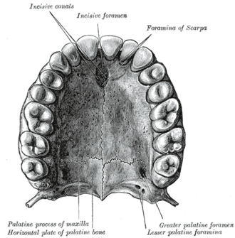
By what age is the palate fully formed?
About 10-12 weeks.
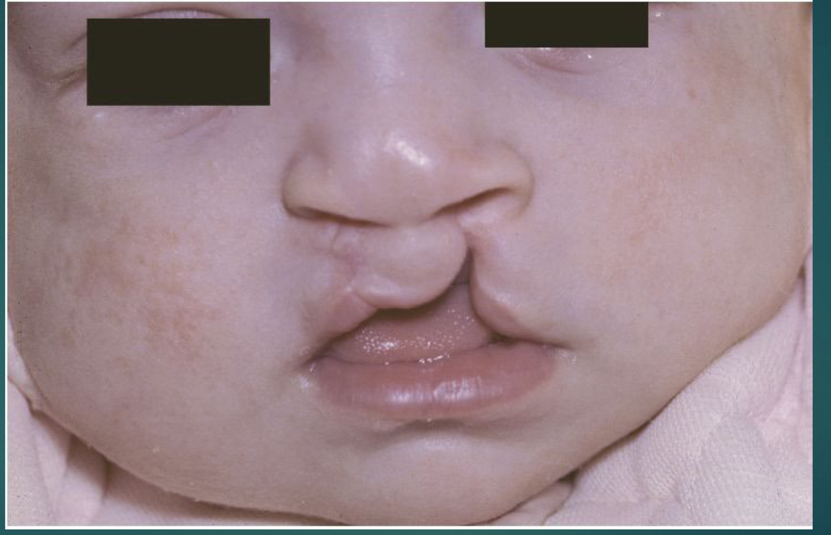
What is the embryologic cause of cleft lip (CL)?
Defective fusion of the medial (median) nasal process with the maxillary process.
What is the embryologic cause of cleft palate (CP)?
Failure of the palatal shelves (secondary palate) to fuse.
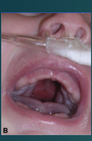
Compare CL±CP vs cleft palate only (CPO): % of clefting and sex predominance.
CL±CP: 70% of clefting, male predominance; CPO: 30% of clefting, female predominance.
What laterality pattern is seen in cleft lip?
80% unilateral; of those, 70% occur on the left side.
Which maternal exposures raise clefting risk, and by how much? (3)
Valproic acid (Depakote) → 10× greater;
maternal smoking → doubles risk; also
maternal alcohol.
What is the MOST COMMON syndrome associated with clefting, and its features?
Van der Woude syndrome: IRF6 mutation, cleft lip ± cleft palate, paramedian lip pits, hypodontia.
What are the three features of Pierre Robin sequence?
Cleft palate, mandibular micrognathia, and glossoptosis.
What is Treacher Collins syndrome (other name, gene, and key features)?
Mandibulofacial dysostosis; TCOF1 mutation; 1st & 2nd branchial arch defects → cleft palate, hypoplastic zygomas, underdeveloped mandible.