ELISA
1/12
There's no tags or description
Looks like no tags are added yet.
Name | Mastery | Learn | Test | Matching | Spaced |
|---|
No study sessions yet.
13 Terms
ELISA
Enzyme-Linked ImmunoSorbant Assay (ELISA)
What is elisa
ELISAs use antibodies to detect proteins in a patient’s
sample.
Many diseases and conditions can be detected and
diagnosed with ELISAs, including:
• Influenza
• HIV
• SARS
• Zika virus
• West Nile virus
• Lyme Disease
Researchers are also developing ELISAs that can be
used to diagnose current or past coronavirus (SARS-CoV-
2) infection.
Antigens
Pollen, bacteria, viruses, and other foreign molecules are
seen by your body as invaders, and they create an
immune response. These foreign invaders are called
antigens.
Antibodies
Your immune system makes antibodies in response to
antigens. The antibodies bind antigens, flagging them for
destruction by immune cells
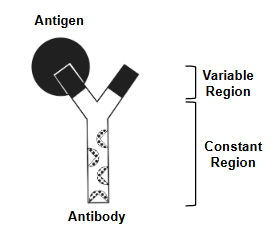
Antigen-Antibody Interactions
Antibodies have two regions: variable and constant. Each
tip of the “Y” in the variable region is highly specific and
binds to only one particular antigen. The constant region is
the same for every antibody of the same type (there are 5
different types of antibodies).
Antibodies can also be produced by injecting an animal
with antigen - disease agents or even antibodies from a
different type of animal
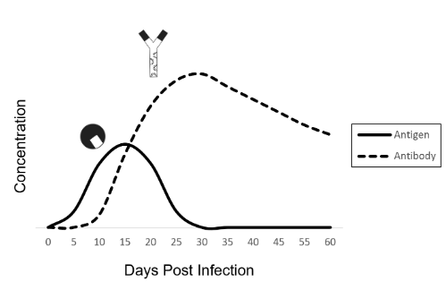
Infection Timeline — Antigens and Antibodies
After infecting a person, a virus multiplies in the body and the
concentration of viral antigens increases.
Soon after infection, the immune system kicks in and starts producing antibodies.
As the antibody concentration increases, antibodies bind to viruses and target them for destruction by the immune system.
Eventually, the viral antigen concentration drops, the infection is cleared, and the person recovers
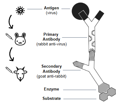
Antigen Detection ELISA Components
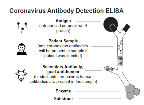
● Antigen — in an antigen detection ELISA, the patient sample is tested for the presence of antigens from viruses, bacteria, etc.
● Primary antibody — binds to the antigen
○ can be produced in a lab by injecting the target antigen into an animal and then harvesting the serum
● Secondary antibody — binds to the constant region of the primary antibody
○ made by injecting the primary antibodies from one animal into a different animal
○ secondary antibodies are attached to an enzyme which catalyzes a color change when substrate is added
● Substrate — changes color in the presence of the enzyme, indicating a positive result
Influenza Antigen Detection
Influenza binds to target cells via the hemagglutinin glycoprotein, HA. Scientists can create antibodies that detect and bind to influenza by injecting HA into an animal
Antigen Detection ELISA – Step 1
Assay Step 1 — Bind antigen to the well
a. Add the patient sample to the well. This sample contains many
different proteins that may or may not contain the antigens that you are trying to detect. These proteins bind non-specifically (adsorb) to the plastic well, due to hydrophobic interactions.
b. Incubate the sample for 5 minutes, then tap out the fluid.
c. Add wash buffer to the well to rinse out anything that isn’t bound, and to block the inner surface of the well. This prevents anything from binding non-specifically in future steps.
d. Repeat the wash step.
Model Step 1
e. Add the antigens around the bottom edge of your blank paper “well”. These represent the proteins / antigens that might be present in any given patient sample
Antigen Detection ELISA – Step 2
Assay Step 2 — Bind primary antibody to antigen
a. Add the primary antibodies to the well. The antibodies bind only to a specific antigen out of the many that may be bound to the well. For example, an anti-HIV antibody would only bind to HIV antigen. This antigen-antibody interaction is specific and strong.
b. Incubate the sample for 5 minutes, then tap out the fluid.
c. Add wash buffer to the well to rinse out anything that isn’t bound. If the primary antibody did not bind to any antigen, then it will be washed away in this step.
d. Repeat the wash step.
Model Step 2
e. Add the primary antibodies into your well. Overlay the antibody so that the black regions align. This represents specific antibody-antigen binding.
f. Model the wash step by removing the unbound primary antibody
Antigen Detection ELISA – Step 3
Assay Step 3 — Bind secondary antibody
a. Add the enzyme-linked secondary antibodies to the well. These
antibodies bind tightly to any primary antibodies that are present. The secondary antibodies are covalently linked to an enzyme, horseradish peroxidase, which will catalyze a reaction with a substrate to produce a color change.
b. Incubate the sample for 5 minutes, then tap out the fluid.
c. Add wash buffer to the well to rinse out any secondary antibodies that did not bind.
d. Repeat the wash step 2 more times (3 times total).
Model Step 3
e. Add the secondary antibodies into your well. Overlay the secondary antibody so that the patterns align with the primary antibody.
f. Model the wash step by removing the unbound secondary antibody
Antigen Detection ELISA – Step 4
Assay Step 4 — Add enzyme substrate for detection
a. Add the substrate to the well, wait 5 minutes, and evaluate results. If the antigen was present, then the primary and secondary antibodies bound and the well will turn blue. If there was no antigen, then no primary or secondary antibodies bound, so the well will remain clear.
Model Step 4
b. Add the substrate into your well. Align the substrate with the enzyme on the secondary antibody.
c. At this point, the enzyme catalyzes a reaction where the substrate turns blue - a positive result
Coronavirus Antibody Detection ELISA