7- Mass Transport in Animals
 Call Kai
 Learn
Learn Practice Test
 Spaced Repetition
 Match
Match Flashcards
 Knowt Play
1/90

There's no tags or description
Looks like no tags are added yet.
Name | Mastery | Learn | Test | Matching | Spaced | Call with Kai | Chat |
|---|
No analytics yet
Send a link to your students to track their progress
91 Terms
Describe the structure of a haemoglobin molecule.
Protein with a quaternary structure
Four polypeptide chains
Each polypeptide chain is associated with a haem group
Each haem group contains an Fe2+ ion
State and explain how many oxygen molecules can be transported by a single haemoglobin molecule in humans.
Four oxygen molecules
Each haemoglobin has four haem groups and therefore four Fe2+ ions
Each Fe2+ ion can combine with a single oxygen molecule
What is the name of the process by which haemoglobin binds with oxygen?
Oxygen loading or
Oxygen association
Where does oxygen loading take place in humans?
In the lungs
What is the name of the process by which haemoglobin releases its oxygen?
Oxygen unloading or
Oxygen dissociation
Where does oxygen unloading take place in humans?
In respiring tissues
What does it mean if haemoglobin has a high affinity for oxygen?
It takes up oxygen more readily
It releases oxygen less readily
What does it mean if haemoglobin has a low affinity for oxygen?
It takes up oxygen less readily
It releases oxygen more readily
Describe and explain the position of the oxygen dissociation curve in the lungs.
Carbon dioxide concentration is low
This means pH is high
Affinity of haemoglobin for oxygen increases
Haemoglobin takes up oxygen more readily
Oxygen dissociation curve shifts to the left
Describe and explain the position of the oxygen dissociation curve in respiring tissues.
Carbon dioxide concentration is high due to respiration
This means pH is low
Affinity of haemoglobin for oxygen decreases
Haemoglobin releases its oxygen more readily
Oxygen dissociation curve shifts to the right
What is the name of the graph showing the relationship between the saturation of haemoglobin with oxygen and the partial pressure of oxygen?
The oxygen dissociation curve
What can the effect of carbon dioxide concentration on the position of the oxygen dissociation curve be described as?
The Bohr effect
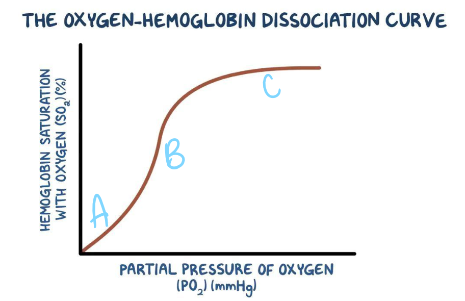
Explain the shape of the oxygen dissociation curve at A
Gradient of the curve is shallow
The shape of haemoglobin makes it difficult for the first oxygen molecule to bind

Explain the shape of the oxygen dissociation curve at B
Steep gradient
Binding of the first oxygen molecules changes the quaternary structure of haemoglobin, causing it to change shape
This makes it easier for the next oxygen molecule to bind
This is known as positive cooperativity

Explain the shape of the oxygen dissociation curve at C
Graph plateaus
Majority of the Fe2+ ions are bound to an oxygen molecule already
This makes it less likely for the final oxygen molecule to find an unbound Fe2+ to bind to
State the relationship between the affinity of haemoglobin for oxygen and the position of the oxygen dissociation curve on the axes.
The further to the left the curve, the greater the affinity of haemoglobin for oxygen
Which types of species would have haemoglobin with a higher affinity for oxygen?
Species living in habitats with a low partial pressure of oxygen
Why is the llama oxygen dissociation curve further to the left than the human oxygen dissociation curve?
Llamas live at high altitudes
Partial pressure of oxygen is lower here
Therefore llamas need haemoglobin with a higher affinity for oxygen than humans
State the relationship between surface area to volume ratio of an organism and its need for a specialised transport system.
The lower the surface area to volume ratio, the greater the need for a transport system
State the relationship between how active an organism is and its need for a specialised transport system
The more active the organism, the greater the need for a transport system
What can the circulatory system of mammals be described as?
A closed, double circulatory system
Why is the circulatory system in mammals described as closed?
Blood is confined to vessels
Why is the circulatory system in mammals described as being a double circulatory system?
Blood passes through the heart twice
For each complete circuit of the body
Why is it an adaptation that the circulatory system in mammals is a double circulatory system?
When blood passes through the lungs, its pressure is reduced to prevent the lungs from bursting
As it is a double circulatory system, blood is returned to the heart
This increases blood pressure again before it is circulated to the rest of the body tissues
This means substances are delivered to the rest of the body quickly
This is necessary because mammals have a high body temperature and therefore a high rate of metabolism
Describe and explain how substances move from blood vessels into cells, after they have moved a long distance via a transport system.
Via diffusion
There is a large surface area
Diffusion distance is short
There is a steep concentration gradient
Therefore, diffusion is rapid enough for cells to obtain the substances they need (no transport system is needed)
What is the function of the pulmonary artery?
To transport deoxygenated blood
From the right ventricle
To the lungs
What is the function of the vena cava?
To transport deoxygenated blood
From the body
To the right atrium
What is the function of the renal vein?
To transport deoxygenated blood
From the kidneys
To the right atrium (through the vena cava)
What is the function of the pulmonary vein?
To transport oxygenated blood
From the lungs
To the left atrium
What is the function of the aorta?
To transport oxygenated blood
From the left ventricle
To the body
What is the function of the renal artery?
To transport oxygenated blood
From the left ventricle (through the aorta)
To the kidneys
How is the renal vein connected to the vena cava?
Via other veins
How is the renal artery connected to the aorta?
Via other arteries
Which type of blood do arteries transport, and which artery is the exception?
Oxygenated blood
The pulmonary artery- transports deoxygenated blood
Which type of blood do veins transport, and which vein is the exception?
Deoxygenated blood
The pulmonary vein- transports oxygenated blood
Which type of blood does each side of the heart deal with?
The right side deals with deoxygenated blood
The left side deals with oxygenated blood
Describe the structure of the atria.
Thin-walled
Elastic so that they can stretch as they collect blood
Describe and explain the structure of the ventricles compared with the atria.
Much thicker muscular walls than the atria
So they can contract strongly to pump blood over a longer distance
What is the difference in structure between the right and left ventricle?
Right ventricle has a thinner muscular wall as it only has to pump blood to the lungs
Left ventricle has a thicker muscular wall as it has to pump blood to the rest of the body
What is the name of the left atrioventricular valve?
Bicuspid valve
What is the name of the right atrioventricular valve?
Tricuspid valve
What are coronary arteries?
Blood vessels which supply the heart with oxygen
What do coronary arteries branch from?
The aorta
Describe the stages leading to a myocardial infarction (heart attack).
Coronary arteries become blocked
Area of the heart muscle is deprived of blood and therefore also oxygen
The muscle cells in this region are unable to aerobically respire
These cells eventually die
What are the three stages of the cardiac cycle?
Diastole
Atrial systole
Ventricular systole
What happens during atrial systole?
Atrial walls contract simultaneously
Atrioventricular valves open as atrial pressure > ventricular pressure
Blood is pushed from the atria into the ventricles
Ventricles remain relaxed
Semi-lunar valves are closed
What happens during ventricular systole?
Atria relax
After ventricles have filled with blood, ventricular walls contract simultaneously
Atrioventricular valves close as ventricular pressure > atrial pressure
Semi-lunar valves open as ventricular pressure > arterial pressure
Blood is pushed from the ventricles into the aorta and pulmonary artery, and then out of the heart
What happens during diastole?
Blood enters atria through pulmonary vein and vena cava
Atria are relaxed and fill with blood
Atrioventricular valves open slightly
Ventricles are relaxed and fill slightly with blood
Semi-lunar valves are closed
Which structures prevent the backflow of blood?
Valves
What are the three types of valve?
Atrioventricular valves
Semi-lunar valves
Pocket valves
Where are atrioventricular valves located?
Between the atria and ventricles
Where are semi-lunar valves located?
In the aorta and pulmonary artery
Where are pocket valves located?
In veins
What equation is used to calculate cardiac output?
Cardiac output = heart rate x stroke volume
What is the cardiac output a measure of?
The volume of blood pumped by one ventricle of the heart in one minute
What is the heart rate a measure of?
The rate at which the heart beats
What is the stroke volume a measure of?
The volume of blood pumped out of the heart at each beat
What is cardiac output usually measured in?
dm3 min-1

What do the labels A-J represent?
A- atrial systole
B- ventricular systole
C- diastole
D- semi-lunar valve opens
E- semi-lunar valve closes
F- atrioventricular valve closes
G- atrioventricular valve opens
H- aortic pressure
I- ventricular pressure
J- atrial pressure
What are the four different types of blood vessels?
Arteries
Arterioles
Capillaries
Veins
What do arteries do?
Transport blood from the heart
Into arterioles
What are arterioles, and what do they do?
Smaller arteries
Transport blood from arteries
Into capillaries
What are capillaries, and what do they do?
Tiny vessels that link arterioles to veins
Exchange metabolic materials (e.g. glucose, carbon dioxide, oxygen) between the blood and cells of the body
What do veins do?
Carry blood from capillaries
Back to the heart
Which type of blood vessel carries out exchange as well as transport?
Capillaries
What are the layers making up arteries, arterioles and veins from the outside inwards?
Tough fibrous outer layer
Muscle layer
Elastic layer
Endothelium
Lumen
What is the structure of an artery?
Thick muscle layer
Relatively thick elastic layer
High overall thickness of the wall
No valves
Why do arteries have a thick muscle layer?
So that they can constrict and dilate in order to control the volume of blood passing through them
Why do arteries have a relatively thick elastic layer?
Elastic layer can stretch and recoil
Maintains a high blood pressure within the arteries
Why do arteries have a thick wall overall?
Prevents the artery from bursting under high pressure
Why do arteries not have valves?
Blood is under high pressure so does not flow backwards
What is the structure of an arteriole?
Muscle layer is thicker than in arteries
Elastic layer is thinner than in arteries
Why do arterioles have a thicker muscle layer than arteries?
Allows constriction of the lumen
Restricts the flow of blood/ controls its movement into capillaries
Why is the elastic layer thinner of arterioles than in arteries?
Blood is under lower pressure in the arterioles than in the arteries
What is the structure of a vein?
Thin muscle layer
Thin elastic layer
Low overall thickness of the wall
Valves
Why is the elastic layer thin in veins?
They do not need to maintain a high pressure
Why do veins have a thin wall overall?
Pressure is low so there is no risk of bursting
Why do veins have valves?
Pressure is low so there is a risk of blood flowing backwards
Valves prevent backflow in veins so that blood only travels towards the heart
What is the structure of a capillary?
Wall only consists of the endothelium
Numerous and highly branched
Narrow lumen
Gaps between endothelial cells
Why is it useful that capillary walls only consist of the endothelium?
They are extremely thin (only one cell thick)
Short diffusion distance
Rapid diffusion between blood and cells
Why is it useful that capillaries are numerous and highly branched?
Large surface area
Rapid diffusion between blood and cells
Why is it useful that capillaries have a narrow lumen?
Cells are close to capillaries
Short diffusion distance
Rapid diffusion between blood and cells
AND
Red blood cells are squeezed flat against the side of the capillary
Brings red blood cells close to the cells requiring oxygen
Short diffusion distance
Why is it useful that capillaries have gaps between endothelial cells?
White blood cells can escape in order to deal with infections within tissues
What is the name of the liquid solution that bathes cells?
Tissue fluid
What does tissue fluid contain?
Water
Glucose
Amino acids
Fatty acids
Ions in solution
Oxygen
Waste products like carbon dioxide
Why is tissue fluid useful for cells?
It provides a mostly constant environment for the cells it surrounds
It supplies important substances to cells
Outline the formation of tissue fluid and how it returns to the circulatory system. (8 marks)
Capillaries are narrower than arteries
Blood passing through capillaries at arterial end has a high hydrostatic pressure
Forces small molecules to move out of the blood plasma (plasma proteins and blood cells are too large so stay in the blood) into the surrounding cells
Known as ultrafiltration
At the venous end of the capillary, hydrostatic pressure is low due to the loss of tissue fluid
Water potential is also low as plasma has lost water but still contains proteins
Tissue fluid is forced back into the capillaries at venous end down a hydrostatic pressure gradient
Water enters the blood plasma from surrounding tissues via osmosis down a water potential gradient
Why would a high concentration of salt in the blood plasma lead to a build-up of tissue fluid?
Tissue fluid has a lower water potential
So less water is returned to the capillary via osmosis
Also, higher salt leads to higher blood pressure
More tissue fluid is forced out of the capillary
What is the movement of tissue fluid out of the blood plasma resisted by?
High hydrostatic pressure of the tissue fluid already outside of the capillaries
The lower water potential of the blood as it moves through the capillaries
What happens to the excess tissue fluid that has not returned to the blood capillaries?
It is transported through lymphatic capillaries as lymph via the lymphatic system
Eventually returns to the bloodstream
What allows lymph to move around the body, despite it not being pumped around by the heart?
The hydrostatic pressure of tissue fluid
Contraction of body muscles which squeeze lymph vessels