Anatomy First exam
 Call Kai
 Learn
Learn Practice Test
 Spaced Repetition
 Match
Match Flashcards
 Knowt Play
1/73

There's no tags or description
Looks like no tags are added yet.
Name | Mastery | Learn | Test | Matching | Spaced | Call with Kai | Chat |
|---|
No analytics yet
Send a link to your students to track their progress
74 Terms
Hierarchical Design
Chemistry of Life
Cell
Tissue
Organ
Organ System
Organism
5 Principal Tissues
Epithelial tissue, Connective/Structural tissue, Muscular tissue, Hematolymphoid Complex (blood), Nervous tissue

Name this
Epithelial
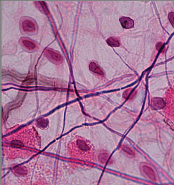
Name this
Connective and Supporting
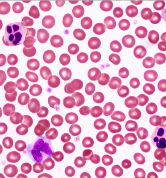
Name this
Hematalymphoid Complex

Name this
Muscle Tissue
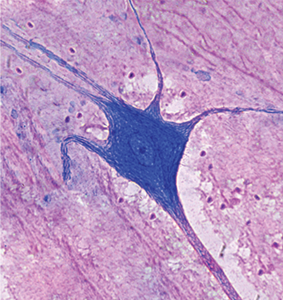
Name this
Nervous Tissue
Epithelial Tissue
Cells held tightly together, found on internal and external surfaces of the body
Connective and Supporting Tissue
Connects and supports tendons, ligaments, bones, and cartilage. Less cells, more “Other Stuff.“
Hematolymphoid Complex
Blood and lymph of the body. Red blood cells, white blood cells, plasma, water.
Muscle Tissue
Proteins that contract
Nervous Tissue
Sends, receives, and processes signals. Control center of the body.
Avascular
Inability to carry blood supply. Epithelial Tissue.
Vascular
Ability to carry blood supply and carry out exchange. Connective Tissue
General Functions of Epithelial Tissues
Absorption
Protection
Secretion
Excretion

Name this
Squamos (flat) epithelial cell

Name this
Cuboidal epithelial cell

Name this
Columnar epithelial cell
“Simple“
1 single layer of cells in a tissue
“Stratified“
2+ layers of cells in a tissue
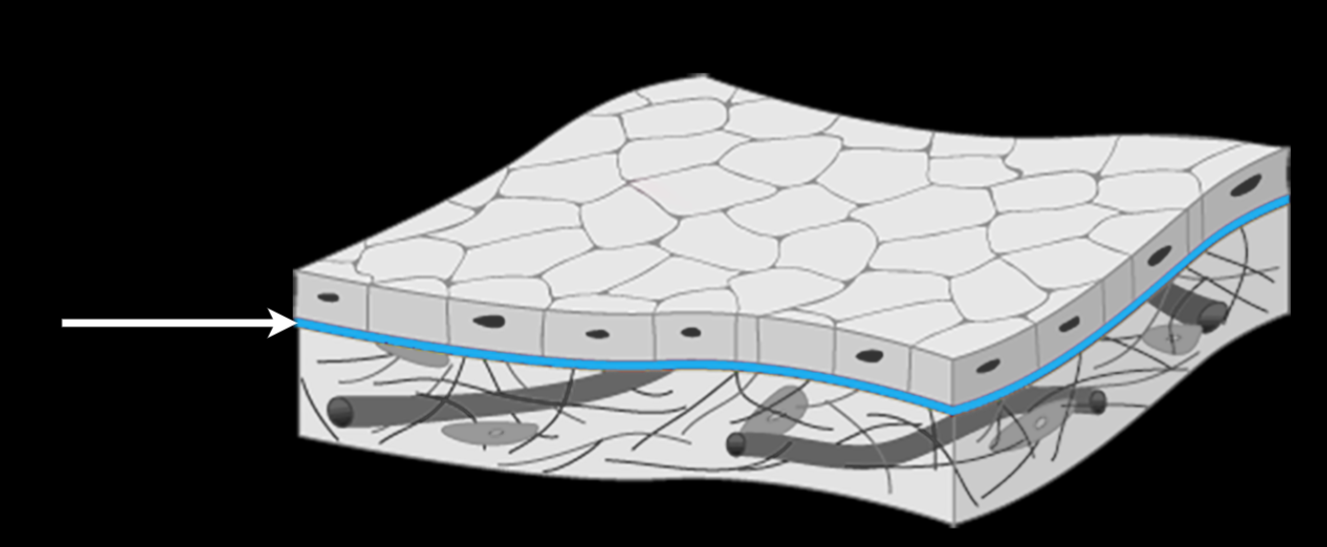
Basal Lamina
Transitional layer between epithelial and connective tissue. Allows for filtration between layers and anchors epithelial to connective tissue.

Name this tissue
Simple squamos epithelium
1 layer thick
flat cells
Endothelium
Simple squamos epithelium layer of specialized cells that lines the interior surface of blood vessels, lymphatic vessels, and the heart
Mesothelium
Simple squamos epithelium layer that serves as a protective membrane made of a single layer of specialized cells that line the body's internal cavities and organs. Reduces friction and allows for easy exchange between systems.

Name this tissue
Simple cuboidal epithelium
1 layer
cube shaped cells
found in lining of glands, function is secretion

Name this tissue
Simple Columnar Epithelium
1 layer
column shaped cells
can be ciliated or nonciliated
Found in inner lining of digestive system, function is absorption and secretion
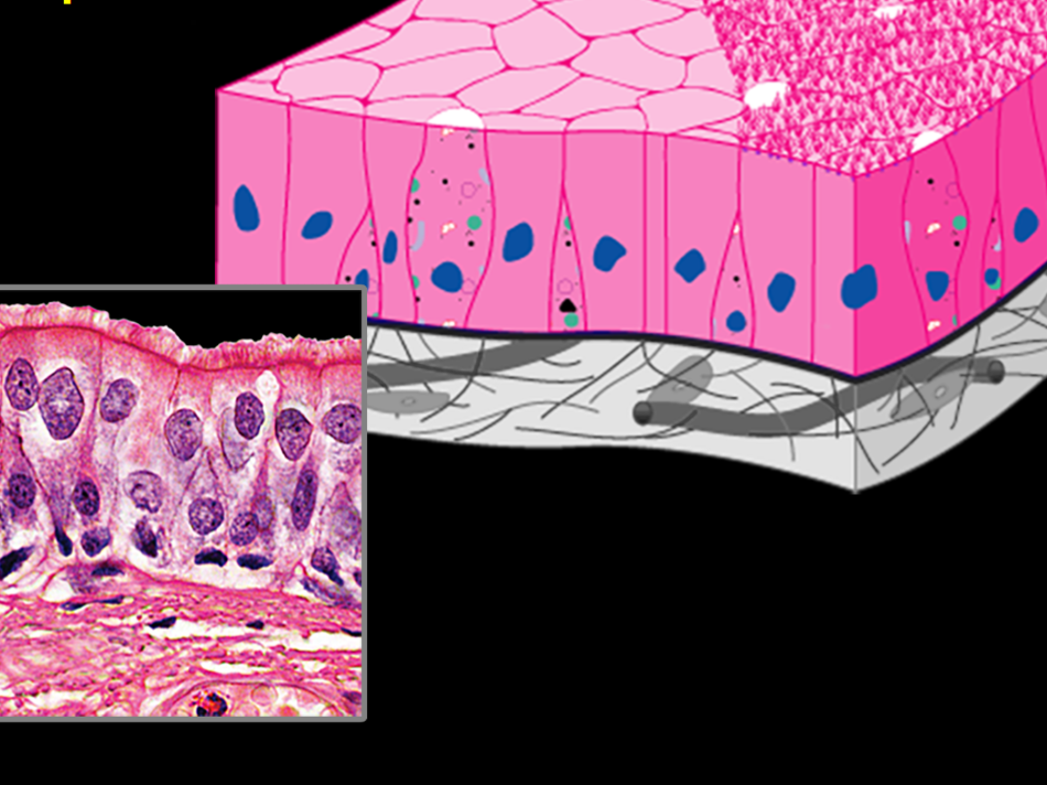
Name this
Psuedostratified columnar epithelium
1 layer technically
can be ciliated or nonciliated, but only focus on ciliated
Found in lining of respiratory system, function is to secrete mucus and protect against air pollution. cilia moves mucus up, to get expelled or swallowed

Name this
Stratified Squamos Epithelium
Multiple layers
Flat squished cells
Keratinized or Nonkeratinized
Outer layer of skin, lining of mouth and esophogus. Function is to protect against abrasion, and prevent waterloss
Keratinized
Stratified squamos epithelium that has a thick, dead, outer layer of skin, filled with protein keratin. Epidermis of skin, palms, and soles of feet.
Non-keratinized
Simple squamos epithelium consists of living, nucleated cells all the way to the surface, and can stay moist. Found in oral cavity, esophagus, vaginal canal, and cornea
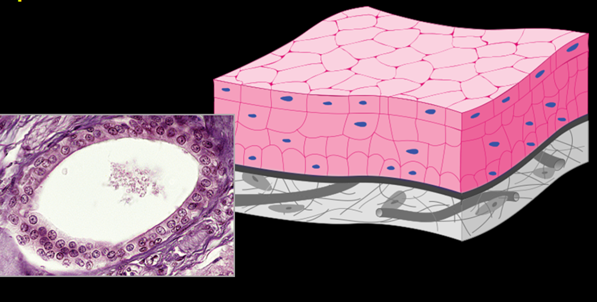
Name this
Stratified cuboidal epithelium
Multi-layer
cube shaped cells
rare (A.K.A. not studied anywhere else in class.)
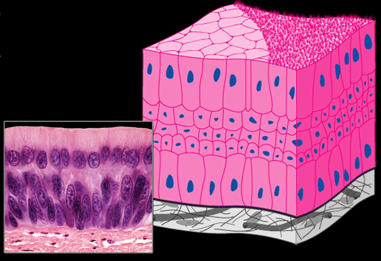
Name this
Stratified Columnar Epithelium
Multi-layer
Top layer is Column shaped cells
Can be cilliated or noncilliated
Rare, but function is protection.
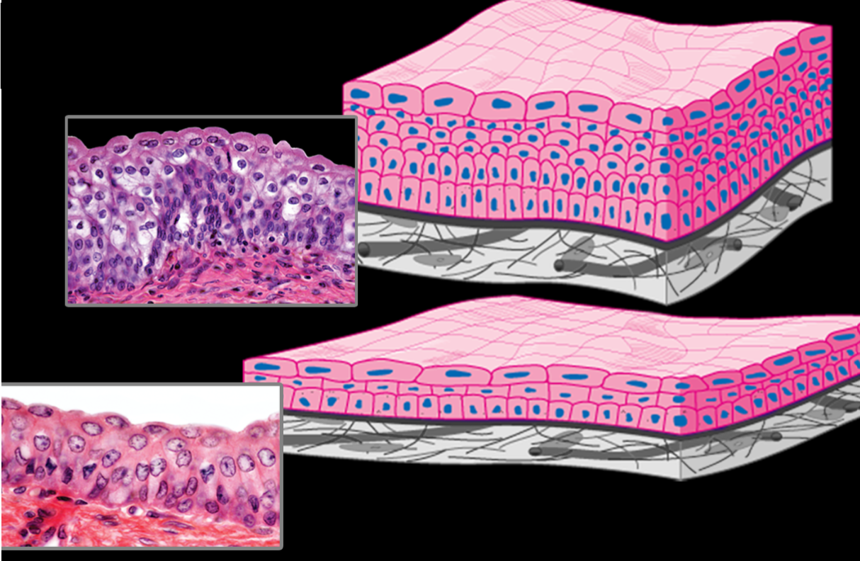
Name this
Urothelium or Transitional Epithelium
changes structure when in use versus not in use
can stretch when full, and contract when empty.
Multi-layered, to prevent exchange/waste passing through
Found in bladder/urinary system, function is to save space and transport urine.
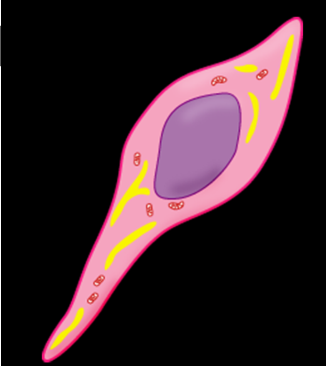
Name this
Mesenchymal cell
found during embryotic/fetal growth periods and can changes into different cells

Name this
Fibroblast and/or Fibrocyte
Blast means “builder.“ These are the main cells or the connective tissue and are responsible for producing fiber components of the extracellular matrix

Name this
Adipocyte
Fat cells. Used for energy storage, contain a lipid droplet.

Collagen Fiber
Non-branching protein fibers
very strong and resistant againts pulling/tension
Not good at resisting compression

Elastic Fiber
Highly branched, thin, stretchy fibers in the extracellular matrix.
Recoil/retain original shape well
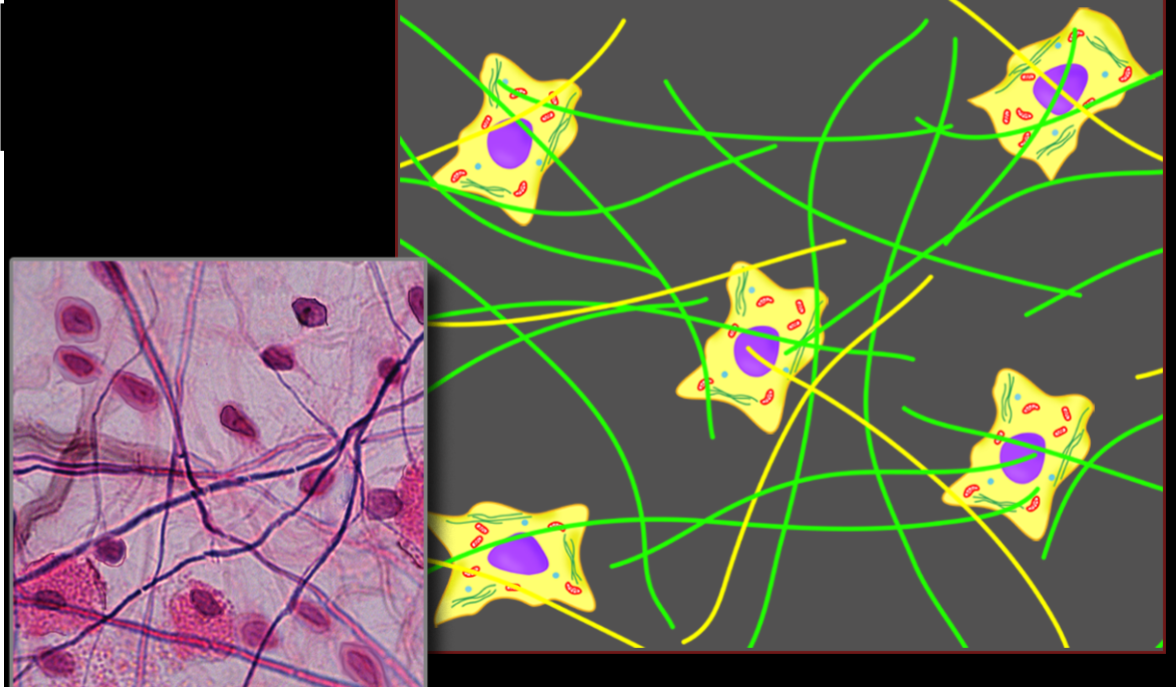
Loose connective tissue
Sparse network of random and loosely arranged fibers
Found everywhere; the most vascular tissue of the whole body, almost ALWAYS found uder epithelial tissue. Function is to hold body together and be vascular

Dense irregular connective tissue
Strong, resists tension from all directions
Found in dermis of skin, strong and resists tearing
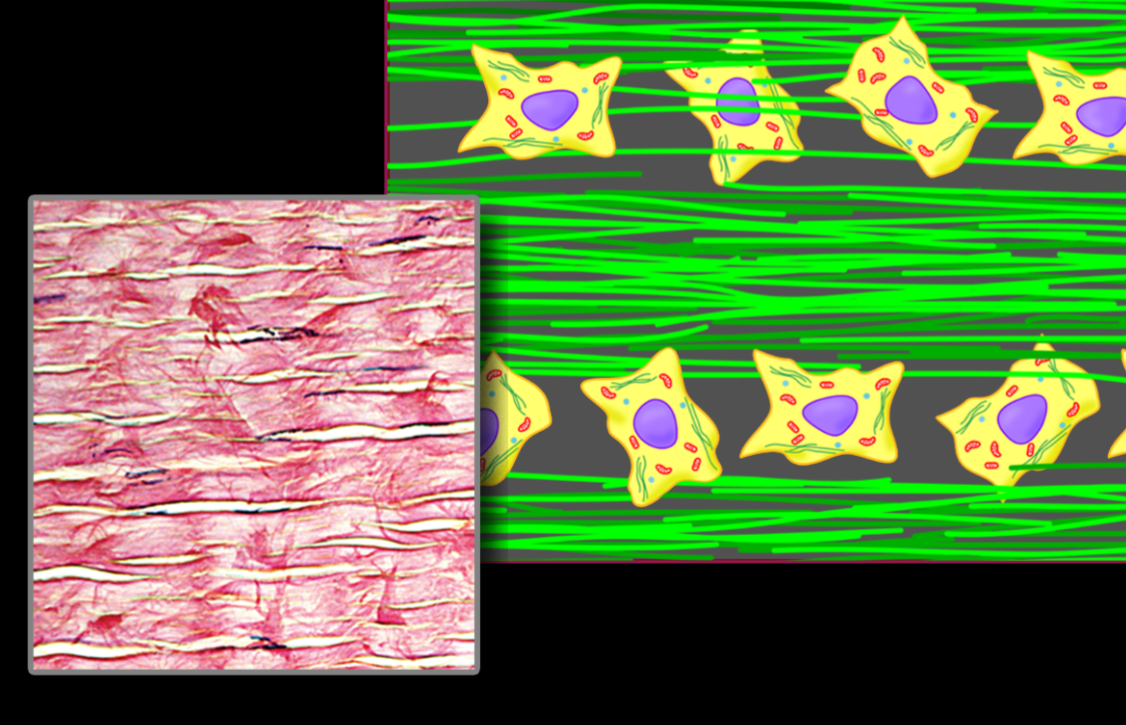
Dense regular connective tissue
Organized layout, straight collagen, so when cells get pulled in one direction, everything is is alligned.
Found in tendons, so when muscle is activated, it only pulls the bone one direction

Elastic Connective tissue
Made completely of elastic fibers, no collagen. Lots of thin, branching fibers.
Found in walls of large blood vessel (Aorta). Expands to fit fast pumping blood and recoils to keep it moving and maintain structure/shape.

Reticular Tissue
Has rare collagen that branches, forming a net of collagen. This lets other cells to get caught.
Found in lymph nodes and red bone marrow. Filters out body, as pathogens get caught in net of lymphocytes in the lymph node, they get filtered out.
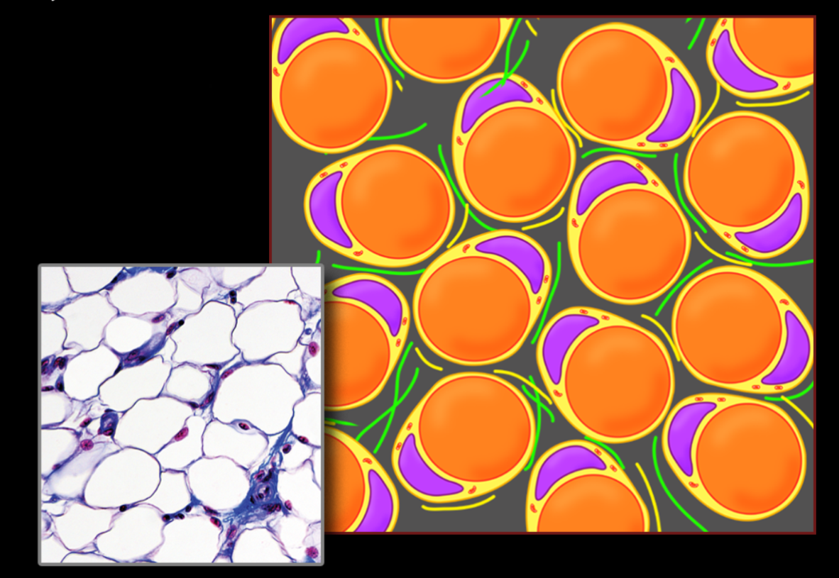
Adipose Tissue
Made up of adypocyte cells. Potentially distributed everywhere/everywhere there is fat. Loose connective tissue can turn into adipose tissue with fat gain. Function as energy storage and protection to the internal organs.
Integument
Covering organ system
Epidermis
Dermis
Subcutaneous Tissue layer
Skin
Organ made up of the epidermis and dermis layer.
Functions of Integument System
Sensory
Excretion
Thermoregulation
Fat metabolism
Exchange surface with environment
Vitamin D production
Sociosecual communication and identification
Epidermis
Type of epithelium tissue, Keratonized stratified squamos epithelium
stratum basale
stratum spinosa
stratum granulosum
stratum luciderm (palms and soles)
Stratum Corneum

Stratum Basale
Lowest part of the Epidermis layer. Contains miotic spindle which divides skin cells and form strata/layers. Closest to connective tissue, so access to vascularity = living cells
Mytotic Spindle
Faciliates cell reproduction in the stratum basale layer of the epidermis
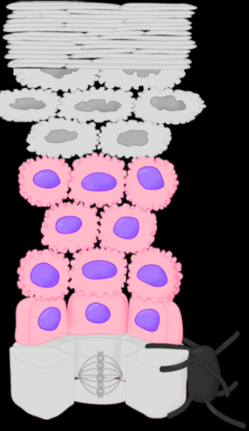
Stratum Spinosum
No longer healthy enough to divide, as they are too far from vascular supply. Cells start to get pushed up by newer dividing cells. Starts to build keratin.

Stratum Granulosum
Actively dying causing them to leak lipids, which creates waterproof barriers/makes skin hydrophobic. Cellas are rapidly flattening, making them take on spiny apprearance, which locks the cell membranes even tighter together.
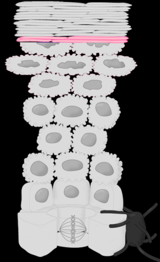
Stratum Lucidum
Only in the thickest skin of the body, palms of hands and soles of feet. Devoid of hairs, and is often included in the stratum corneum
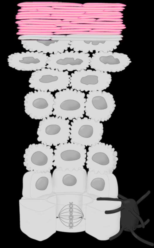
Stratum Corneum
The most outer layer of the epidermis, forms closely packed layers of dead cells. This allows for protection from abrasion.
Dermis
Second layer of the integument system. Has a superior layer of loose connective tissue, that brings vascularity to epidermis, and a dense irregular connective layer that creates toughness.

Papillary layer
Loost connective tissue, that is highly vascular. Papilla (nipple bumps) increase surface area, increasing the epidermis’ access to blood supply. Papilla also create finger prints.

Reticular Layer
Dense irregular connective tissue layer of the dermis. Creates very tough “leather“ layer of skin from thick nets of collagen. Creates cleavage lines in the skin that cause tears and stretch marks.

Subcutaneous Tissue
The lowest layer of the integument system, not technically apart of the skin organ however. This can be composed of adipose tissue or loos connective tissue, depending on fat content. Made up of a Fatty layer and Fibrous layer of collagen.

Hair Follicle
The space where the bulb of hair, root, and shaft form. This layer is conitnuous with epidermis, but not considered the same structure.

Arrector Pili Muscle
Smooth muscle that attaches epidermis to base of hair follicule, creating goose bumps
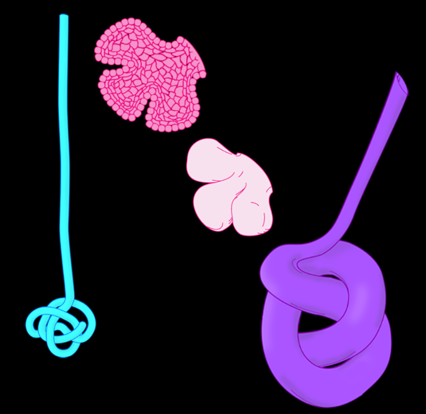
Skin glands
Sebaceous holocrine gland - Sebum, oily natural lotion substance
Eccrine Sweat gland - Produces sweat to cool your body
Apocrine Sweat Gland - Armpits, anal, and genital regious. Peremonal sweat for mating.