TOPIC 1: HEAD AND NECK
1/35
There's no tags or description
Looks like no tags are added yet.
Name | Mastery | Learn | Test | Matching | Spaced |
|---|
No study sessions yet.
36 Terms
SKULL
FRAMEWORK OF THE HEAD
DIVIDED INTO TWO SUBSECTIONS:
CRANIUM AND THE FACE
CRANIUM
HOUSE AND PROTECHTS THE BAIN AND MAJOR SENSORY ORGANS
FACIAL BONES
immovable except for the MANDIBLE
TEMPORAL ARTERY
major artery, is located between the eye and the top of the ear.
TWO IMPORANT STRUCTURES LOCATED IN THE FACIAL REGION
parotid
submandibular salivary glands
PAROTID GLANDS
located on each side of the face
anterior and inferior to the ears and behind the mandible
SUBMANDIBULAR GLANDS
located inferior to the mandible, underneath the base of the tongue.
THE NECK
composed of muscles, ligaments, and the cervical vertebrae.
STERNOMASTOID (STERNOCLEIDOMASTOID) AND TRAPEZIUS MUSCLES
two of paired muscles that allow movement and provide support to the head and neck.
STERNOMASTOID MUSCLE
ROTATES AND FLEXES THE HEAD
TRAPEZIUS MUSCLE
EXTENDS THE HEAD AND MOVES THE SHOULDERS
ELEVETH CRANNIAL NERVE
RESPONSIBLE FOR MUSCLE MOVEMENT THAT PERMIT SHRUGGING OF THE SHOULDERS BY THE TRAPEZIES MUSCLES.
TWMO MAJOR MUSCLES THAT FORM TWO TRIANGLE THAT SERVES AS A LANDMARK FOR ASSESSMENT
ANTERIOR TRIANGLE
POSTERIOR TRIANGLE
ANTERIOR TRIANGLE
LOCATED UNDER MANDIBLE
POSTERIOR TRIANGLE
LOCATED BETWEEN THE TRAPEZIUS AND STERNOMASTOID MUSCLES
C1 TRHOUGH C7
POSTERIOR NECK AND SUPPORT THE CRANIUM
C7
VERTEBRA PROMINENT
CAN SEASILY BE PALPATED WHEN THE NECK IS FLEXED
USING C7 WILL HELP YOU TO LOCATE OTHER VERTEBRAE.
INTERNAL JUGULAR VEINS AND CAROTID ARTERIES
LOCATED BILATERALLY, PARALLEL AND ANTERIOR TO THE STERNOMASTOID MUSCLES.
EXTERNAL DUGULAR VEIN
DIAGONALLY OVER THE SURFACE OF THESE MUSCLES.
NEED TO KNOW THE LOCATION OF CAROTID ARTERIS
TO AVOID BILATERAL COMPRESSION OF THE CESSEL
CAN REDUCE THE BLOOD SUPPLY TO THE BRAIN
THYROID GLAND
LARGEST ENDOCRINE GLAND IN THE BODY
PRODUCED HORMONES THAT INCREASE THE METABOLIC RATE OF MOST BODY CELLS
KINDS OF CHARACTERISTICS OF HEADACHE
MIGRAINE
CLUSTER
TENSION
TUMOR RELATED
MIGRAINE
acoompoanied by nausea, vomiting and sensitiviy to noise or light
location: around eyes, temples, cheecks, or forehead
duration: last up to 3 days
severity: throbbing. severe, recurring
pattern: rest may bring relief
associated factors: migraines occur more often in women
CLUSTER
accompanied by tearing, eyelid drooping, reddened eye, or runny nose.
sudden onset
may be precipitated by ingesting alcohol
localized in the eye and orbit and radiating to the facial and temporal region
typically occurs in the late evening or night
intense and stabbing
movement or walking back and forth may relieve the discomfort
cluster headaches oocur more in young male
Tension
symptoms of anxiety, tension, and depression may be present
no prodromal stage
may occur with stress, anxiety or depression
usually located in the frontal, temporal or occipital region
lasts dyas, months or years
dull, aching, tight or diffuse
symptomatic relief may be obtained by local heat, massage, analgesics, anti-depressants, must be relaxants
tesnion headached affect women more ofen than men.
Tumor Related
neurologic and mental symptoms and nausea and vomiting may develop
no prodromal stage
amy be aggravated by coughing, sneezing, or sudden movements of head
varies with location of tumor
commonly occurs in the morning and lasts for several hourse
aching, steady, variable in intensity
headahce usually, subsides later in the days.
EQUIPMENT
GLOVE
SMALL CUP OF WATER
STETHOSCOPE
INSPECT THE NECK
OBSERVE THE CLIENT’S SLIGHTLY EXTENDED NECK FOR POSITION, SYMMETRY AND LUMPS OR MASSES. SHINE A LIGHT FROM THE SIDE OF THE NECK ACROSS TO HIGHLIGHT ANY SWLLING.
INSPECT MOVEMENT OF THE NECK STRUCTURES
ASK CLIENT TO SWALLO A SMALL SIP OF WATER. OBSERVE THE MOVEMENT OF THE THYROID CARTILAGE.
INSPECT THE CERVICAL VERTEBRAE
ASK THE CLIENT TO FLEX THE NECK CHIN TO CHEST, EAR TO SHOULDER, TWIST LEFT TO RIGHT AND RIGHT TO LEFT, AND BACKWARD AND FORWARD.
INSPECT RANGE OF MOTION
ASK THE CLIENT TO TURN THE HEAD TO THE RIGHT AND TO THE LEFT CHIN TO SHOULDER, TOUCH EACH TO THE SHOULDER, TOUCH CHIN TO CHEST, AND LIFT THE CHIN TO THE CEILING.
PALPATE FTHE TRACHEA
PLACE YOUR FINGER IN THE STERNAL NOTCH
PALPATE THE THYROID GLAND
LOCATE KEY LANDMARKS WITH YOUR INDEX FINGER AND THUMBS
PALPATING THE TRACHEA
PLACE YOUR FINGER ALONG ONE SIDE OF THE TRACHEA. ASSESS THE DISTANCE BETWEEN THE TRACHEA’S OUTER EDGE AND THE STERNOCLEIDOMASTOID MUSCLES. THEN ASSESS THE DISTANCE ON THE OTHER SIDE, AND COMPARE THE TWO DISTANCE. THEY SHOULD BE THE SAME.
AUSCULTATION
AUSCULTATE THE THYROOID ONLY IF YOU FIND AN ENLARGED THYROID GLAND DURING INSPECTION OR PALPATION.
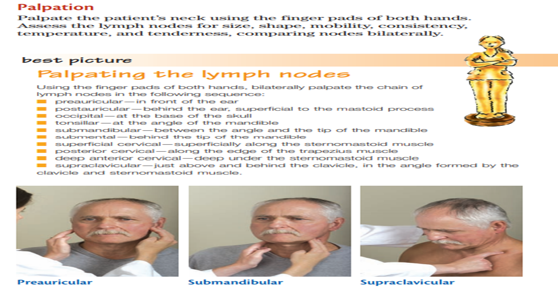
PALPATION