Chapter 5 - The Integumentary System
1/14
There's no tags or description
Looks like no tags are added yet.
Name | Mastery | Learn | Test | Matching | Spaced |
|---|
No study sessions yet.
15 Terms
Describe the functions of skin
Skin is first and foremost a barrier
Protection
Body temperature regulation
Cutaneous sensation
Metabolic functions
Blood reservoir
Excretion
List the two layers of skin
EpidermisEpithelial cells, outermost layer
DermisBulk of skin; tough and leathery layer, mostly dense connective tissue
Resting on Subcutaneous tissue
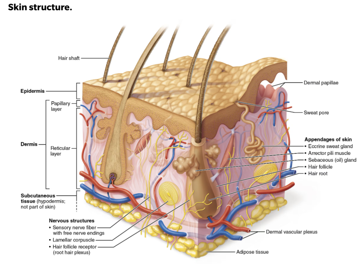
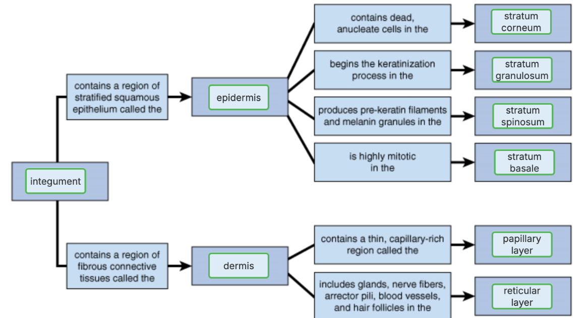
Define the Epidermis
Superficial layer of the skin
Keratinized sheet of stratified squamous epithelium
Avascular but innervated
Epidermis - Layers
Stratum corneum (Horny Layer)
Most superficial
20-30 layers of dead cells
Stratum lucidum (Clear Layer)
Only in thick skin
Few rows of flat, dead keratinocytes identical to stratum corneum
Stratum granulosum (Granular Layer)
1-5 layers of flattened cells, organelles deteriorating; keratinocytes that are undergoing keratinization
Keratohylaine granules → help to form keratin in the upper layers by providing the “glue” that binds pre-keratin intermediate filaments together to form keratin
Lamellar granules → contain a water-resistant glycolipid that is secreted into the extracellular space
Stratum spinosum (Prickly Layer)
Several layers of keratinocytes unified by desmosomes
Stratum basale (Basal Layer)
Deepest layer
Single row of actively mitotic stem cells
Epidermis - Cell Types & Functions
Keratinocytes
LOCATION: Predominat cells of the epidermis → Stratum basale (deepest part of the epidermis)
DESCRIPTION: Tightly connected by demosomes
Millions slough off every day
FUNCTION:Produce keratin (protein that gives skin its protective properties)
Melanocytes
LOCATION: Stratum basale (deepest part of the epidermis
DESCRIPTION:Spider-shaped cells located in deepest epidermis
Melanosomes are transferred to keratinocytes, where they protect the nucleus from UV damage
FUNCTION: Produce pigment melanin, which is packaged into melanosomes
Dendritic (Langerhans) cells
LOCATION: Arise from bone marrow and migrate to the epidermis
DESCRIPTION: Star-shaped macrophages that patrol deep epidermis
FUNCTION: key activators of immune system
Tactile (Merkel) cells
DESCRIPTION: Spiky, hemi-spheral epidermal cells
LOCATION: Epidermal-dermal junction
FUNCTION: Sensory receptors that sense touch; associated with free nerve endings
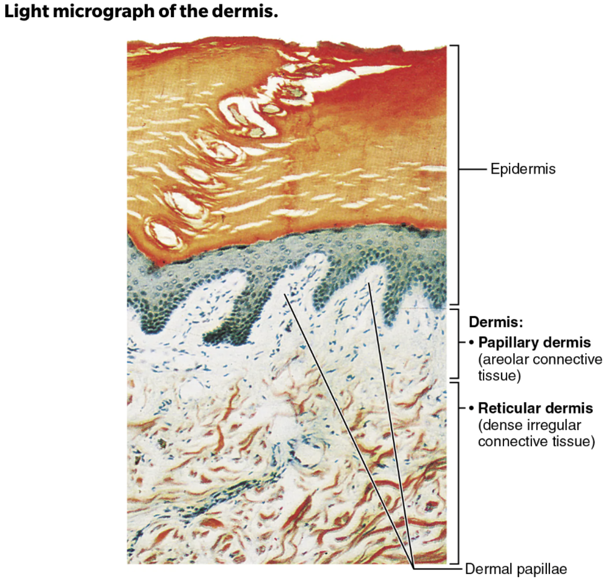
Define Dermis
Layer of skin deep to the epidermis
Composed mostly of dense irregular connective tissue
Strong, flexible connective tissue
Cells include fibroblasts, macrophages, and occasionally mast cells and white blood cells
Fibers in matrix bind body together
Makes up the “hide” that is used to make leather
Dermis - Layers
Papillary dermis
Thin, superficial layer
Areolar connective tissue with interlacing collagen and elastic fibers
Many small blood vessels
Reticular Layer
Deeper layer; Makes up ~80% of dermal thickness
Dense irregular connective tissue
Many elastic fibers provide stretch-recoil properties
Collagen fibers provide strength and resiliency
Bind water, keep skin hydrated
Dermis - Function
Dense irregular connective tissue
Supplied with:
Blood vessels
Lymphatic vessels
Reside within the derms:
Epidermal hair follicles
Oil glands
Sweat glands
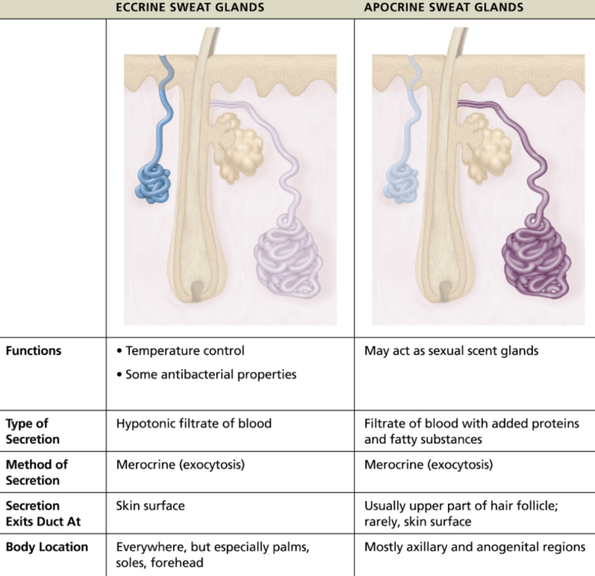
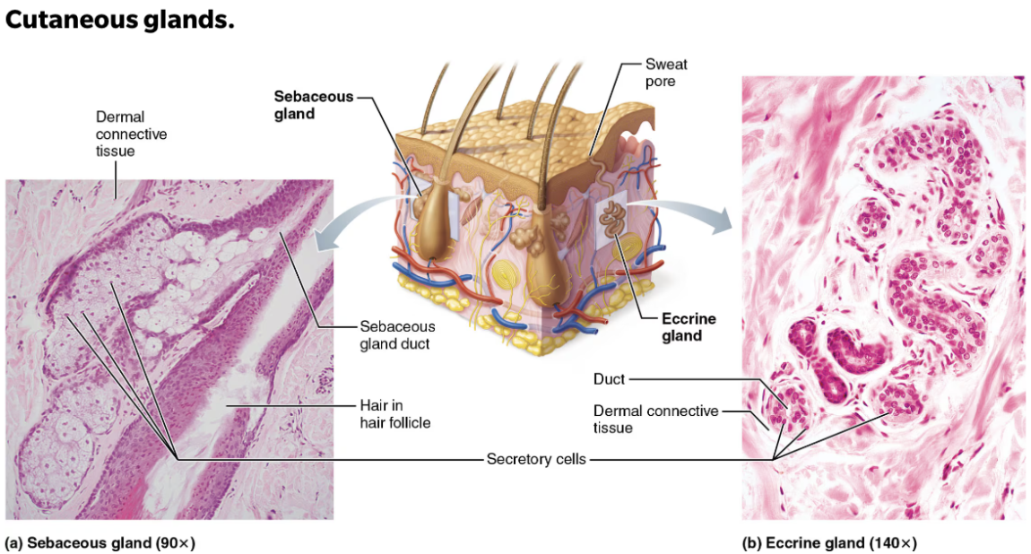
Define Sweat Glands
Also called sudoriferous glands
FUNCTION:
Epidermal glandthat produces sweatLOCATION: All skin surfaces except nipples and parts of external genitalia contain sweat glands
About 3 million per person
Compare and contrast eccrine and apocrine glands
Define Sebaceous (oil) Glands
LOCATION: Widely distributed, EXCEPT for thick skin of palms and soles
Most develop from hair follicles and secrete into hair follicles
Relatively inactive until puberty
Stimulated by hormones, especially androgens
FUNCTION:
Epidermal glandsthat produce an oily secretion called sebum
Define Sebum
STRUCTURE: Oily holocrine secretion
Bactericidal (bacteria-killing) properites
FUNCTION: Softens hair and skin
Increase their activity during puberty and are controlled by androgens.
What can influence skin color?
Skin color reflects the amount of pigments (melanin and carotene) in the skin and the oxygenation level of hemoglobin in blood
Melanin
Dark pigment formed by cells called melanocytes
Responsible for color to skin and hair
Produced in the stratum basale of the
epidermis
Carotene
Yellow to orange pigment that accumulates in the stratum corneum
epidermal layerand in fatty tissue of thesubcutaneous tissue
Hemoglobin
Reddish color of red blood cells in blood vessels that can be seen in Caucasian skin due to translucent skin
Circulating through the
dermalcapillaries
Examples how alterations in skin color can indicate disease
Cyanosis
Blue skin color → low oxygenation of hemoglobin
Pollar (blanching or pale color)
Anemia, low blood pressure, fear, anger
Erythema
Fever, hypertension, inflammation, allergy
Jaundice (yellow cast)
Liver disorders
Newborns
Bruises (black and blue marks)
Also referred to as ecchymoses or hematomas, are a result of clotted blood beneath skin
As clot is broken down, color of bruise changes
Disease and ailments that can affect skin color
Basal cell carcinoma
DESCRIPTION
Least malignant and most common
Stratum basale cells (
epidermis) proliferate and slowly invadedermisandhypodermis
TREATMENT
Cured by surgical excision in 99% of cases
Squamous cell carcinoma
DESCRIPTION
Second most common type; can metastasize
Involves keratinocytes of stratum spinosum (
epidermis)Usually is a scaly-reddened papule on scalp, ears, lower lip, or hands
TREATMENT
Good prognosis if treated by radiation therapy or removed surgically
Melanoma
DESCRIPTION
Cancer of melanocytes
Is most dangerous type because it is HIGHLY metastatic and resistant to chemotherapy
TREATMENT
Treated by wide surgical excision accompanied by immunotherapy
Key to survival is early detection