Ophtho pics
1/63
There's no tags or description
Looks like no tags are added yet.
Name | Mastery | Learn | Test | Matching | Spaced |
|---|
No study sessions yet.
64 Terms
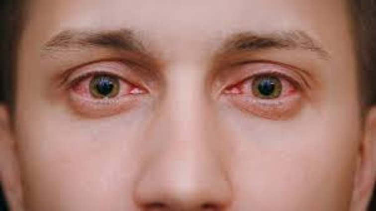
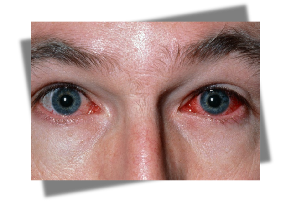
allergic conjunctivitis
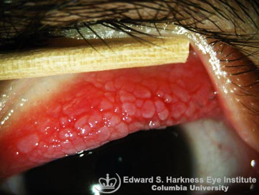
vernal keratoconjunctivitis
not copious
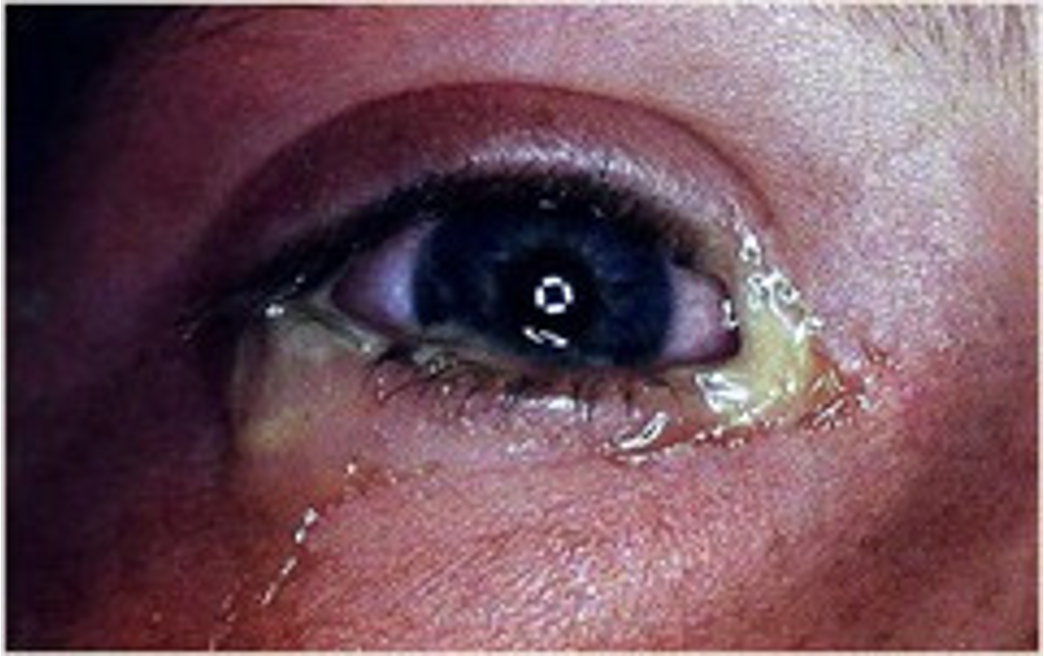
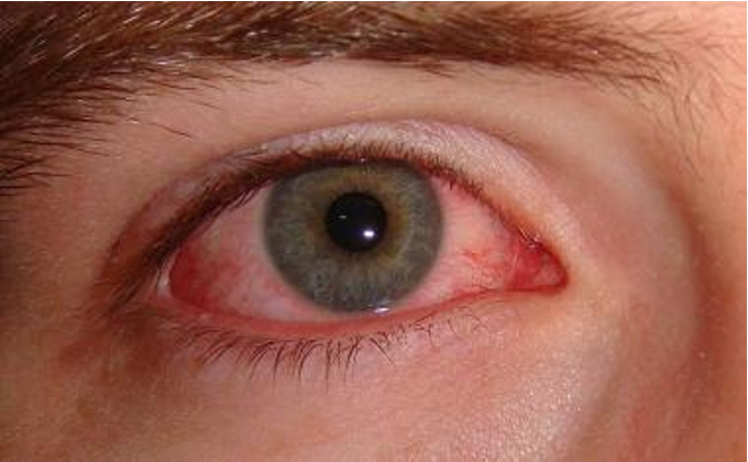
bacterial conjunctivitis
copious
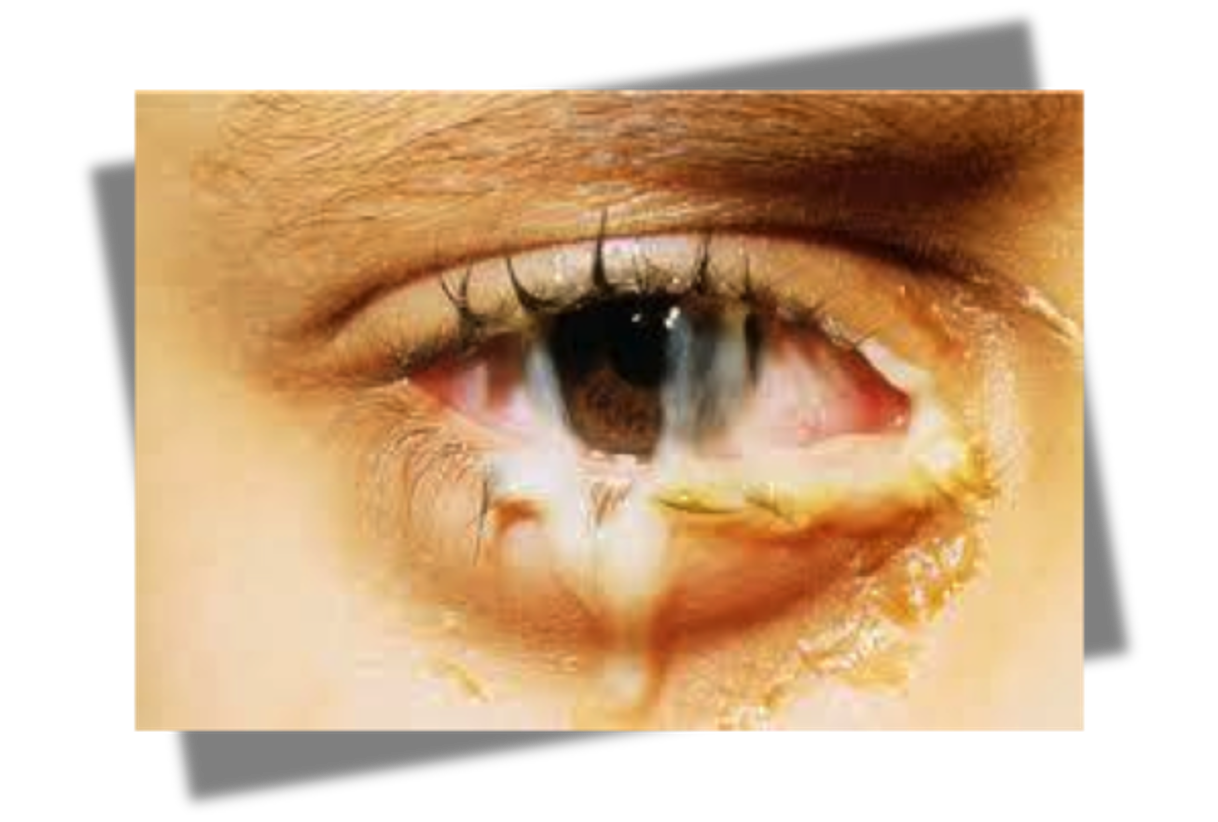
Bacterial gonorrhoeae conjunctivitis
gradual onset 1-4 weeks, clear to mucopurulent discharge, highly contagious
bacterial chlamydia conjunctivitis

slept with contacts in
contact lens conjunctivitis

(this pic has nothing to do with it, there wasn’t one to go off of)
not cleaning contacts or exposure to fungus
fungal conjunctivitis
viral (non herpetic) conjunctivitis or herpetic Idek the pics are the fkn same
watery discharge, contact with COVID
viral (COVID) conjunctivitis
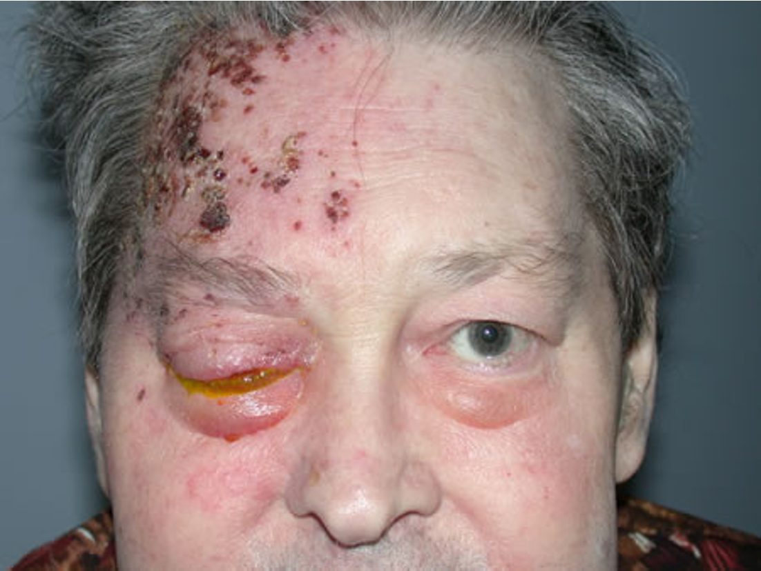
herpetic zoster ophthalmicus
chronic bilateral dry eyes and dry mouth
keratoconjunctivitis sicca (dry eye disease)
red, painful eye, corneal opacity
keratitis
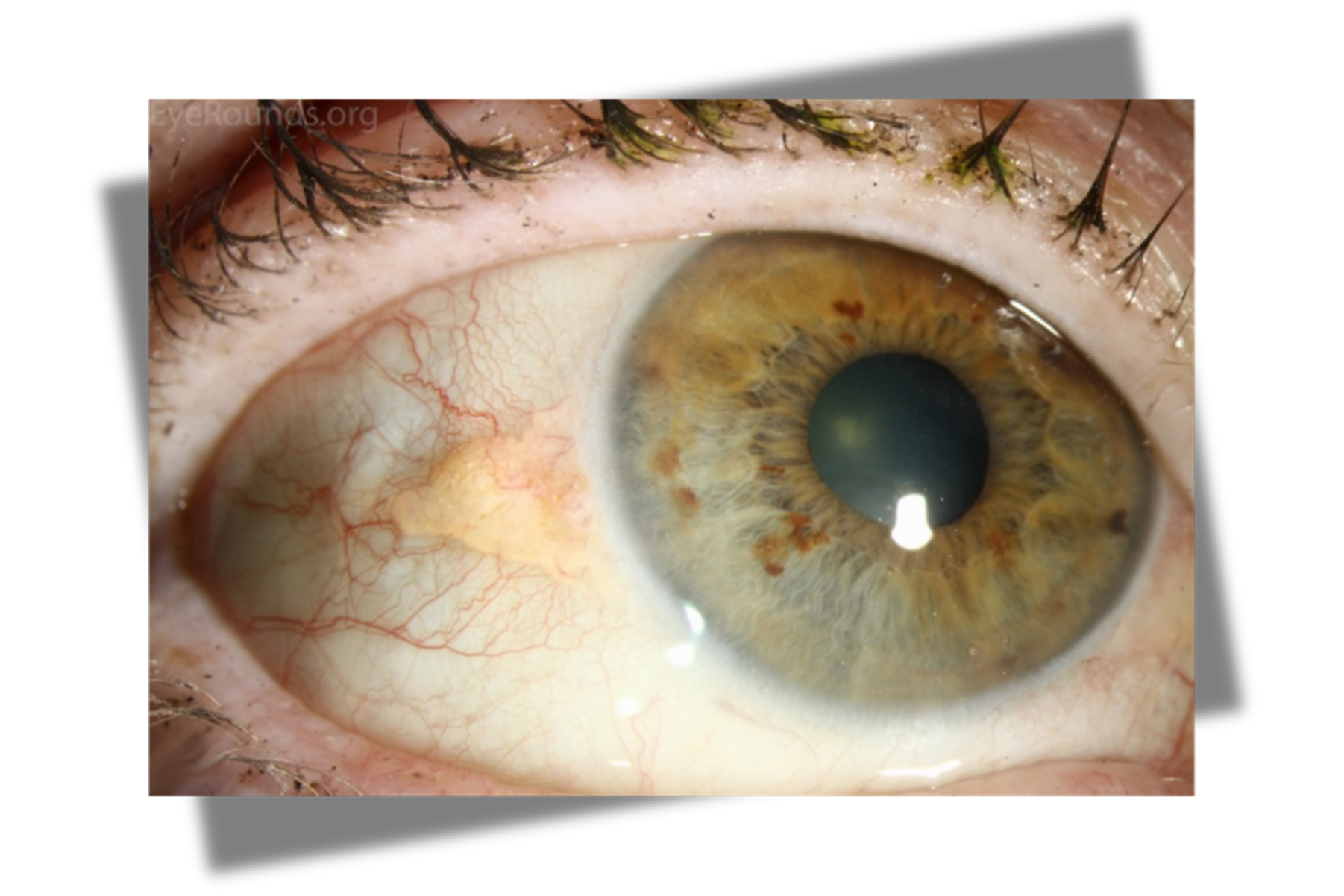
pinguecula
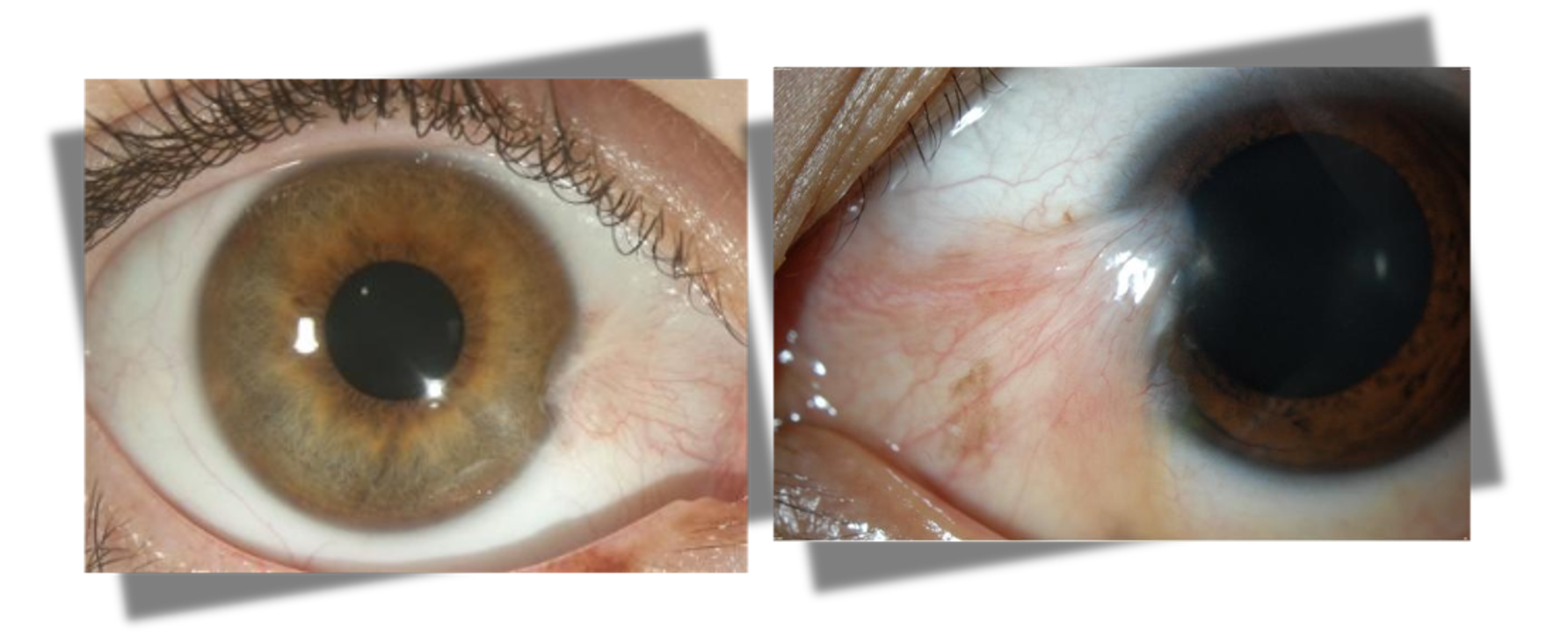
pterygium
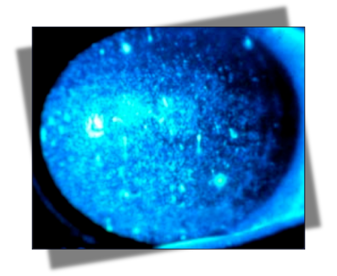
positive uptake w/ cobalt blue light; FB sensation, severe pain, photophobia
corneal abrasion
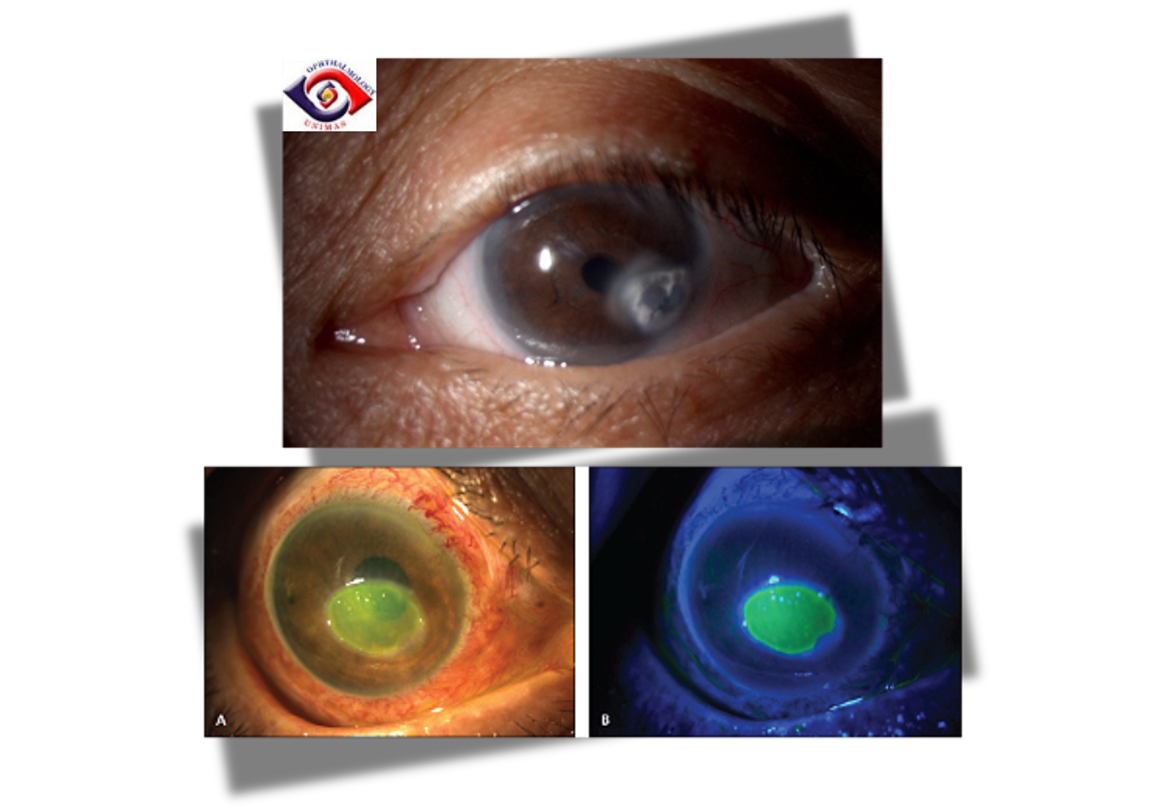
fluorescein stain shows pooling, cornea is hazy
corneal ulcer
burns to cornea
actinic keratitis / UV keratitis
dacryocystitis
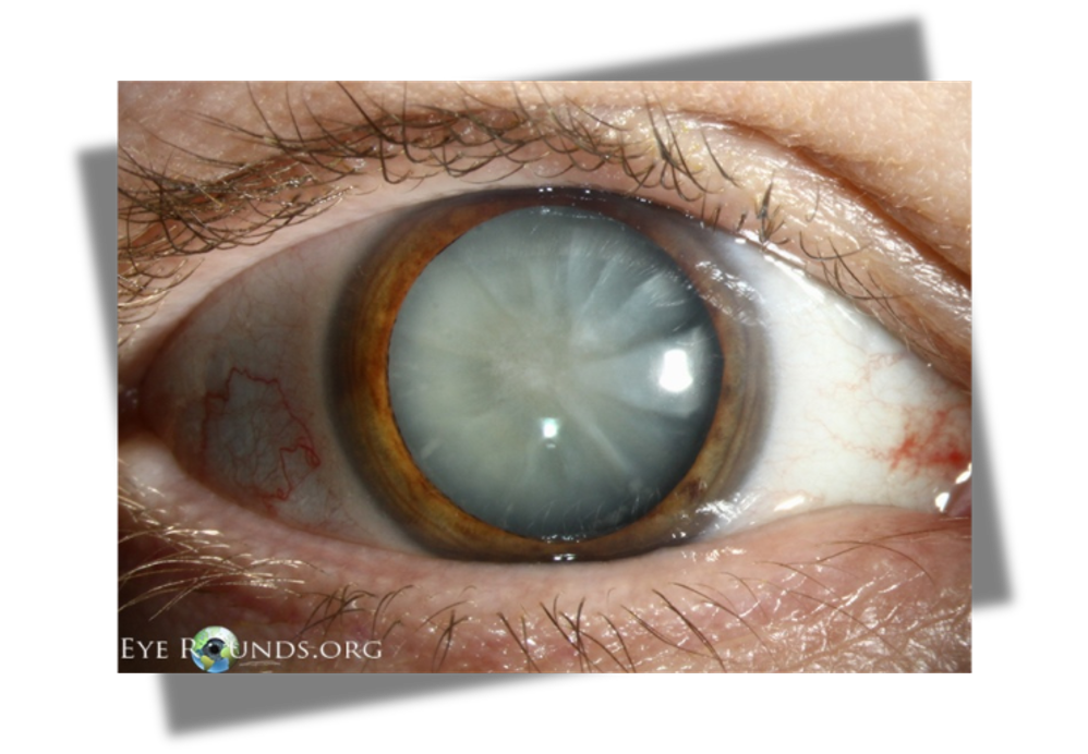
gradual progression of blurred vision more common in > 60 y.o
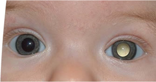
non traumatic cataract
blurred vision secondary to blunt or penetrating ocular trauma; young people
traumatic cataract
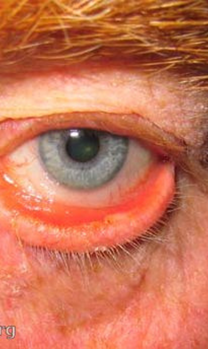
chronic bilateral inflammation of lid margins
anterior blepharitis
chronic bilateral inflammation of meibomian gland
posterior blepharitis
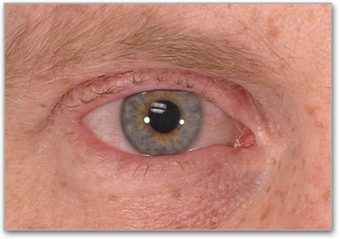
ectropion
entropion
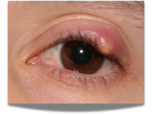
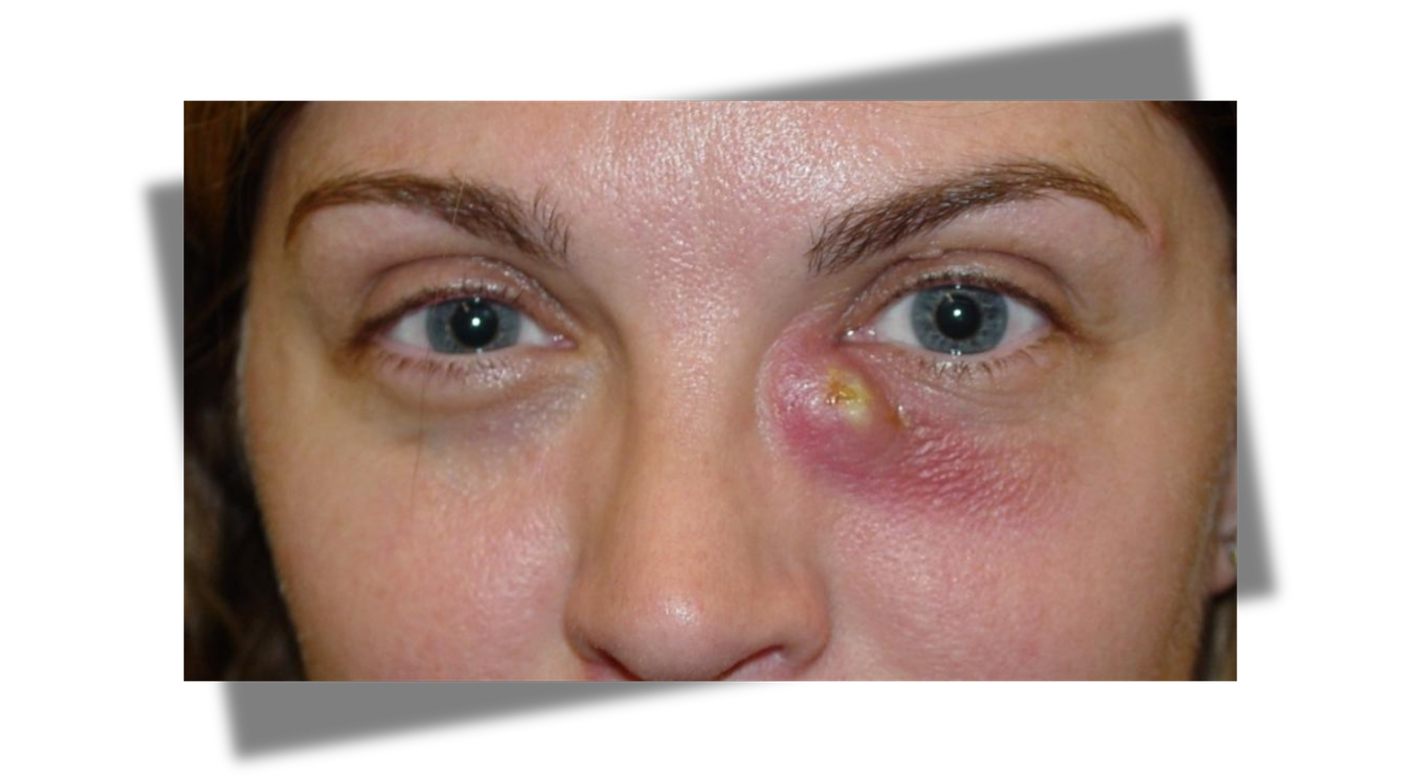
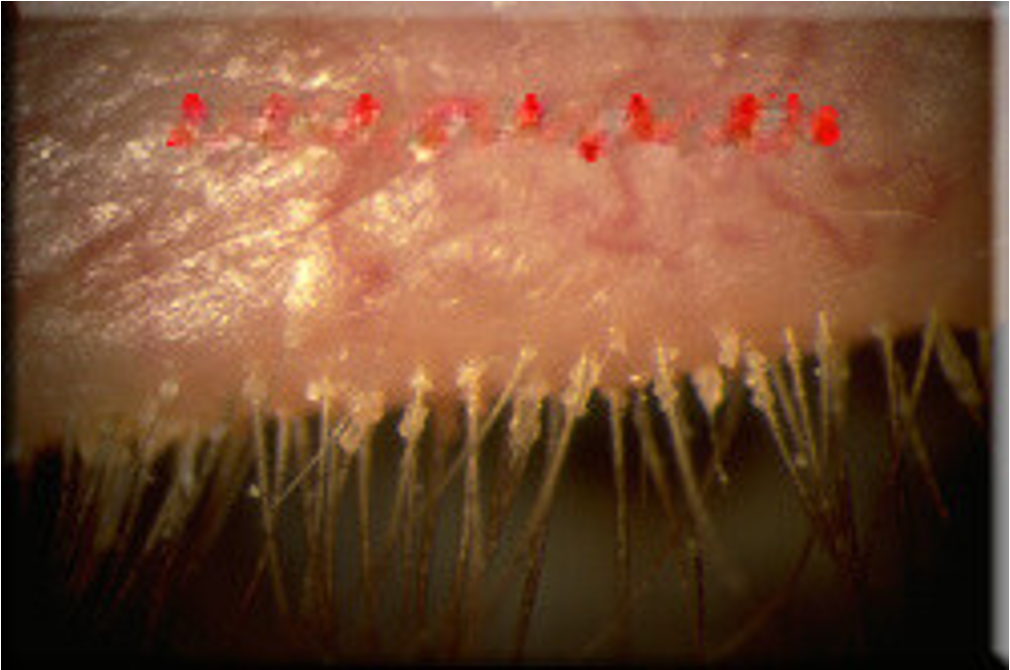
blockage of meibomian sebaceous gland in eyelid; painful
hordeolum (stye)
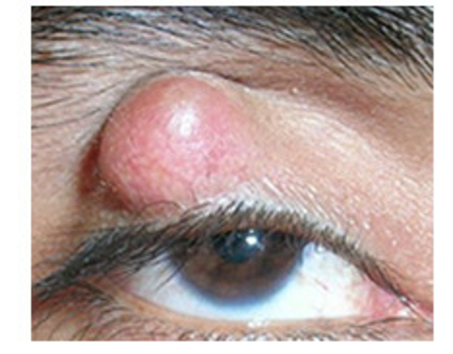
obstruction of meibomian gland; nontender; firm
chalazion
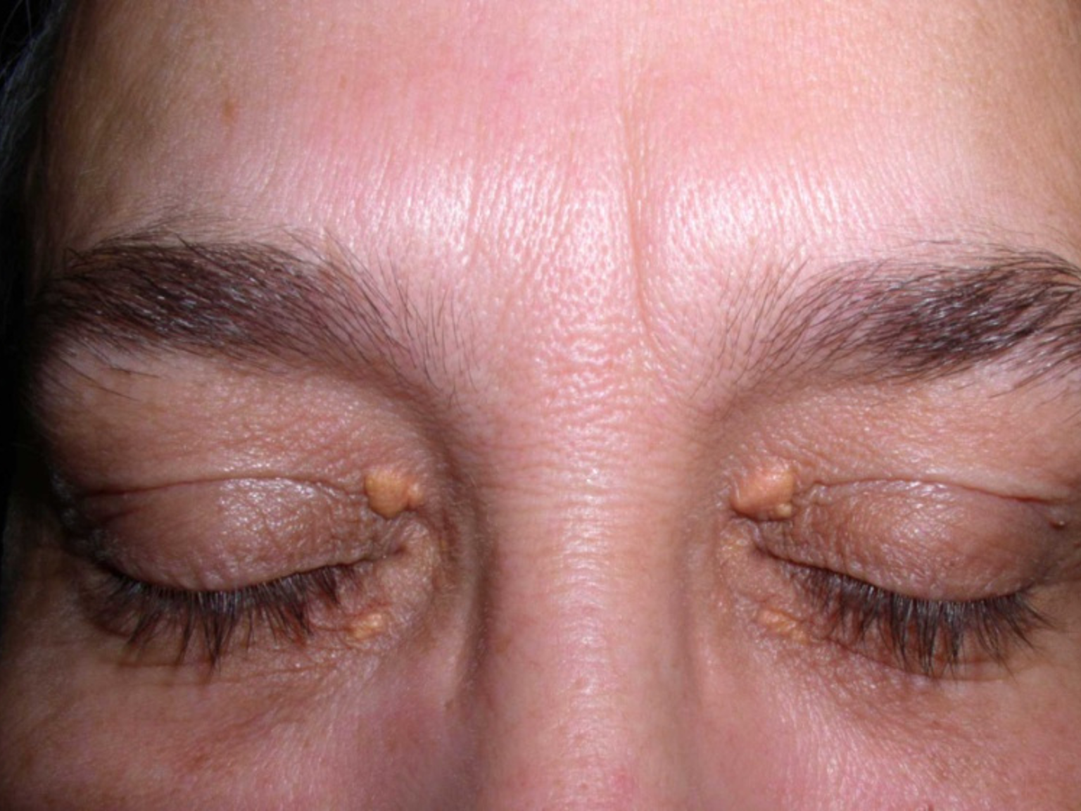
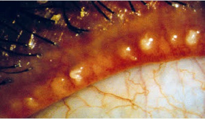
(yellow lesions)
xanthelasma
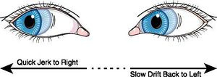
nystagmus

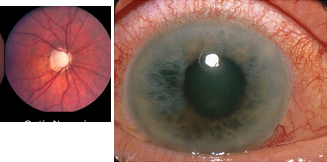
unilateral central loss of vision
optic neuritis
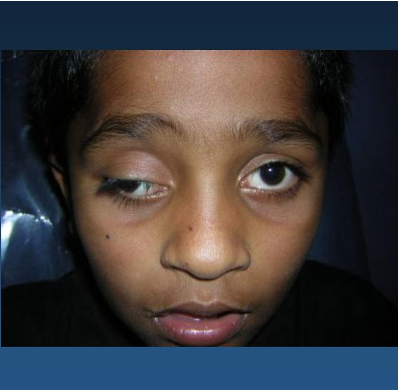
sudden dysfunction with oculomotor nerve
third nerve paralysis

diplopia, lack of superior oblique
fourth nerve paralysis
diplopia, lack of lateral rectus movement
sixth nerve paralysis
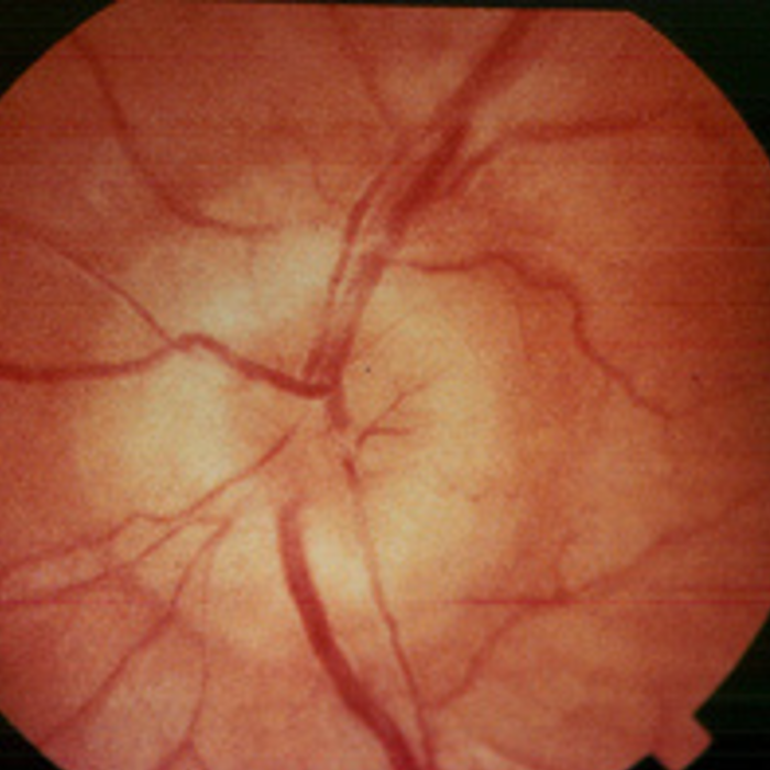
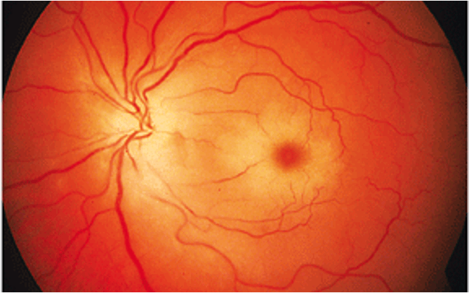
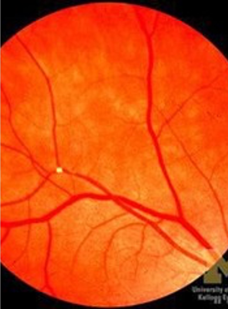
disc swelling due to severe HTN; inc intracranial pressure
papilledema
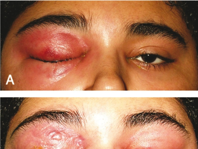
infection of anterior portion of eyelid
anterior periorbital cellulitis
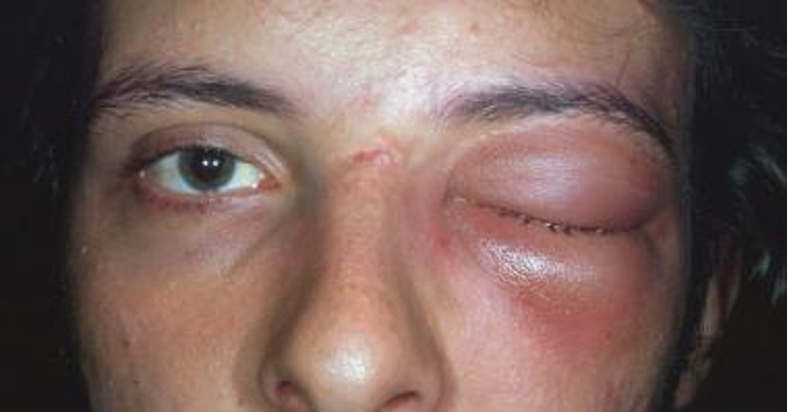
infection of orbit posterior to orbital septum
posterior orbital cellulitis / septal cellulitis
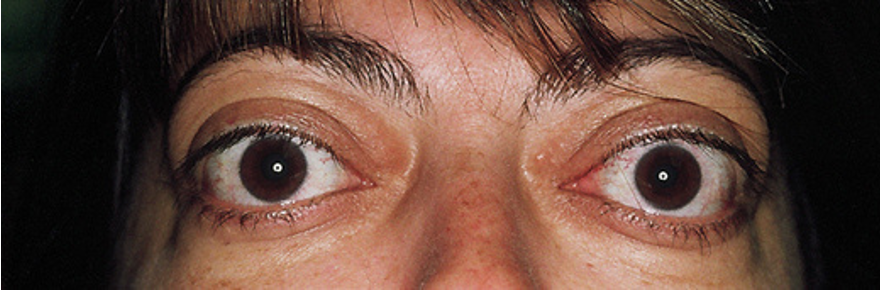
bilateral proptosis
thyroid eye disease / exophthalmos
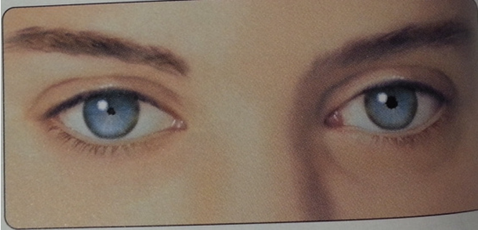
small pupils constrict on accommodation but not to light
argyll robertson pupil
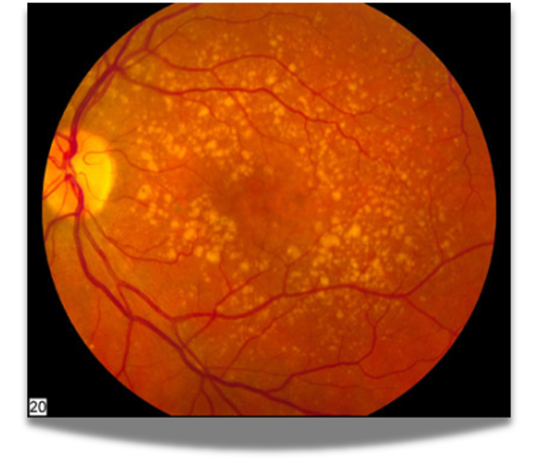
drusen
age related macular degeneration (ARMD)
slow breakdown of cones/rods; patchy focal atrophy and extensive confluent atrophy
dry ARMD
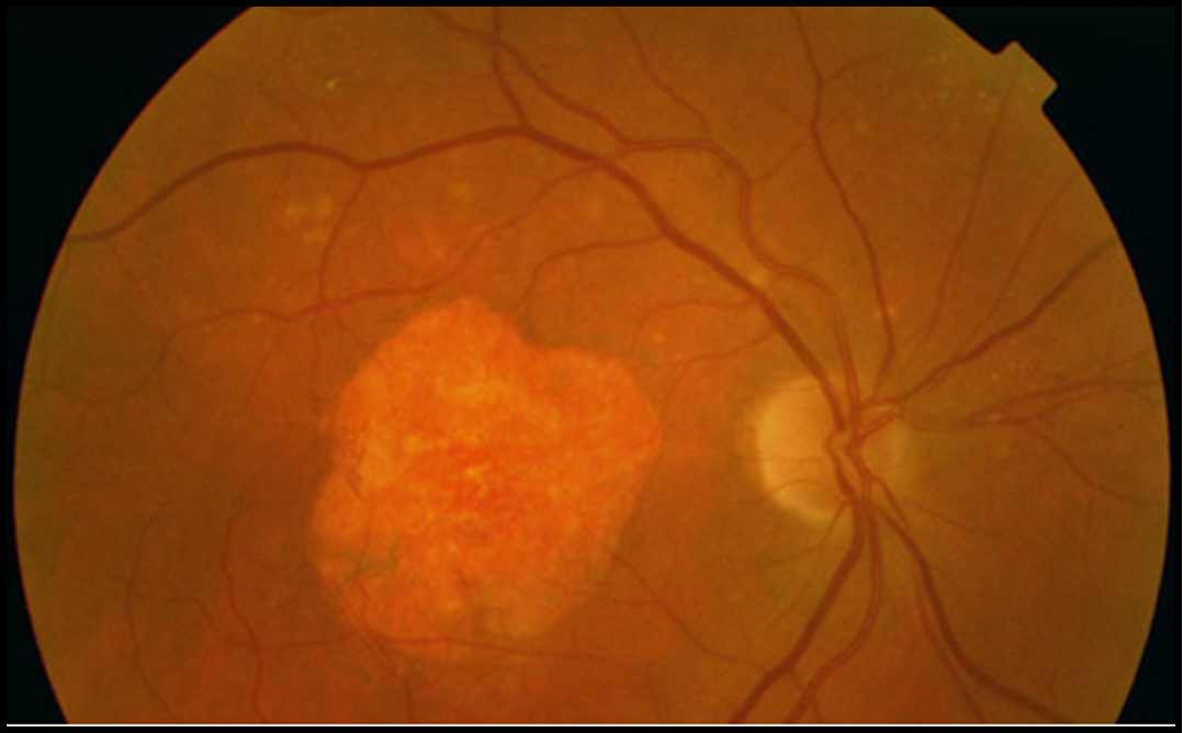
macular sub retinal fibrosis and hemorrhages; sudden vision loss
wet (neovascular) ARMD
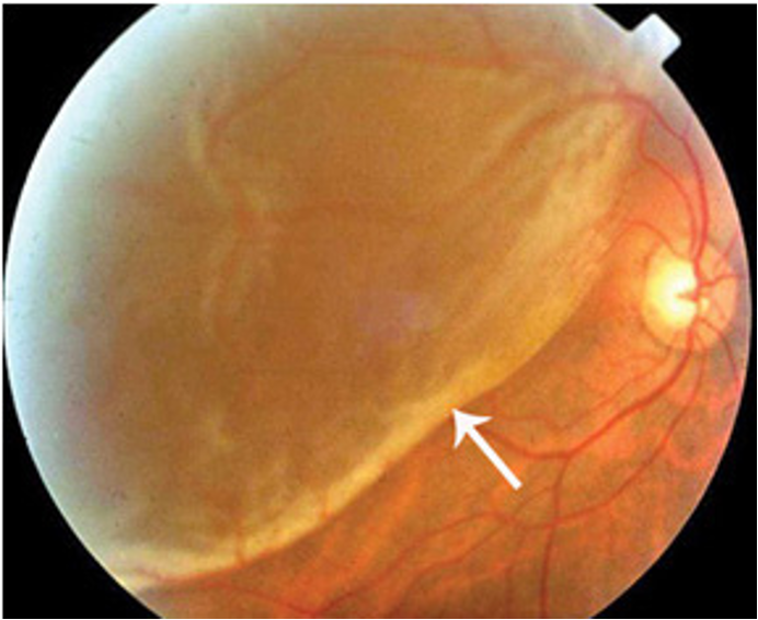
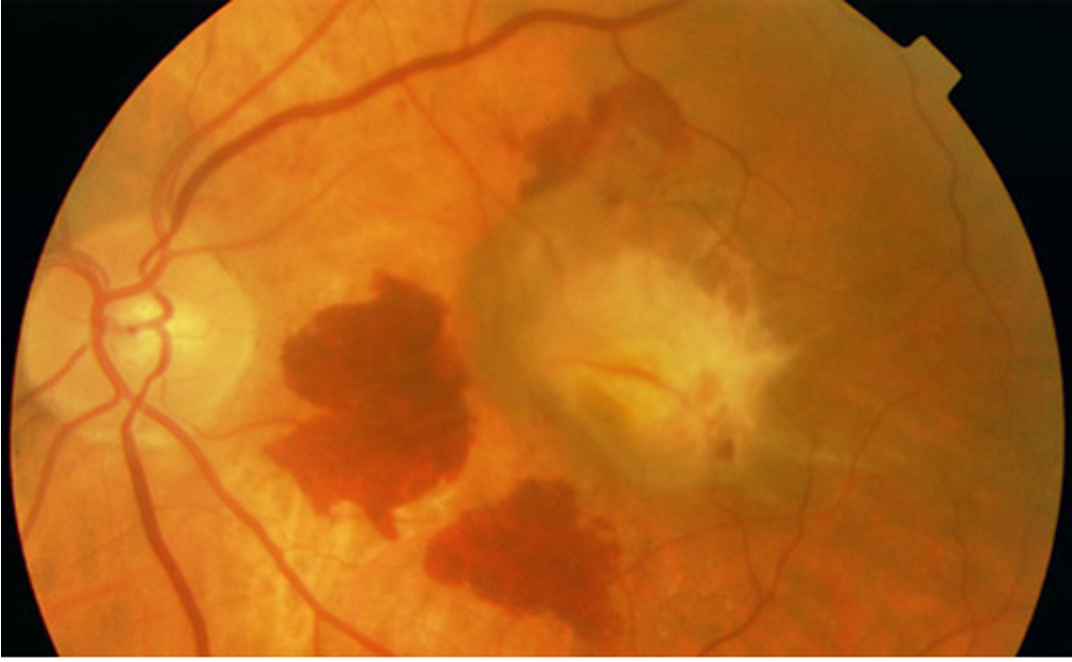
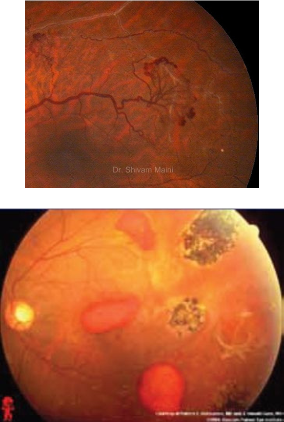
retinal detachment
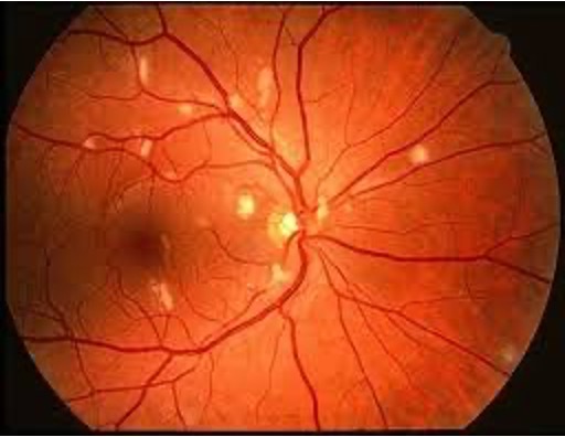
(yellow white patches)
retinopathy CMV
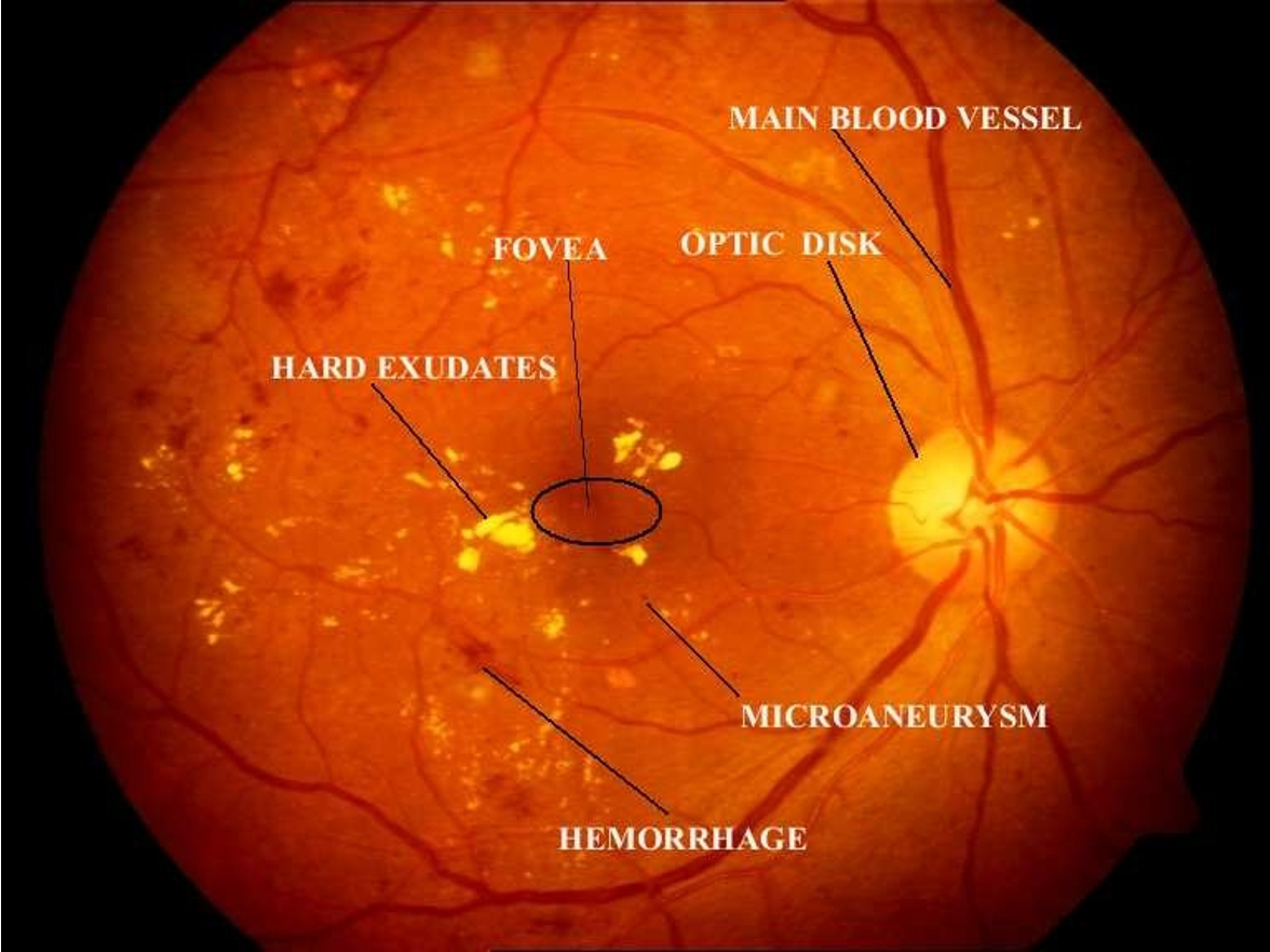
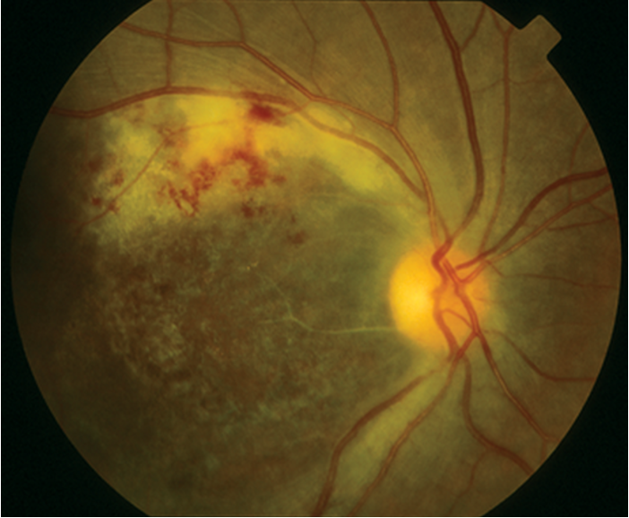
leading cause of blindness; proliferative + non proliferative
retinopathy- diabetic
+HIV
retinopathy- HIV
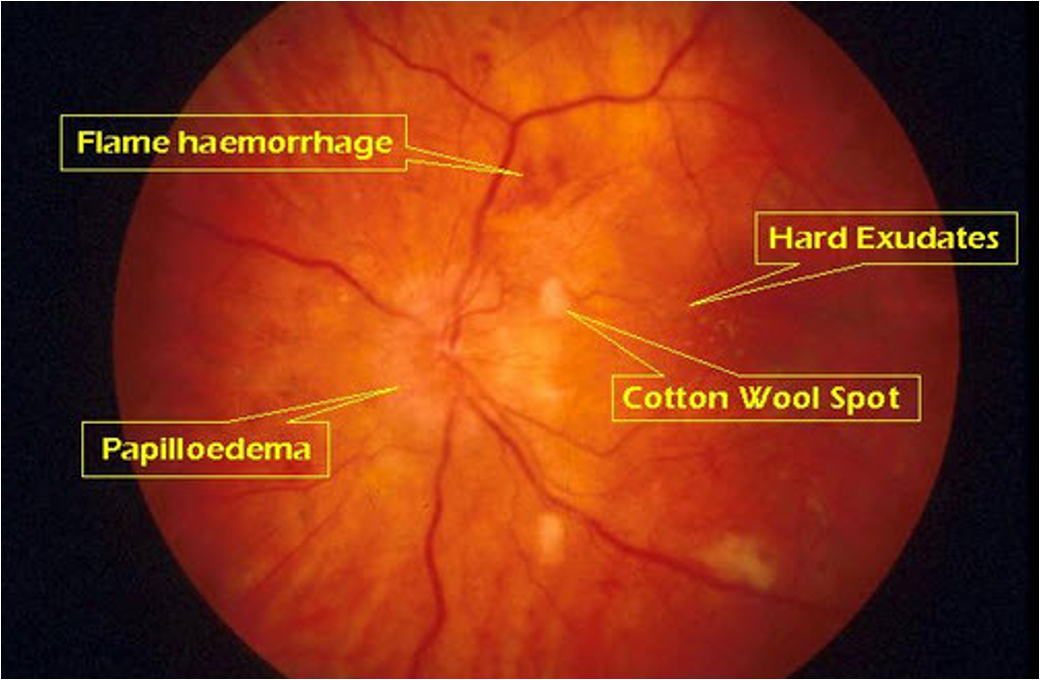
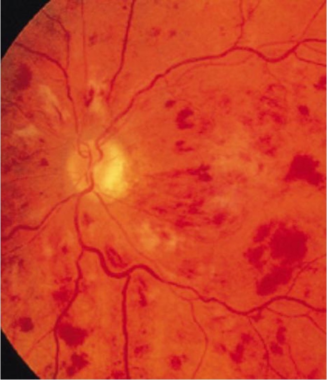
opaque arterial wall, AV nicking, flame hemorrhage, papilledema, cotton wool, hard exudates, silver/copper wire
retinopathy- hypertension
sea fan, salmon patches, black sunburst
retinopathy- sickle cell
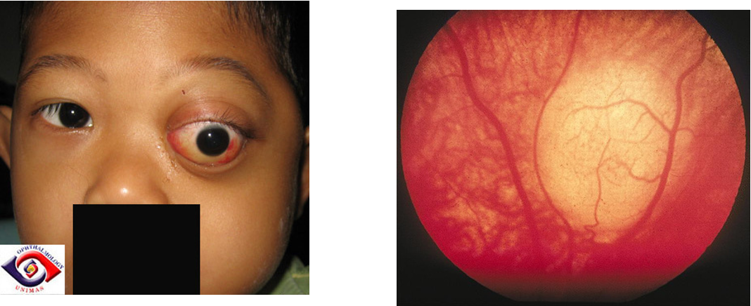
retinoblastoma
rhabdomyosarcoma
enophthamlos, positive Seidel’s sign
blowout fracture
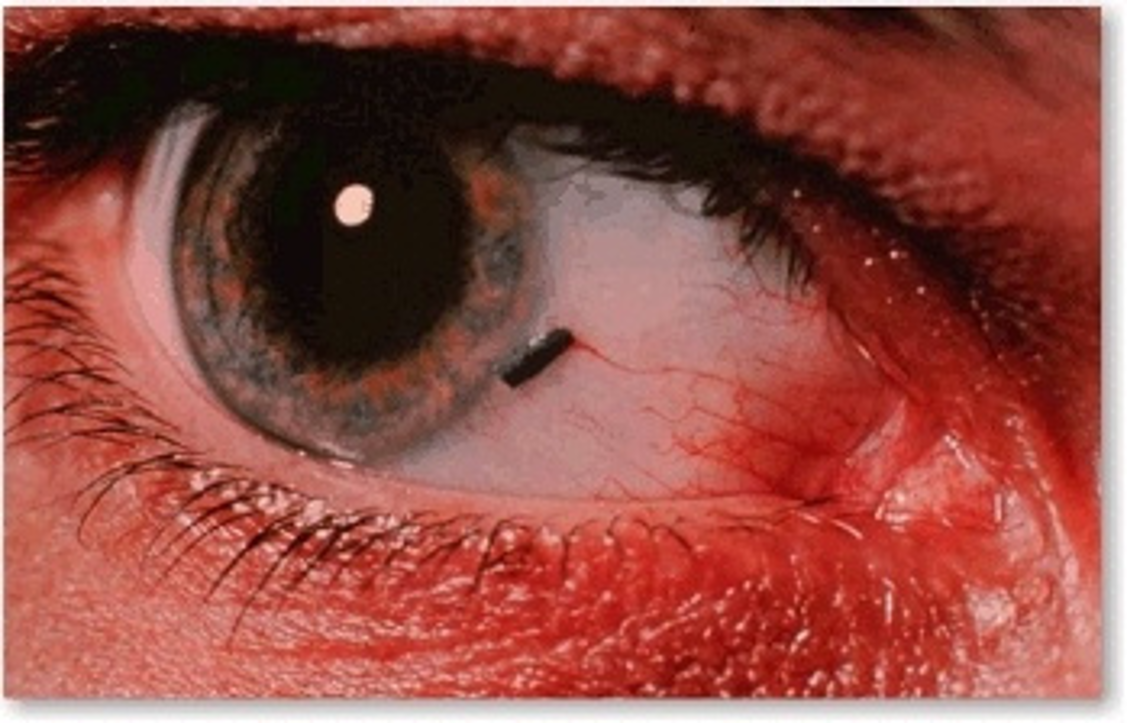
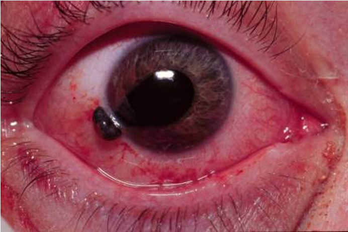
foreign body
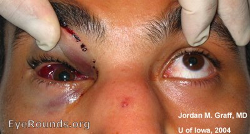
globe rupture
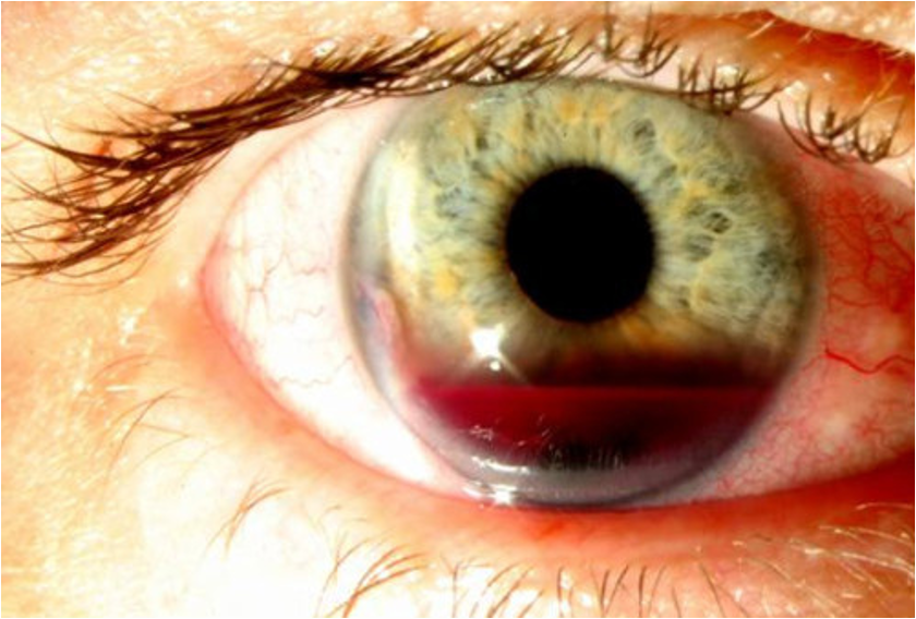
hyphema
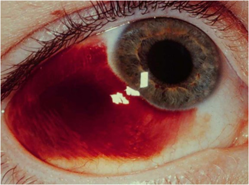
subconjunctival hemorrhage
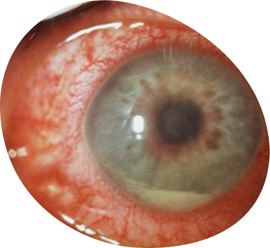
inflammation of uvea
acute uveitis (iritis)
central retinal artery occlusion (CRAO)
central retinal vein occlusion
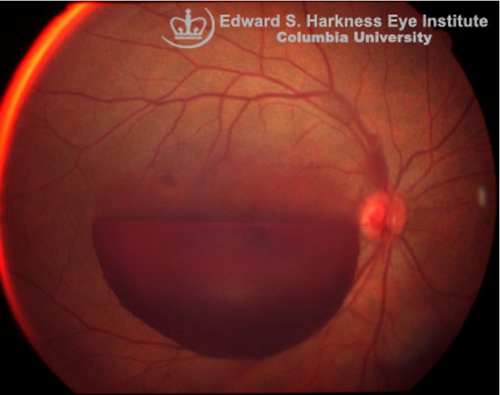
vitreous hemorrhage
abrupt monocular loss of vision lasting only a few minutes
transient amaurosis fugax / ocular TIA

>2 line difference between eyes; most common cause of pediatric visual impairment
amblyopia
obstruction to drainage of aqueous humor
Glaucoma
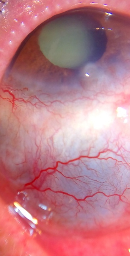
painful, sclera edema
scleritis
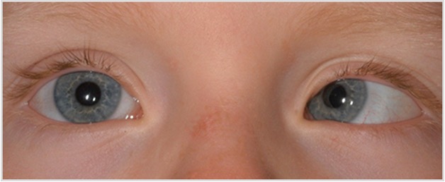
strabismus
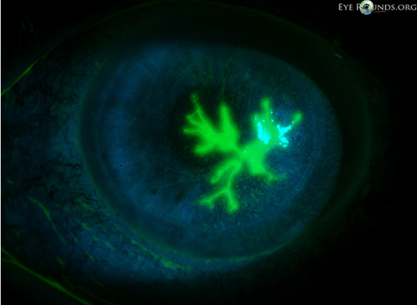
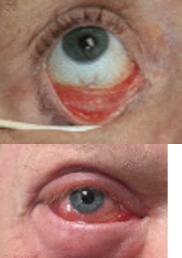
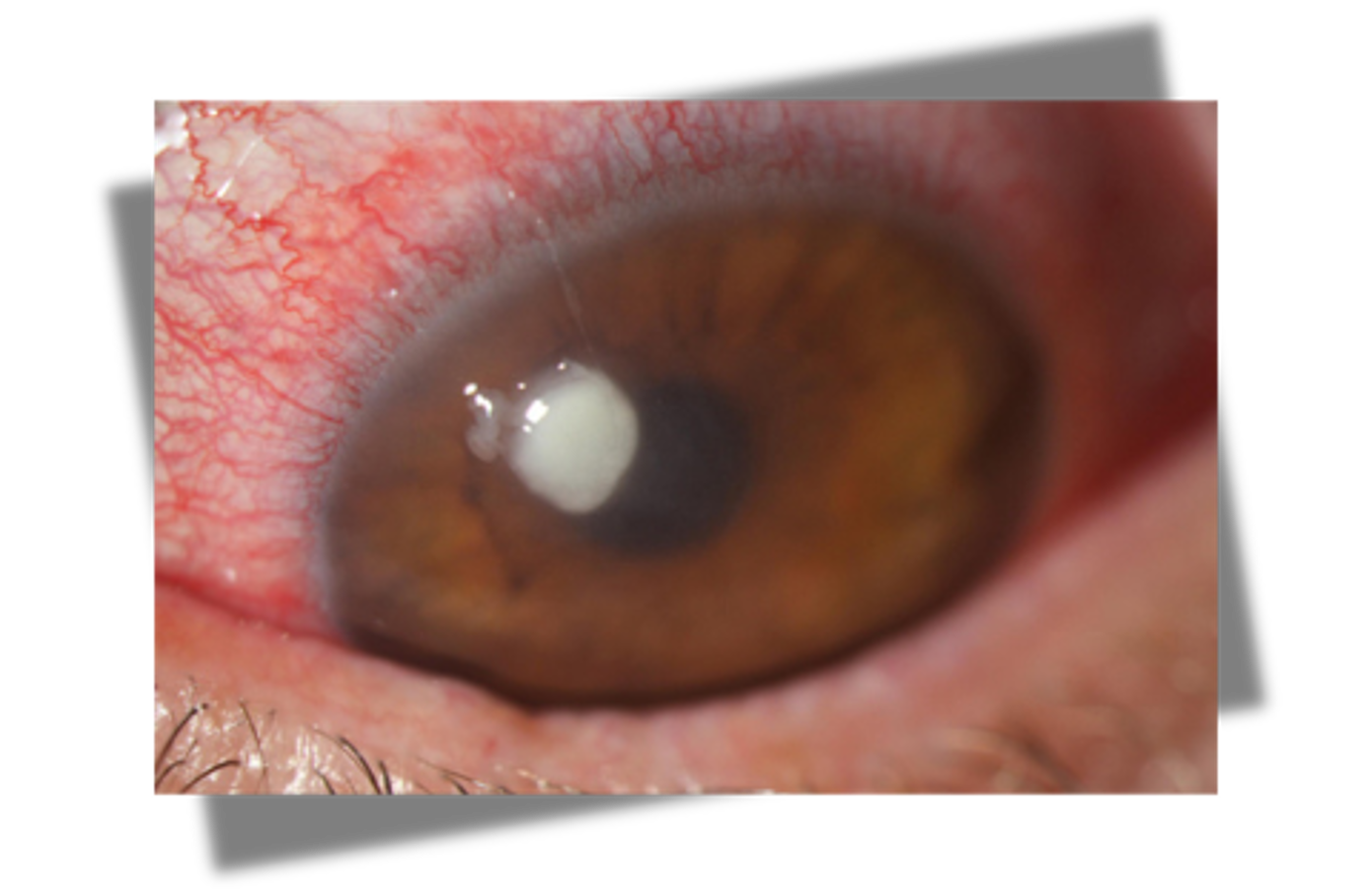
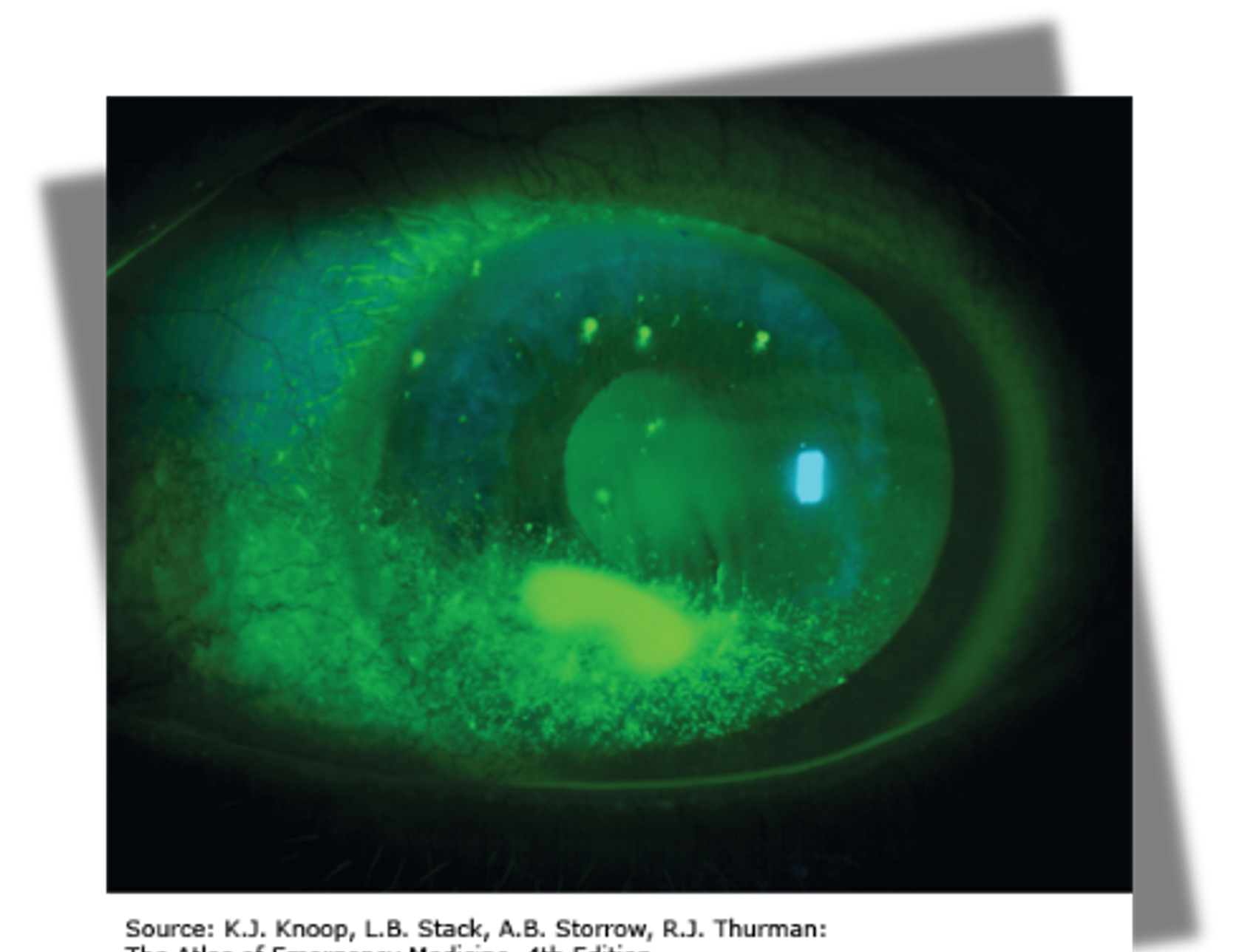
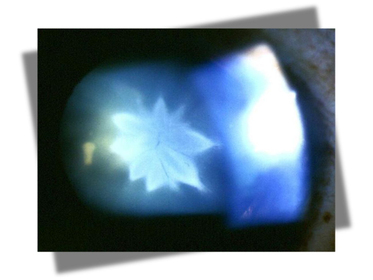
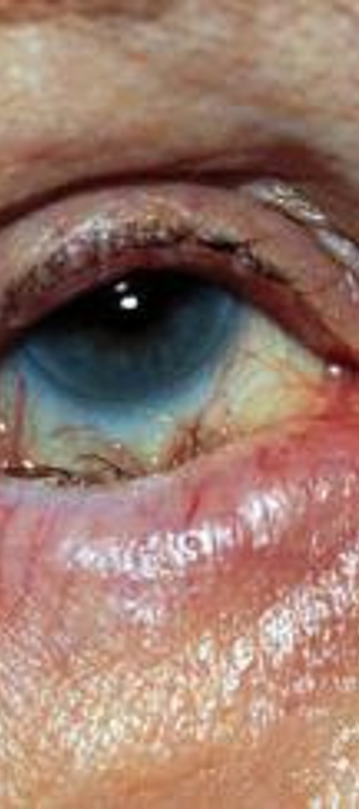
What is this fluorescein stain showing?
dendritic lesions (seen in herpetic viral conjunctivitis)
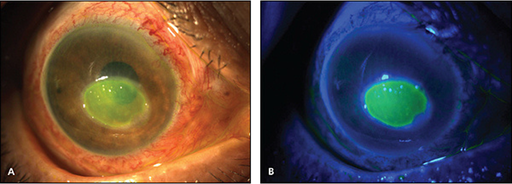
What is this fluorescein stain showing?
pooling (seen in corneal ulcers)