The Skeletal System
1/46
There's no tags or description
Looks like no tags are added yet.
Name | Mastery | Learn | Test | Matching | Spaced | Call with Kai | Chat |
|---|
No analytics yet
Send a link to your students to track their progress
47 Terms
Functions of the Skeletal System (6)
Support body & provide shape
Provide attachment and leverage for muscles
Protect internal organs - brain skull, heart lungs ribs
Site and protection for sensory organs
Store minerals - e.g. calcium and phosphorus
Produce new blood cells (haematopoiesis)
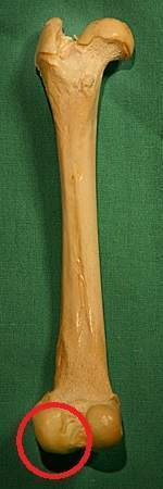
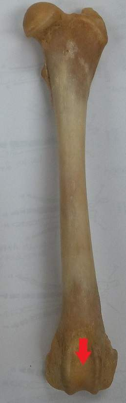
Condyle
Round protuberance (bulge/knob) at end of bone
Crest
Raised area of bone
Foramen
Hole/opening within bone
Fossa
Depression within bone where another structure is found
Groove
Depression in bone
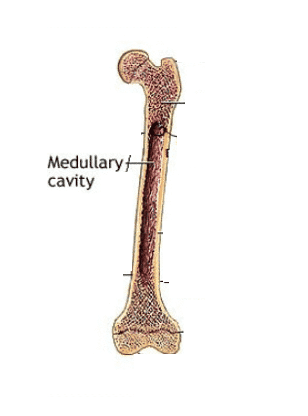
Medullary Cavity
Centre of long bones, often containing bone marrow
Periosteum
Outer covering of bone
Process
Thin, elongated projection
Sinus
Narrow hollow cavity
Spine
Central part bone
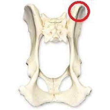
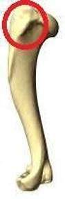
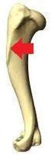
Trochanter
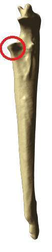
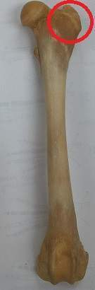
Prominent area of femur that lies behind head of femur
Tubercle
Small elevation on surface of bone
Tuberosity
Area of tubercle where tendons attach
Bones are … , made of …
Connective tissue, matrix of collagen fibres, filled with cells to build and modify bone. Minerals e.g. Ca, P (hyrdroxyapatite), stored within matrix, presence gives bone hardness and rigidity
Osteoblast
Immature bone cell responsible for developments and repair of bone
Osteocyte
Mature bone cells, main function is bone maintenance
Able to divide and convert into osteoblasts when necessary
Osteoclast
Bone cell responsible for breakdown of bone matrix
Sections of bone
Epiphysis, Metaphysis and Diaphysis
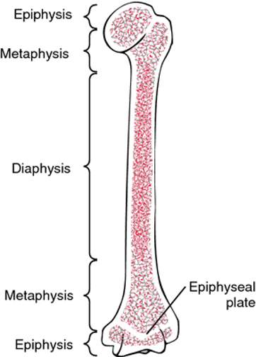
Epiphysis
Ends of long bone. Develop separately, initially separated by cartilage from shaft
Metaphysis
Site allows growth of bone (growth plate), between epiphysis and diaphysis. Visible on xrays as cartilage (epiphyseal line), area becomes calcified once bone growth complete
Diaphysis
Central shaft of bone, contains medullary cavity
Periosteum
Tough, fibrous membrane covers bone and provide ligament and tendon attachment. Covers all bone except articulate surfaces

Articular cartilage
Hyaline cartilage covers bone area within joints, provide cushioning and shock-absorption
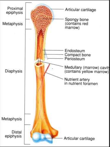
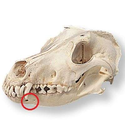
Nutrient foramen
Hole provides entry for blood supply, ensure oxygen and nutrients reach bone
Bone marrow
Spongy tissue, within medullary cavity (yellow marrow) and epiphyses (red marrow). Primary site of haematopoiesis (RBCs, WBCs, platelets)
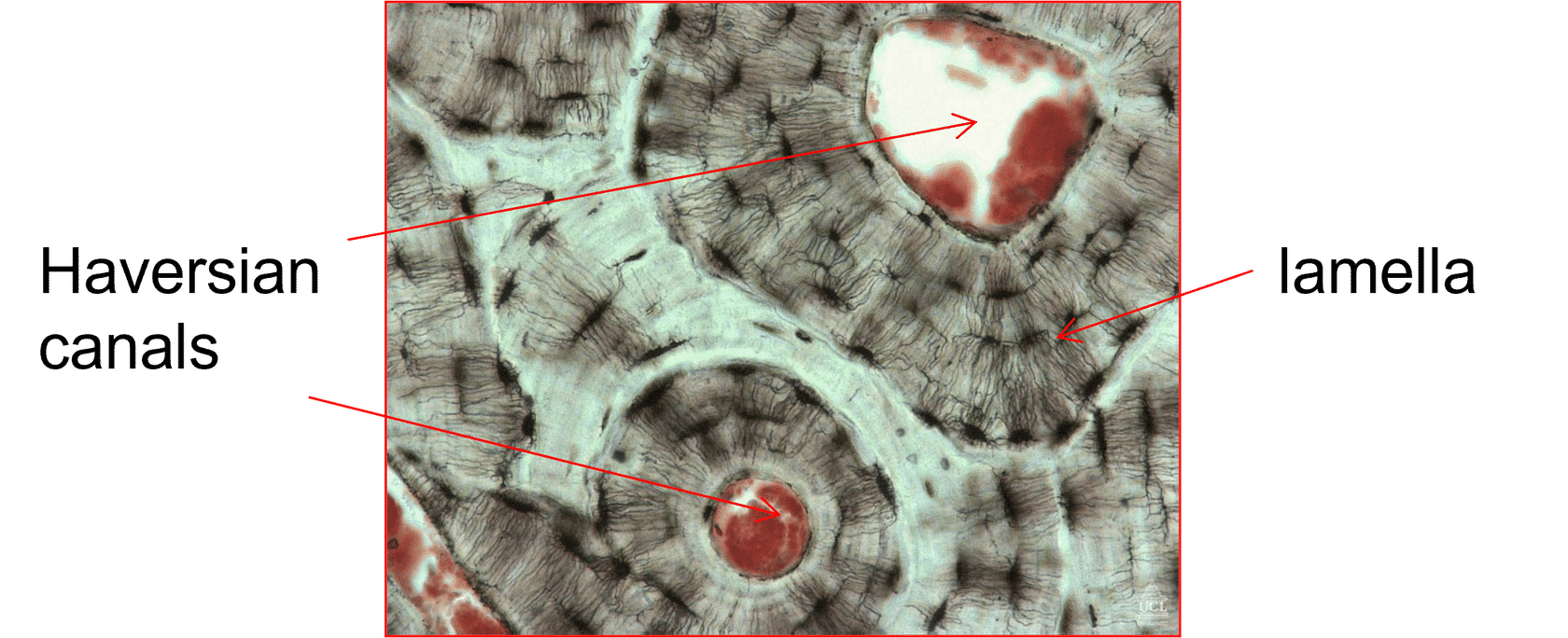
Cortical (compact) bone
Dense, regular structure, high mineral content for strength
Outer surfaces, covered in periosteum membrane
Close arrangement of canals (haversian system).
Cells form in circular patterns (lamellae) around central canal (Haversian canal)
Contained within - blood, lymph and nerves supplying bone
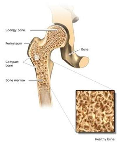
Cancellous (spongy) bone
Less dense than compact,
Honeycomb pattern
Less strength, commonly ends of long bones/smaller bones e.g. vertebrae
Less structured network - Loose network of fibres - trabeculae
Production of blood cells (haemopoiesis) occur predominantly
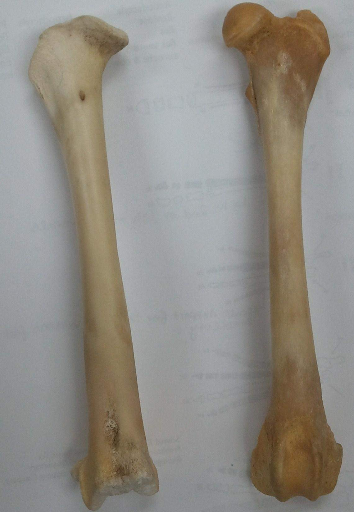
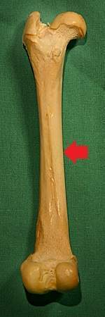
Long bones
Cylindrical, longer than wide, describes shape not size
Small long bone - toe bone (phalanges)
Long bones contain outer cortex (cortical bone), medullary cavity in main shaft (diaphysis), and cancellous bone at ends (epiphyses)
Majority of appendicular skeleton (limbs) are long bones, function as levers against which muscles contract and create movement
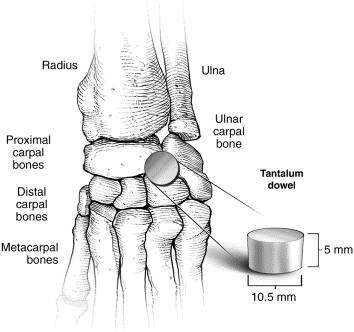
Short Bones
Cube-shaped approximately equal length, width, thickness
One section, no medullary cavity, develop from one area of ossification
Provide stability and support, limited motion.
Typically found within carpus and tarsus
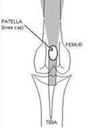
Sesamoid Bones
Small, round, seed
Form in tendons, great pressure generated.
Provide extra support to resist strong forces created when extending/flexing joint
Sesamoid bones vary in number/placement between species, typically involve patellae (sitting over stifle), and fabellae (sitting behind stifle)
Flat bones
Thin, curved, consist layer of cancellous bone within layer of compact, without medullary cavity
Flat bones serve as points of attachment for muscles, e.g. scapula or pelvis, specific protection of internal organs e.g. ribs or skull
Irregular bones
Complex shaped, e.g. vertebrae or provide specific structural features e.g. jaw or sinuses
Pneumatic bones
Medullary cavity in these bones largely replaced by air to reduce weight
Bones confined to skull and paranasal sinuses, which communicated with nasal cavities. e.g. sphenoid bone in skull
More significant in birds, most of skeleton pneumatic and connected to air sacs (avian respiratory system). Skeleton lighter, adapted for flight
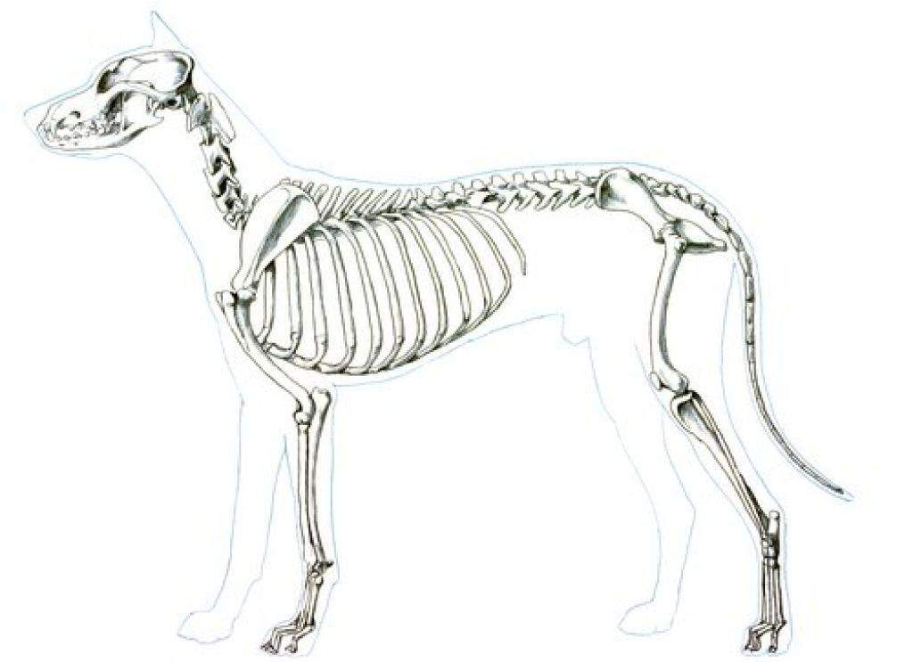
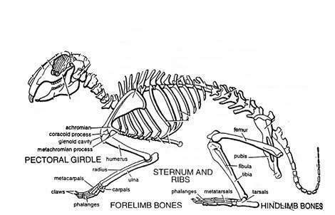
Appendicular Skeleton Consists of
Forelimb, scapula, hindling, pelvis
(Scapula, clavicle (collar bone), humerus, radius ulna, carpus, metacarpals, phalanges, pelvis, femur, stifle, tibia and fibula, tarsus, metatarsus)
Forelimb
No bony connection to body
Attached by muscles
Shock absorber that use all 4 limbs, most weight bearing occur
Hindlimb
Bony attachment at pelvis
Limb provides attachment for powerful muscles used during running, jumping
Axial Skeleton
Bones of skull, vertebral column, ribs and sternum, hyoid apparatus and middle ear
Skull: mandible, skull cranium, hyoid apparatus, atlas axis, vertebral column, ribs, sternum, sacrum,
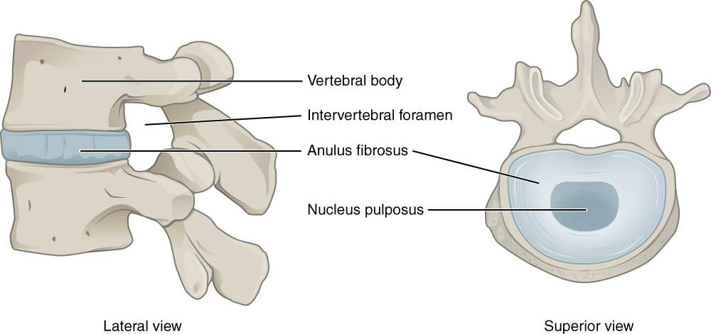
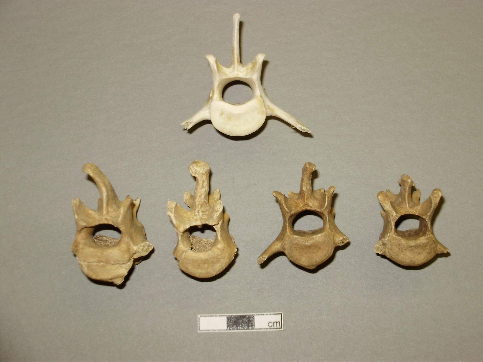
Vertebral Column and Intervertebral Discs:
Intervertebral disc: Central portion (nucleus pulposus) jelly-like material providing shock absorption, outer layer (anulus fibrosus) more fibrous to provide support and stability
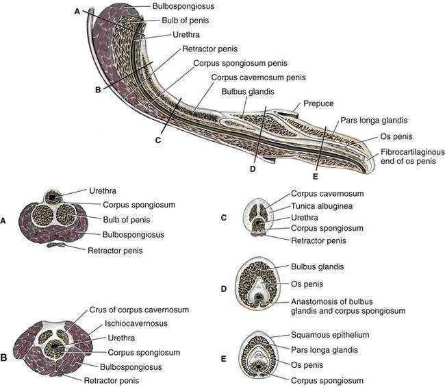
Splanchnic skeleton
Develop in soft tissue remote from rest of skeleton
os penis/baculum
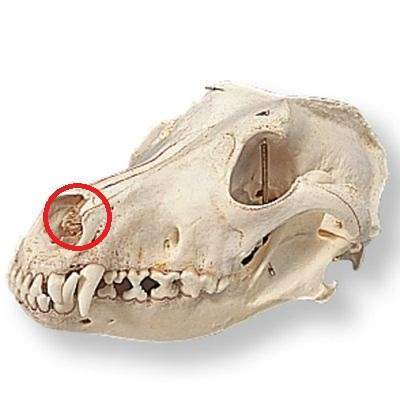
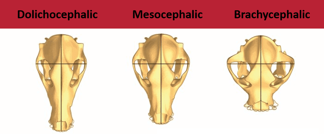
Dog skulls
Dolichocephalic, Mesocephalic, Brachycephalic
Greyhound, Labrador and pug
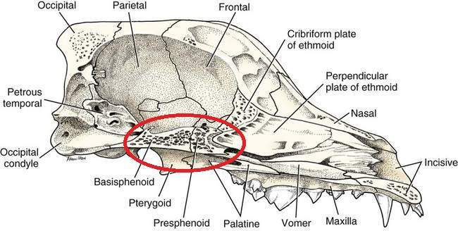
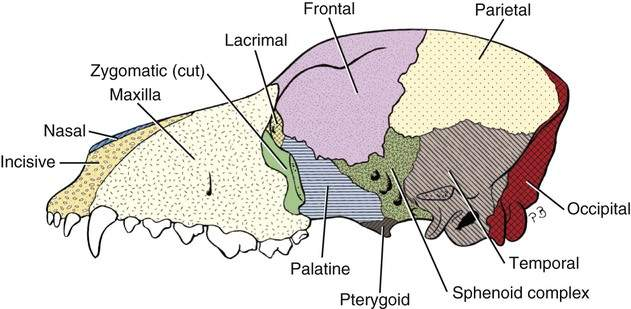
Bones within skull Lateral
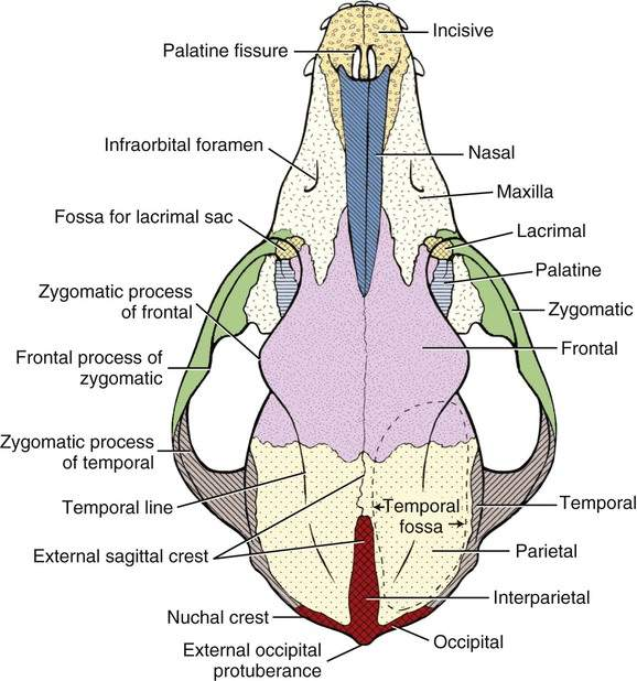
Ventral skull
Rabbit skeletal system
Rabbit skeleton 6-7% BW, prone to fractures esp. spine and hindlimbs due to powerful musculature hindlimbs, kicking out
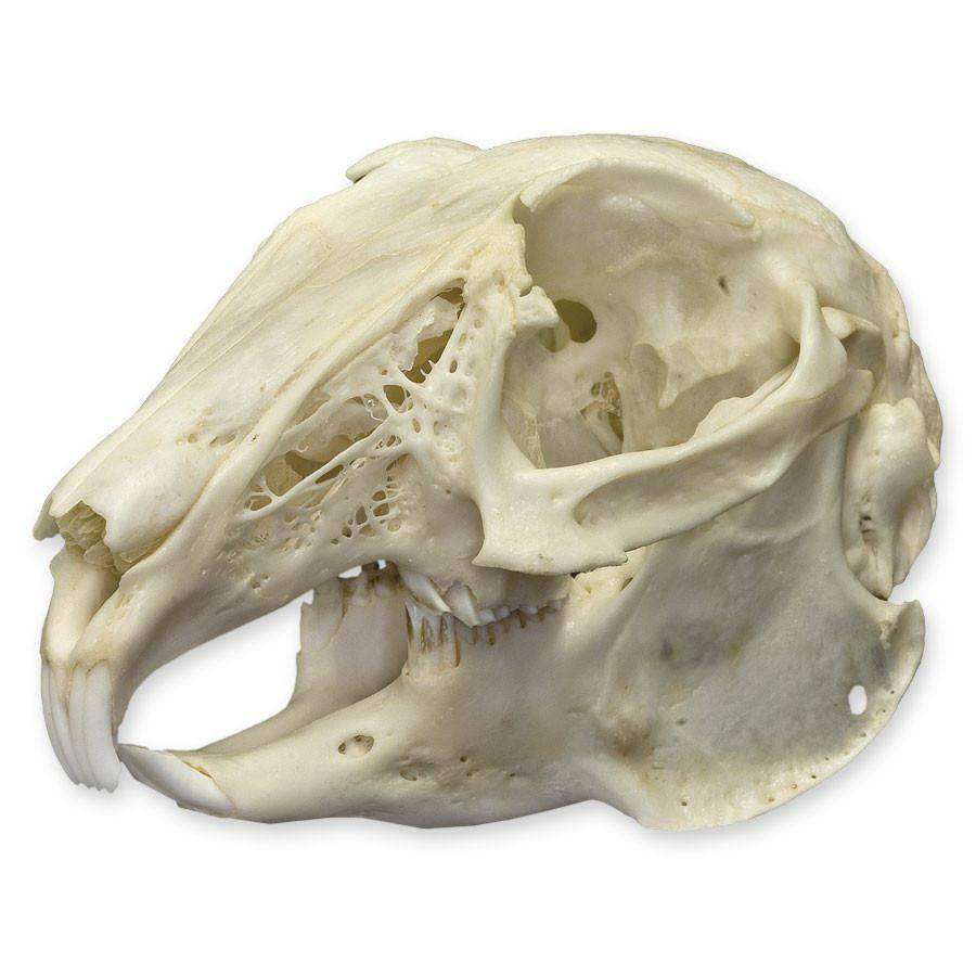
Rabbit skull
Mandible narrower than maxilla. Temperomandibular joint has wide SA allow lateral movement of mandible in relation to maxilla
Rabbit Axial skeleton
Cervical vertebrae box-like, small, great mobility.
Thoracic vertebrae possess attachment to 12 paired ribs
Pelvis narrow, positioned vertically, ilial wings meet ischium and pubic at acetabulum, accessory bone unique to rabits - os acetabuli
Pubis forms floor of pelvis and borders obturator foramen, oval in rabbits
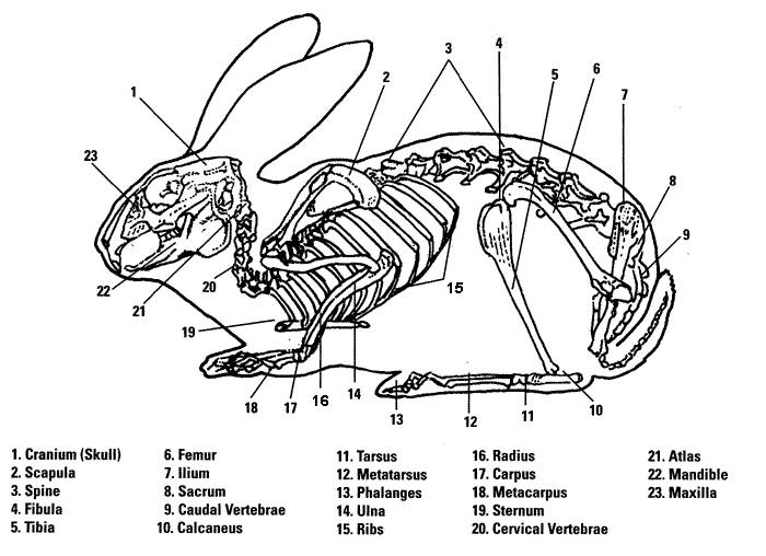
Rabbit Appendicular skeleton
Ulna fuses to radius in older animals, two bones deeply bowed
Hindlimbs similar to cats, femur flatter and tibia, fibula fused
Calcaneous of tarsal bone prominent