1750 Bony Thorax IA
 Call Kai
 Learn
Learn Practice Test
 Spaced Repetition
 Match
Match Flashcards
 Knowt Play
1/28

There's no tags or description
Looks like no tags are added yet.
Name | Mastery | Learn | Test | Matching | Spaced | Call with Kai | Chat |
|---|
No analytics yet
Send a link to your students to track their progress
29 Terms
PA Oblique Projection; Sternum; RAO Position (CR & SS)
Perpendicular to the IR. The central ray enters the elevated side of the posterior thorax at the level of T7 and approximately 1 inch (2.5 cm) lateral to the midsagittal plane
This image shows a slightly oblique projection of the sternum. The detail depends largely on the technical procedure employed. If breathing motion is used, the pulmonary markings are obliterated.
NOTE: SID 30”, Pt rotated 15-20 degrees, Make sure pt body is equally rotated (shoulders and hips) Use breathing technique
PA Oblique Projection; Sternum; RAO Position (Eval Criteria)
• Evidence of proper collimation
• Entire sternum from jugular notch to tip of xiphoid process
• Sternum projected over the heart, but free of superimposition from the thoracic spine
• Minimally rotated sternum and thorax, as shown by the following:
o Sternum projected just free of superimposition from vertebral column
o Minimally oblique vertebrae to prevent excessive rotation of the sternum
o Lateral portion of manubrium and sternoclavicular joint free of superimposition by the vertebrae
• Blurred pulmonary markings, if a breathing technique was used
• Exposure technique sufficient to demonstrate the sternum through the thorax
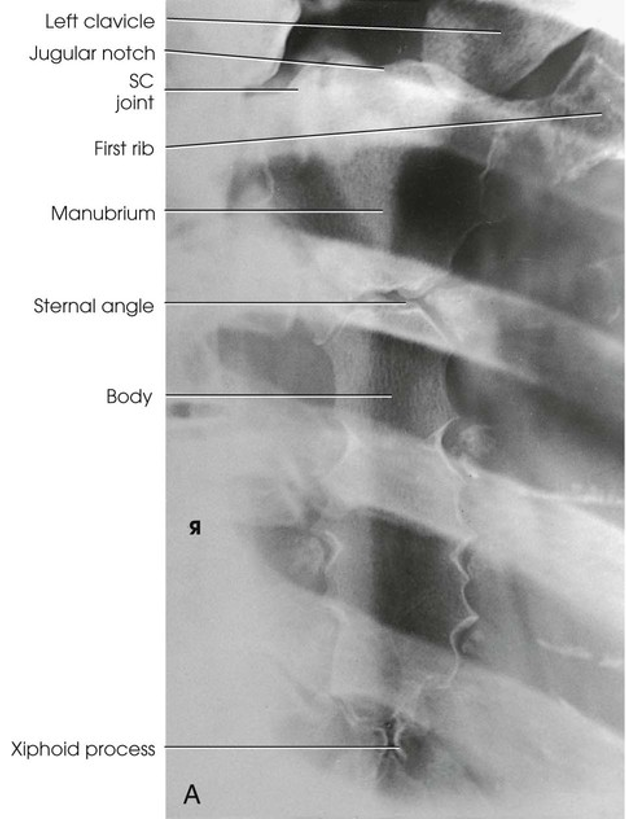
PA Oblique Projection; Sternum; RAO Position
Lateral Projection; Sternum; Right or Left Position (CR & SS)
Perpendicular to the center of the IR and entering the lateral border of the mid-sternum
A lateral image of the entire length of the sternum shows the superimposed sternoclavicular joints and medial ends of the clavicles
(NOTE: 72” SID - To reduce magnification and distortion caused by increased OID. Positioning: Rotate shoulders posteriorly with hands behind back if upright or above head if recumbent; Large breasted women should pull breast to side and apply ace bandage to keep in place. May need second radiograph with increased penetration. IR: Place 1.5” above jugular notch)
Lateral Projection; Sternum; Right or Left Position (Eval Criteria)
• Evidence of proper collimation
• Sternum in its entirety
• Manubrium free of superimposition by the soft tissue of the shoulders
• Sternum free of superimposition by the ribs
• Lower portion of the sternum unobscured by the breasts of a female patient (a second radiograph with increased penetration may be necessary)
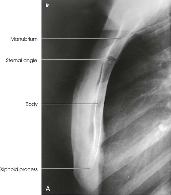
Lateral Projection; Sternum; Right or Left Position
If patient is hurt or injured on Anterior side, what projection is done?
PA
If Patient is hurt or injured on posterior side, what projection is done?
AP
For upper ribs what breathing instructions are used?
Inspiration
For lower ribs what breathing instructions are used?
Expiration
Are upper ribs done erect or recumbent
Erect
Are lower ribs done erect or recumbent
Recumbent
PA Projection; Upper Anterior Ribs (CR & SS)
Perpendicular to the center of the IR. If the IR is positioned correctly, the central ray is at the level of T7
PA Projection best shows the anterior ribs above the diaphragm. Although the posterior ribs are seen, the anterior ribs are shown with greater detail because they are closer to the IR
PA Projection; Upper Anterior Ribs (Eval Criteria)
• Evidence of proper collimation
• First through ninth ribs in their entirety, with posterior portions lying above the diaphragm
• First through seventh anterior ribs from both sides, in their entirety and above the diaphragm
• In a unilateral examination, ribs from the opposite side possibly not included in their entirety
• Ribs visible through the lungs with sufficient contrast
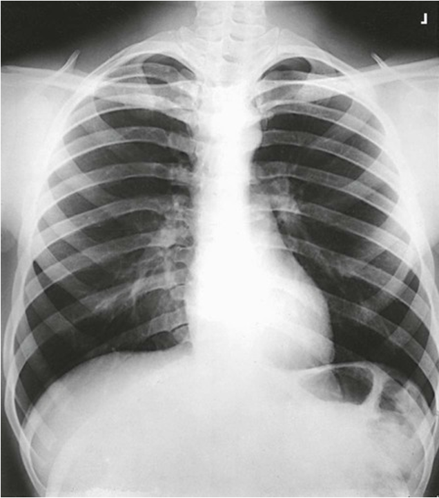
PA Projection; Upper Anterior Ribs
AP Projection; Posterior Ribs (CR & SS)
Perpendicular to the center of the IR
Upper: IR 1.5” above relaxed shoulders. Shoulders in same transverse plane and rotated forward to move out of ribs. Inspiration: 1-10 ribs
Lower: lower edge of IR at the level of Crest. Expiration: 8-12 ribs
AP Projection best shows the posterior ribs above or below the diaphragm, according to the region examined. Although the anterior ribs are seen, the posterior ribs are shown in greater detail because they are closer to the IR
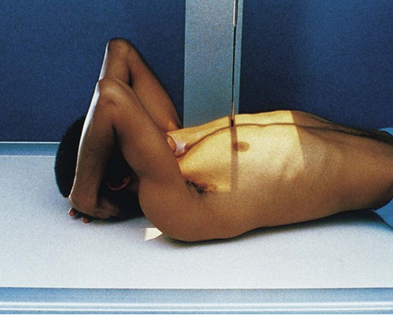
Positioning: AP Projection; Posterior Ribs (Ribs above diaphragm)
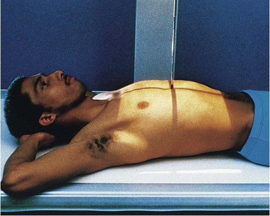
Positioning: AP Projection; Posterior Ribs (Ribs below diaphragm)
AP Projection; Posterior Ribs (Eval Criteria)
• Evidence of proper collimation
• For ribs above the diaphragm, first through tenth posterior ribs from both sides in their entirety
• For ribs below the diaphragm, eighth through twelfth posterior ribs on both sides in their entirety
• Ribs visible through the lungs or abdomen, according to the region examined
• In a unilateral examination, ribs from the opposite side possibly not included in their entirety
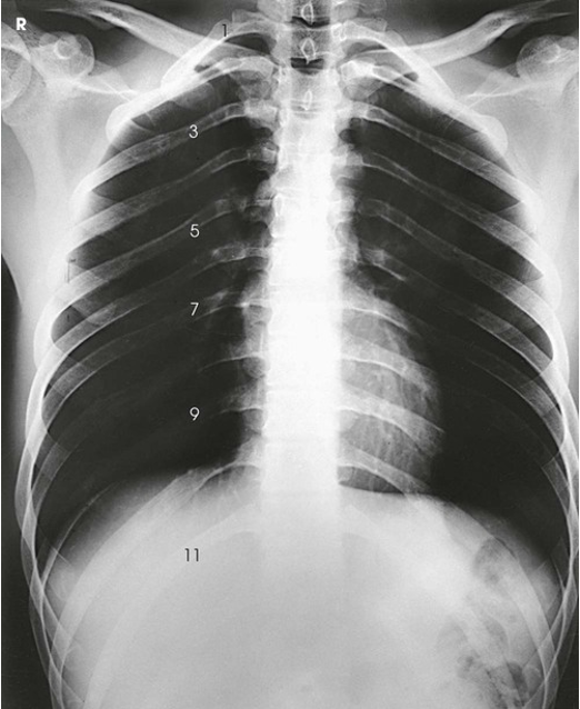
AP Projection; Posterior Ribs (Ribs above diaphragm)
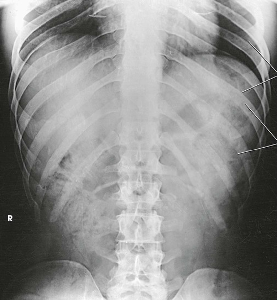
AP Projection; Posterior Ribs (Ribs below diaphragm)
AP Oblique Projection; Axillary Ribs: RPO or LPO Position (CR & SS)
Perpendicular to the center of the IR
In these images, the axillary portion of the ribs closest to the IR is projected free of superimposition with the thoracic spine. The posterior ribs closest to the IR are also well shown
(NOTE: 45 degree rotation. Upright recommended for ribs above diaphragm. Recumbent patients require radiolucent support

Positioning: AP Oblique Projection; Axillary Ribs: RPO or LPO Position (Upright AP oblique ribs LPO position)
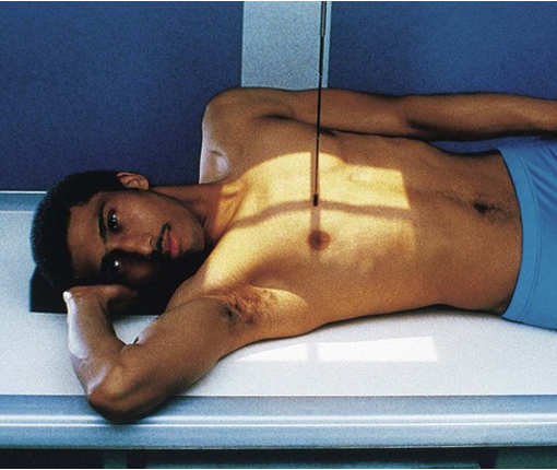
Positioning: AP Oblique Projection; Axillary Ribs: RPO or LPO Position (Recumbent AP oblique ribs RPO position)
AP Oblique Projection; Axillary Ribs: RPO or LPO Position (Eval Criteria)
• Evidence of proper collimation
• Approximately twice as much distance between the vertebral column and the lateral border of the ribs on the affected side as is present on the unaffected side
• Axillary portion of the ribs free of superimposition with the thoracic spine
• First through tenth ribs visible above the diaphragm for upper ribs
• Eighth through twelfth ribs visible below the diaphragm for lower ribs
• Ribs visible through the lungs or abdomen according to the region examined
(AP)
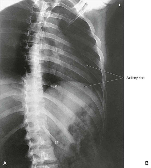
AP Oblique Projection; Axillary Ribs: RPO or LPO Position
PA Oblique Projection; Axillary Ribs; RAO or LAO Position (CR & SS)
Perpendicular to center of IR
In these images, the axillary portion of the ribs farthest from the IR is projected free of bony superimposition with the thoracic spine. The anterior ribs farthest from the IR are also well shown
PA Oblique Projection; Axillary Ribs; RAO or LAO Position (Eval Criteria)
• Evidence of proper collimation
• Approximately twice as much distance between the vertebral column and the lateral border of the ribs on the affected side as is present on the unaffected side
• Axillary portion of the ribs free of superimposition with the thoracic spine
• First through tenth ribs visible above the diaphragm for upper ribs
• Eighth through twelfth ribs visible below the diaphragm for lower ribs
• Ribs visible through the lungs or abdomen according to the region examined
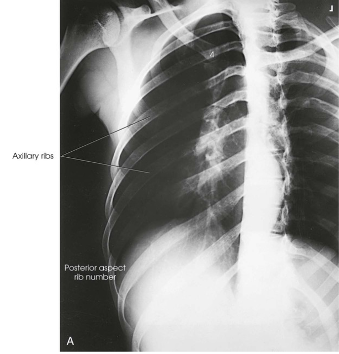
PA Oblique Projection; Axillary Ribs; RAO or LAO Position