Finger/Hand/Wrist IA
 Call Kai
 Learn
Learn Practice Test
 Spaced Repetition
 Match
Match Flashcards
 Knowt Play
1/55

There's no tags or description
Looks like no tags are added yet.
Name | Mastery | Learn | Test | Matching | Spaced | Call with Kai | Chat |
|---|
No analytics yet
Send a link to your students to track their progress
56 Terms
PA Projection; Digits 2-5 (CR & SS)
Perpendicular to the PIP joint of the affected digit
PA projection of the appropriate digit is visualized
PA Projection; Digits 2-5 (Eval Criteria)
Evidence of proper collimation
Entire digit from fingertip to distal portion of adjoining metacarpal
No soft tissue overlap from adjacent digits
No rotation of the digit, demonstrated by concavity of the phalangeal bodies and an equal amount of soft tissue on both sides of the phalanges
Fingernail, if visualized and normal, centered over the distal phalanx
Open IP and MCP joint spaces without overlap
Soft tissue and bony trabecular
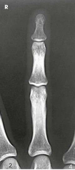
PA Projection; Digits 2-5
PA Oblique Projection; Digits 2-5; Lateral Rotation (CR & SS)
Perpendicular to the PIP joint of the affected digit
PA Oblique projection of the bones and soft tissue of the affected digit
PA Oblique Projection; Digits 2-5; Lateral Rotation (Eval Criteria)
• Evidence of proper collimation
• Entire digit, including the distal portion of the adjoining metacarpal
• Digit rotated at 45 degrees, demonstrated by concavity of the elevated side of the phalangeal bodies
• No superimposition of the adjacent digits over the proximal phalanx or MCP joint
• Open IP and MCP joint spaces
• Soft tissue and bony trabecular detail
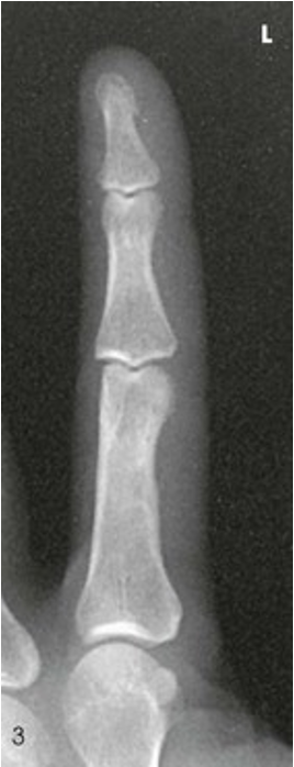
PA Oblique Projection; Digits 2-5; Lateral Rotation
Lateral Projection; Digits (2-5); Lateromedial/Mediolateral (CR & SS)
Perpendicular to the PIP joint of the affected digit
A lateral projection of the affected digit is shown
Lateral Projection; Digits (2-5); Lateromedial/Mediolateral (Eval Criteria)
• Evidence of proper collimation
• Entire digit in a true lateral position
• Fingernail in profile, if visualized and normal
• Concave, anterior surfaces of the phalanges
• No rotation of the phalanges
• No obstruction of the proximal phalanx or MCP joint by adjacent digits
• Open IP joint spaces
• Soft tissue and bony trabecular detail
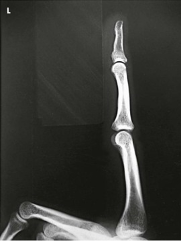
Lateral Projection; Digits (2-5); Lateromedial/Mediolateral
AP/PA Projections; Thumb (CR & SS)
Perpendicular to the MCP joint for AP and PA projections
AP, PA, projections of the thumb are shown
AP/PA Projections; Thumb (Eval Criteria)
• Evidence of proper collimation
• Area from the distal tip of the thumb to the trapezium
• No rotation
• Concavity of the phalangeal and metacarpal bodies
• Equal amount of soft tissue on both sides of the phalanges
• Thumbnail, if visualized, in the center of the distal thumb
• Overlap of soft tissue profile of the palm over the mid-shaft of the first metacarpal
• Open IP and MCP joint spaces without overlap of bones
• Soft tissue and bony trabecular detail
• PA thumb projection magnified compared with AP projection
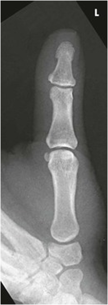
AP/PA Projections; Thumb
PA Oblique Projection; Thumb (CR & SS)
Perpendicular to the MCP joint
PA oblique projection of thumb
PA Oblique Projection; Thumb (Eval Criteria)
• Evidence of proper collimation
• Area from the distal tip of the thumb to the trapezium
• Proper rotation of phalanges, soft tissue, and first metacarpal
• Concave surface of elevated side of the proximal phalanx
• Open IP and MCP joint spaces
• Soft tissue and bony trabecular detail
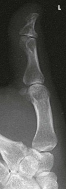
PA Oblique Projection; Thumb
Lateral Projection; Thumb (CR & SS)
Perpendicular to the MCP joint
Lateral projection of the thumb
Lateral Projection; Thumb (Eval Criteria)
• Evidence of proper collimation
• Area from the distal tip of the thumb to the trapezium
• First digit in a true lateral projection
• Thumbnail, if visualized and normal, in profile
• Concave, anterior surface of the proximal phalanx
• No rotation of the phalanges
• Open IP and MCP joint spaces
• Soft tissue and bony trabecular detail
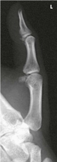
Lateral Projection; Thumb
PA Projection; Hand (CR & SS)
Perpendicular to the third MCP joint
PA projections of the carpals, metacarpals, phalanges (except the thumb), interarticulations of the hand, and distal radius and ulna, also a PA oblique of the first digit
PA Projection; Hand (Eval Criteria)
• Evidence of proper collimation
• Anatomy from fingertips to distal radius and ulna
• Slightly separate digits with no soft tissue overlap
• No rotation of the hand
• Equal concavity of the metacarpal and phalangeal bodies on both sides
• Equal amount of soft tissue on both sides of the phalanges
• Fingernails, if visualized, in the center of each distal phalanx
• Equal distance between the metacarpal heads
• Open MCP and IP joints, indicating that the hand is placed flat on the IR
• Soft tissue and bony trabecular detail
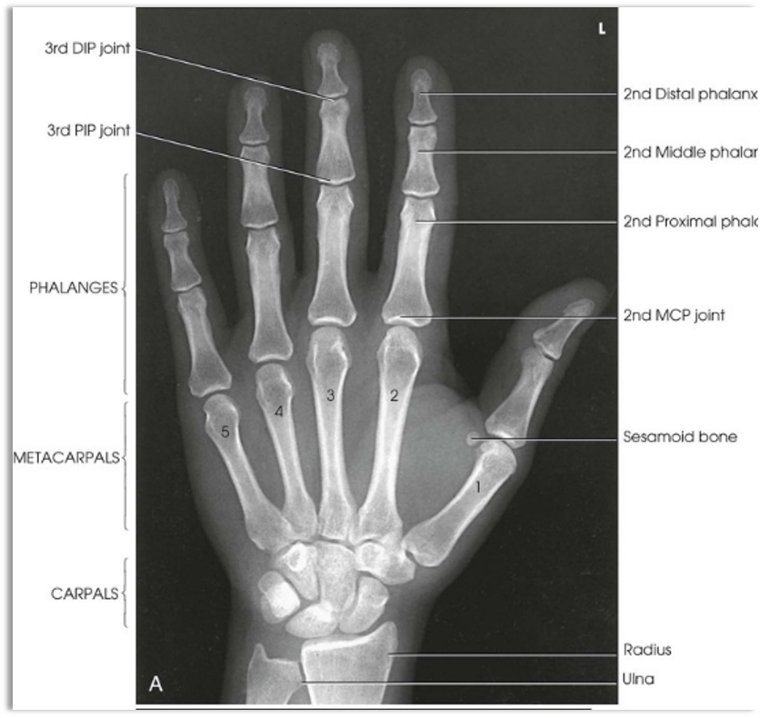
PA Projection; Hand
PA Oblique projection; Hand (CR & SS)
Perpendicular to the third MCP joint
PA oblique projection of the bones and soft tissues of the hand
PA Oblique projection; Hand (Eval Criteria)
• Evidence of proper collimation
• Anatomy from fingertips to distal radius and ulna
• Digits separated slightly with no overlap of their soft tissues
• 45 degrees of rotation of anatomy
• Minimal overlap of the third, fourth, and fifth metacarpal bodies
• Slight overlap of the metacarpal bases and heads
• Separation of the second and third metacarpals
•Open IP and MCP joints
•Soft tissue and bony trabecular detail
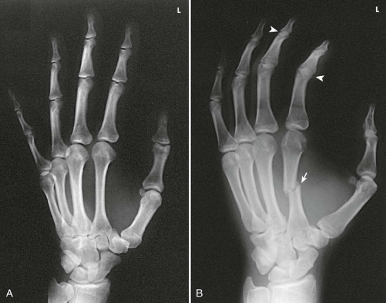
PA Oblique Projection; Hand
Lateral Projection; Hand (Mediolateral, Lateromedial, Extension, Fan Lateral) (CR & SS)
Perpendicular to the second digit MCP joint
The image, which shows a lateral projection of the hand in extension, presents the customary position for localizing foreign bodies and metacarpal fracture displacement. The exposure technique depends on the foreign body. The fan lateral superimposes the metacarpals but shows almost all of the individual phalanges. The most proximal portions of the proximal phalanges remain superimposed
Lateral Projection; Hand (Mediolateral, Lateromedial, Extension, Fan Lateral) (Eval Criteria)
• Evidence of proper collimation
• Anatomy from fingertips to distal radius and ulna
• Extended digits
• Hand in a true lateral position
• Superimposed phalanges (individually seen on fan lateral)
• Superimposed metacarpals
• Superimposed distal radius and ulna
• Thumb free of motion and superimposition
• Soft tissue and bony trabecular detail
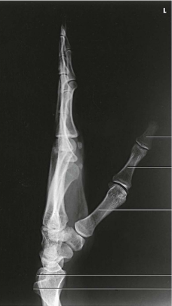
Lateral Projection; Hand (lateromedial)
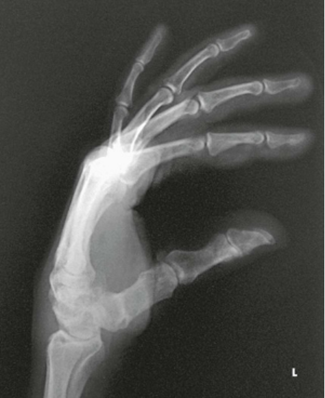
Lateral Projection; Hand (Fan Lateral)
PA Projection; Wrist (CR & SS)
Perpendicular to the mid carpal area
A PA projection of the carpals, distal radius and ulna, and proximal metacarpals is shown. The projection gives a slightly oblique rotation to the ulna. When the ulna is under examination, an AP projection should be taken.
PA Projection; Wrist (Eval Criteria)
• Evidence of proper collimation
• Distal radius and ulna, carpals, and proximal half of metacarpals
• No excessive flexion of digits to overlap and obscure metacarpals
• No rotation in carpals, metacarpals, radius, and ulna
• Open radioulnar joint space
• Soft tissue and bony trabecular detail 1
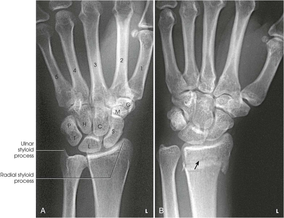
PA Projection; Wrist
AP Projection; Wrist (CR & SS)
Perpendicular to the mid carpal area
The carpal interspaces are better shown in the AP image than in the PA image. Because of the oblique direction of the interspaces, they are more closely parallel with the divergence of the x-ray beam
AP Projection; Wrist (Eval Criteria)
• Evidence of proper collimation
• Distal radius and ulna, carpals, and proximal half of metacarpals
• No excessive flexion of digits to overlap and obscure metacarpals
• No rotation in carpals, metacarpals, radius, and ulna
• Open radioulnar joint space
• Soft tissue and bony trabecular detail
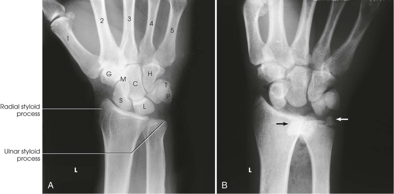
AP Projection; Wrist
PA Oblique Projection; Wrist; Lateral Rotation (CR & SS)
Perpendicular to the mid carpal area; in enters just distal to the radius
This projection shows the carpals on the lateral side of the wrist, particularly the trapezium and the scaphoid. The scaphoid is superimposed on itself in the direct PA projection
PA Oblique Projection; Wrist; Lateral Rotation (Eval Criteria)
• Evidence of proper collimation
• Distal radius and ulna, carpals, and proximal half of metacarpals
• 45-degree rotation of anatomy
• Slight interosseous space between the third, fourth, and fifth metacarpal bodies
• Slight overlap of the distal radius and ulna
• Carpals on lateral side of wrist
• Trapezium and distal half of the scaphoid without superimposition
• Open trapeziotrapezoid and scaphotrapezial joint space
• Soft tissue and bony trabecular detail
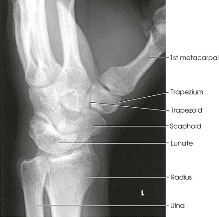
PA Oblique Projection; Wrist; Lateral Rotation
AP Oblique Projection; Wrist; Medial Rotation (CR & SS)
Perpendicular to the midcarpal area; it enters between the anterior surface of the wrist midway between its medial and lateral borders
This position separates the pisiform from adjacent carpal bones. It also provides a more distinct radiograph of the triquetrum and hamate; carpals on medial side
AP Oblique Projection; Wrist; Medial Rotation (Eval Criteria)
• Evidence of proper collimation
• Distal radius and ulna, carpals, and proximal half of metacarpals
• Carpals on medial side of wrist
• Triquetrum, hook of hamate, and pisiform free of superimposition and in profile
• Soft tissue and bony trabecular detail
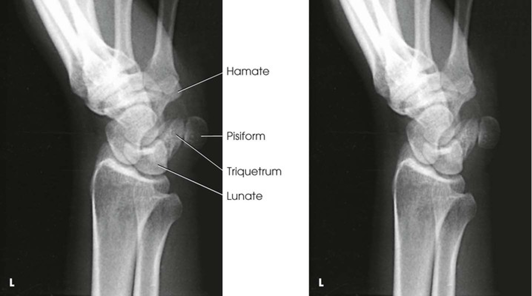
AP Oblique Projection; Wrist; Medial Rotation
Lateral Projection; Wrist (CR & SS)
Perpendicular to the wrist joint
This image shows a lateral projection of the proximal metacarpals, and the distal radius and ulna. This position can also be used to shown anterior and posterior displacement in fractures
Lateral Projection; Wrist (Eval Criteria)
• Evidence of proper collimation
• Distal radius and ulna, carpals, and proximal half of metacarpals
• Superimposed distal radius and ulna
• Superimposed metacarpals
• Soft tissue and bony trabecular detail
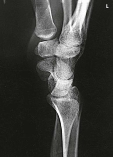
Lateral Projection; Wrist
PA Projection; Wrist; Ulnar Deviation (CR & SS)
Perpendicular to the scaphoid
This position reduces foreshortening of the scaphoid, which occurs with a perpendicular central ray. It also opens the spaces between adjacent carpals
PA Projection; Wrist; Ulnar Deviation (Eval Criteria)
• Evidence of proper collimation
• Distal radius and ulna, carpals, and proximal half of metacarpals
• Scaphoid with adjacent articulations open
• No rotation of wrist
• Maximum ulnar deviation, as revealed by the angle formed between the longitudinal axis of the ulna and the longitudinal axis of the fifth metacarpal
• Soft tissue and bony trabecular detail
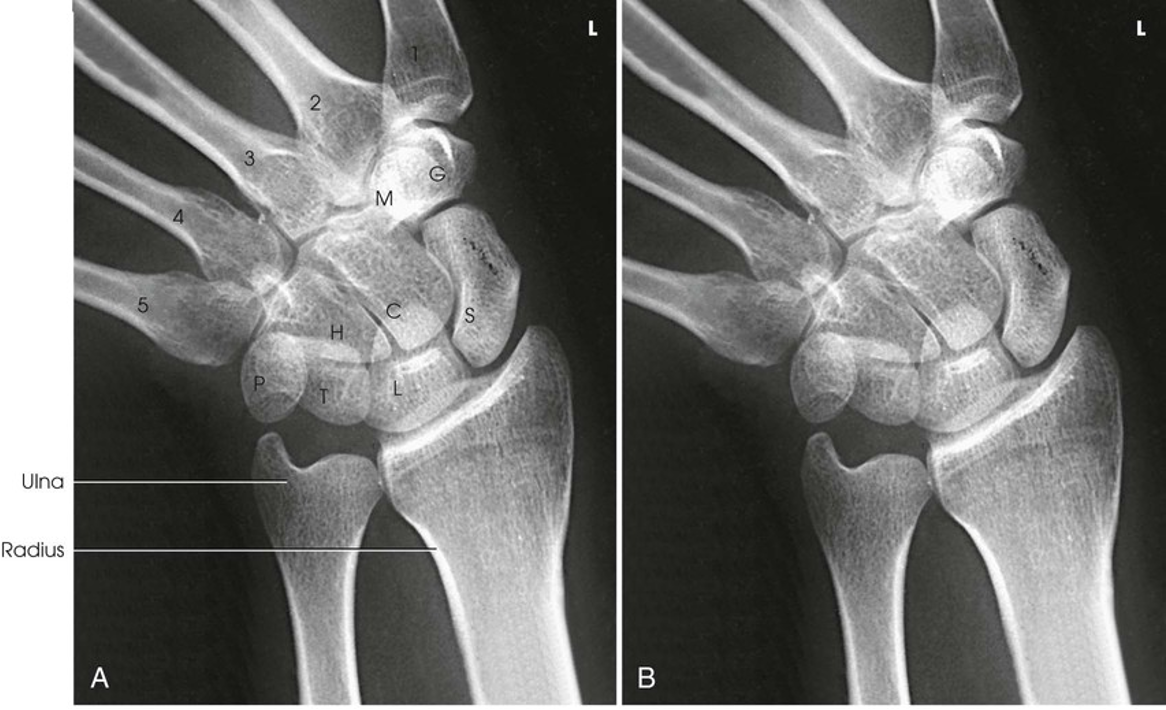
PA Projection; Wrist; Ulnar Deviation
PA Axial Projection; Wrist/Scaphoid; Stecher Method (CR & SS)
Perpendicular to the table and direct to enter the scaphoid
The 20- degree angulation of the wrist places the scaphoid at right angles to the central ray, so that it is projected with minimal superimposition
PA Axial Projection; Wrist/Scaphoid; Stecher Method (Eval criteria)
• Evidence of proper collimation
• Distal radius and ulna, carpals, and proximal half of the metacarpals
• Scaphoid with adjacent articulations open
• No rotation of wrist
• Soft tissue and bony trabecular detail
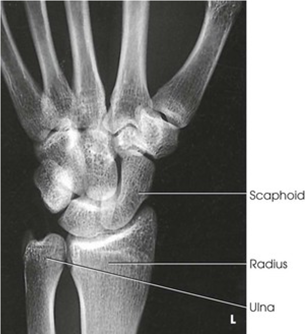
PA Axial Projection; Wrist/Scaphoid; Stecher Method
Tangential Projection (Inferosuperior); Carpal Canal; Gaynor-Hart Method (CR & SS)
Directed to the palm of the hand at a point approximately 1 inch (2.5cm) distal to the base of the third metacarpal and at an angle of 25 to 30 degrees to the long axis of the hand
This image of the carpal (carpal tunnel) shows the palmar aspect of the trapezium; the tubercle of the trapezium; and the scaphoid, capitate, hook of hamate, triquetrum, and entire pisiform
Tangential Projection; Carpal Canal (Eval Criteria)
• Evidence of proper collimation
• Carpals in an arch arrangement
• Pisiform in profile and free of superimposition
• Hamulus of hamate
• Soft tissue and bony trabecular detail
Tangential Projection (superoinferior); Carpal Canal (CR)
Tangential to the carpal canal at the level of the midpoint of the wrist
Angled toward the hand approximately 20-35 degrees from the long axis of the forearm
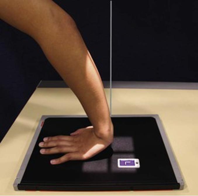
POSITIONING: Tangential Projection (superoinferior); Carpal Canal
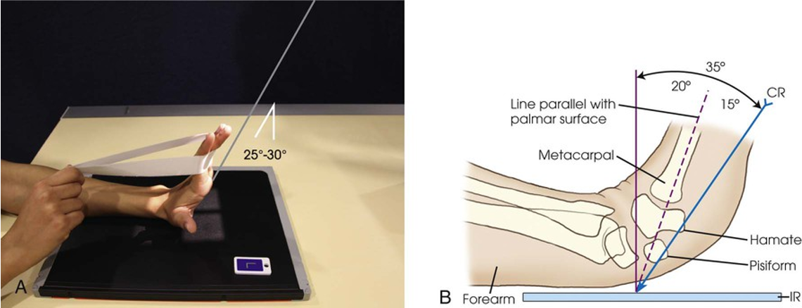
POSITIONING: Tangential Projection (Inferosuperior); Carpal Canal; Gaynor-Hart Method
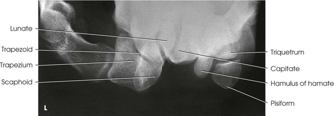
Tangential Projection (Inferosuperior); Carpal Canal; Gaynor-Hart Method
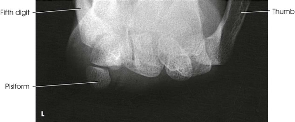
Tangential Projection (superoinferior); Carpal Canal