APEX Brain
 Call Kai
 Learn
Learn Practice Test
 Spaced Repetition
 Match
Match Flashcards
 Knowt Play
1/305

There's no tags or description
Looks like no tags are added yet.
Name | Mastery | Learn | Test | Matching | Spaced | Call with Kai | Chat |
|---|
No analytics yet
Send a link to your students to track their progress
306 Terms
The brain is made up of 2 types of cells. What are they?
Neurons and Glial cells
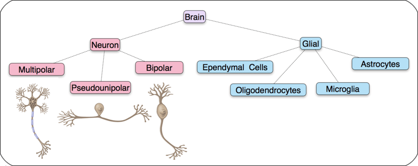
What is the functional unit of the nervous system?
-Primary role?
Neuron - receives and sends information
The cell bodies form what kind of matter?
Grey matter
The axons form what kind of matter?
White matter d/t myelin
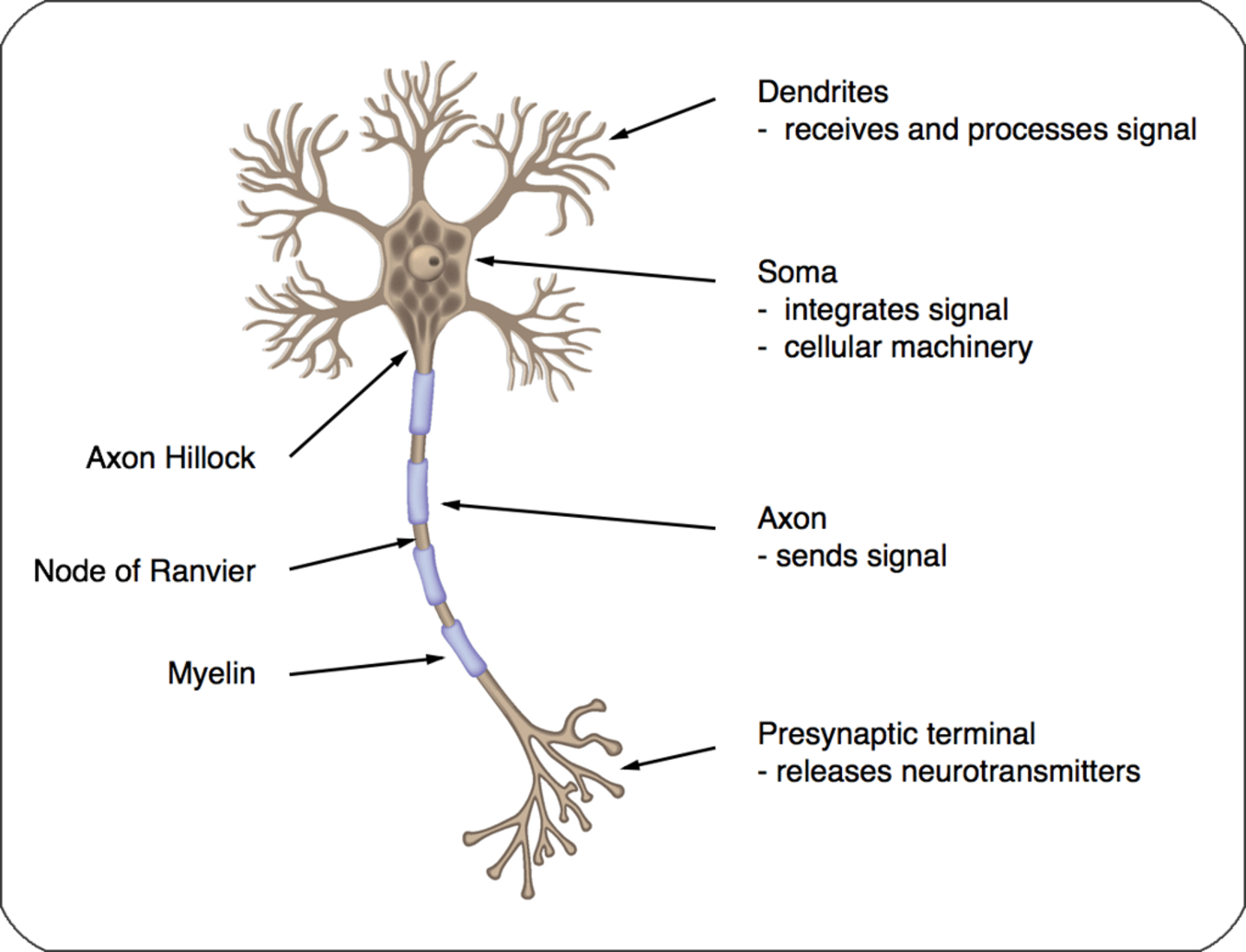
Which part of the neuron receives and processes signals?
Dendrites
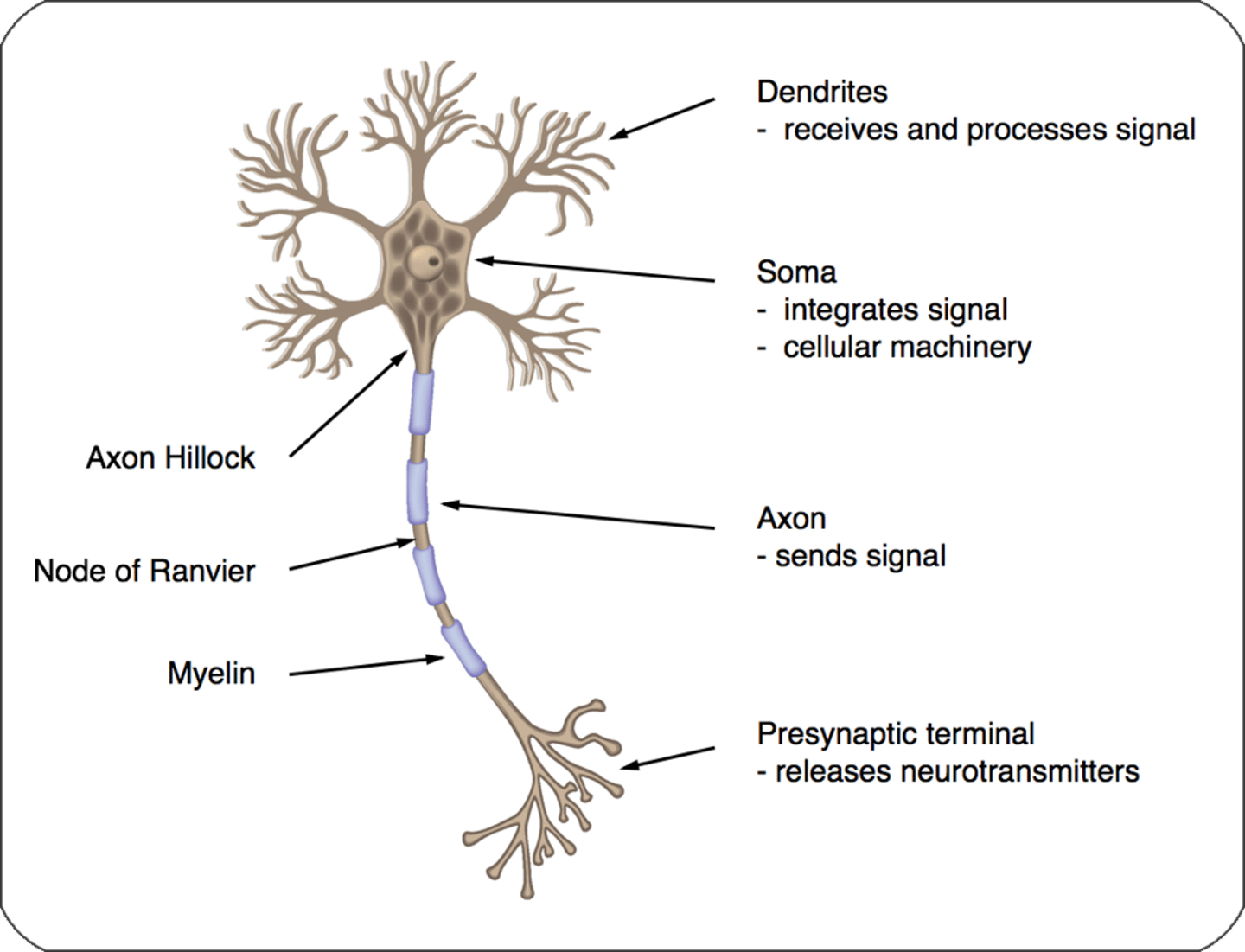
Which part of the neuron integrates the signal and has the cellular machinery?
Soma
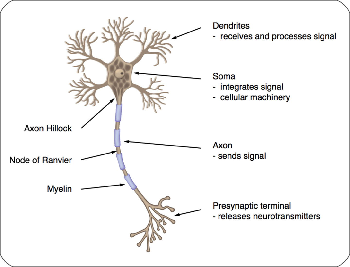
Where is the axon hillock located?
What does the axon do?
-Between soma and axon of a neuron
-Sends signals
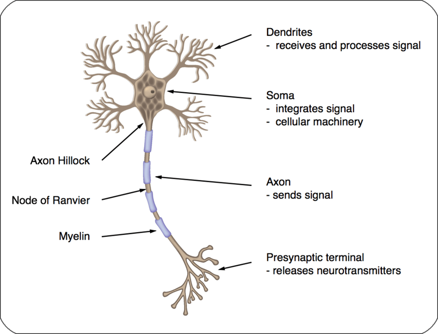
What does the presynaptic terminal do?
Releases neurotransmitters
What are the 3 types of neurons found in the CNS?
Multipolar
Pseudounipolar
Bipolar
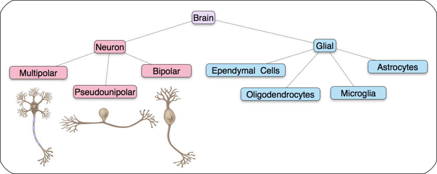
Most of the CNS neurons are what type?
Multipolar
Where are pseudounipolar neurons found?
Dorsal root ganglion and cranial ganglion
Where are bipolar neurons found?
Retina and ear
You have 2 eyes and 2 ears
Functions of the glial cells (nerve glue) (4)
Support neuronal function by:
-Creating a healthy ionic environment
-Modulate nerve conduction
-Controlling reuptake of neurotransmitters
-Repairing neurons following neuronal injury
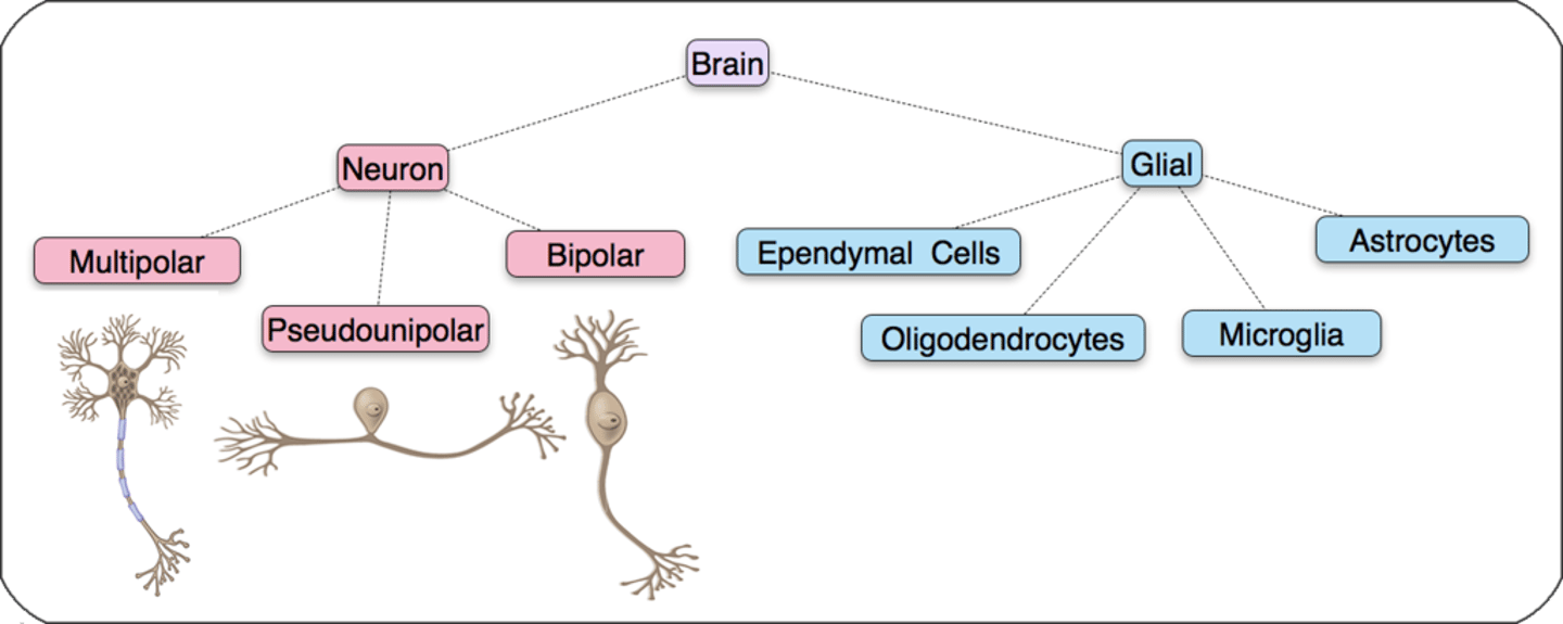
4 Types of glial cells
-Astrocytes
-Oligodendrocytes
-Microglia
-Ependymal cells
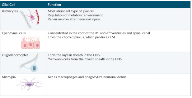
Which type of glial cell is most abundant?
Astrocytes
Astrocytes function
-Regulation of metabolic environment
-Repair neuron after neuronal injury
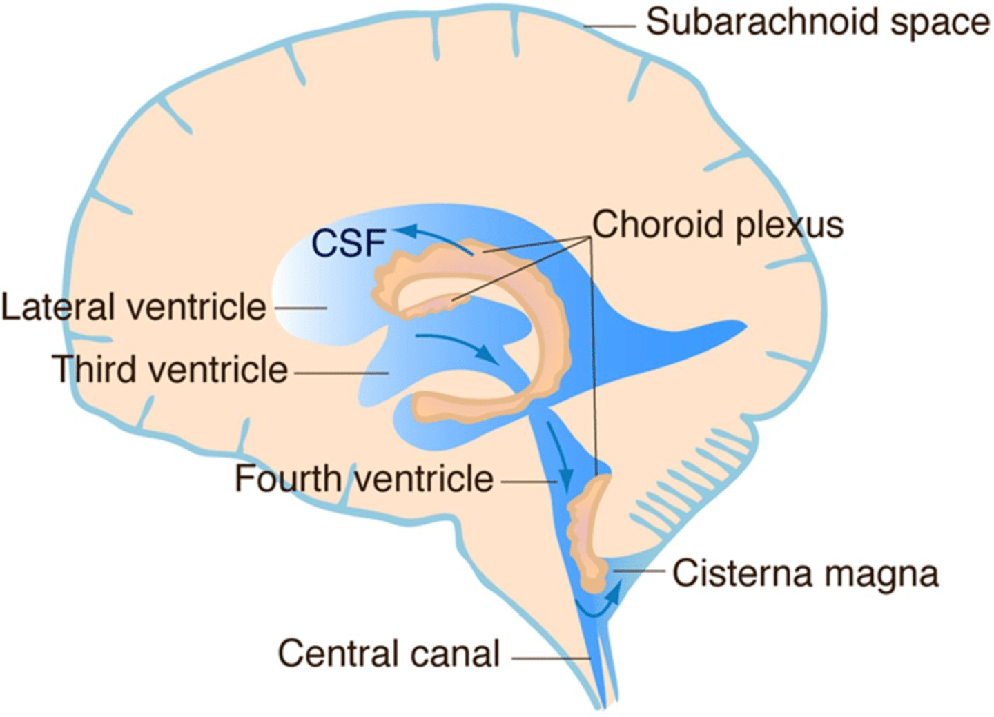
Where are ependymal cells located?
-Roof of the 3rd and 4th ventricles and spinal canal
*Involved in CSF production
Which cells form the choroid plexus?
Ependymal cells
Oligodendrocytes function
Form the myelin sheath in the CNS
Which cells form the myelin sheath in the PNS?
Schwann cells (myelinated beans that get skipped- in periphery)
Microglia function
-Act as macrophages and phagocytize neuronal debris
Where do most CNS tumors arise from?
The glia
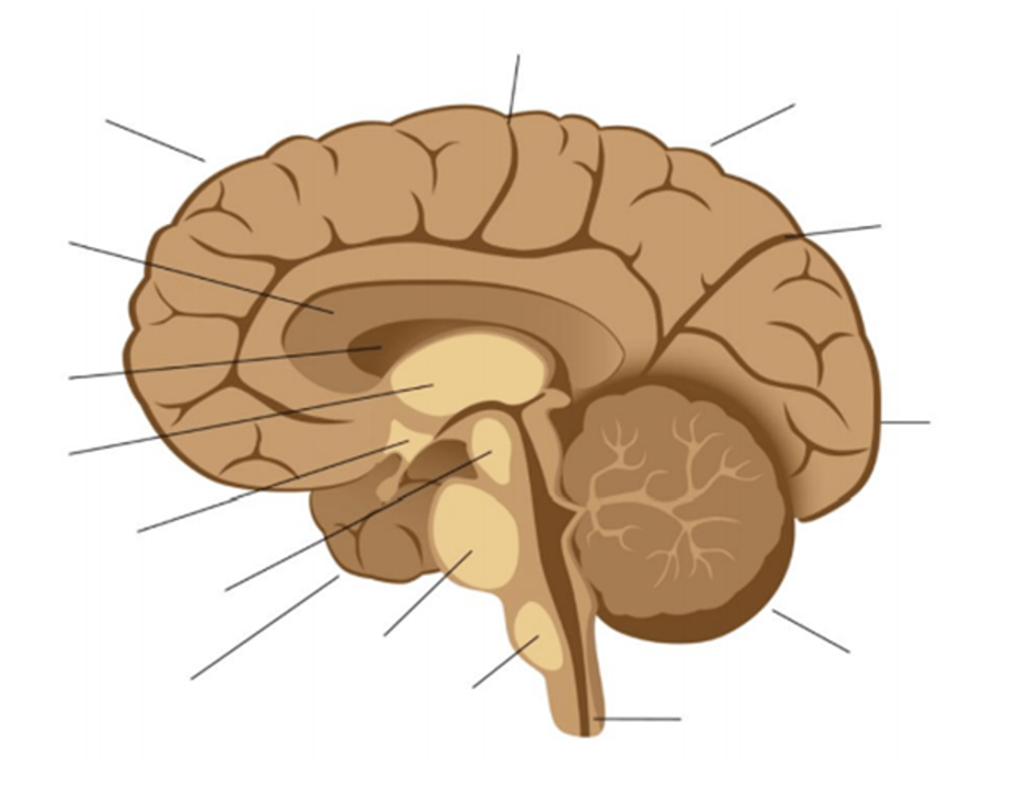
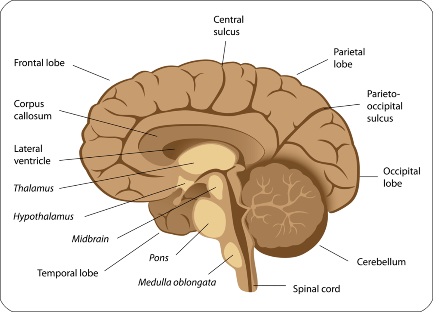
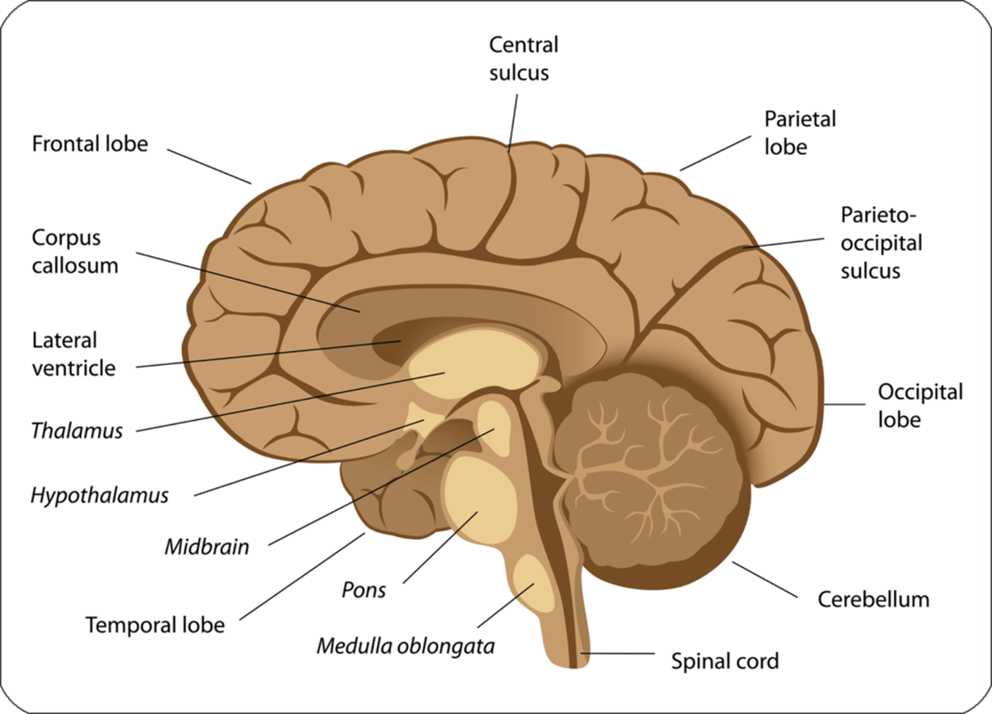
Label the brain
Pons
Medulla oblongata
Midbrain
Temporal lobe
Hypothalamus
Spinal cord
Cerebellum
Occipital lobe
Parietal lobe
Parietooccipital sulcus
Frontal lobe
Central sulcus
Thalamus
Corpus callosum
Lateral ventricle
The brain can be divided into 4 areas:
-Cerebral hemispheres
-Diencephalon
-Brainstem
-Cerebellum
What connects the hemispheres of the brain?
Corpus callosum
-Located deep in the longitudinal fissure
What are the 4 lobes of each cerebral hemisphere
-What do they contain?
-Frontal - contains motor cortex
-Parietal - contains somatic sensory cortex
-Occipital - contains vision cortex
-Temporal - contains auditory cortex and speech centers
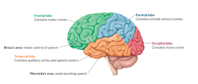
Wernicke's area location and function
Located in temporal lobe
Understanding speech
Broca's area location and function
Located in temporal lobe
Motor control of speech
Cerebral cortex functions
Cognition, sensation, movement
-Structure of the cerebral hemisphere
Hippocampus functions
Memory and learning
-Structure of the cerebral hemisphere
"University campus for memory and learning"
Amygdala functions
Emotion, appetite, responds to pain and stressors
-Structure of the cerebral hemisphere
Basal ganglia functions
Fine control of movement
-Structure of the cerebral hemisphere
"fine control when adding basal to food"
Diencephalon contains...
Thalamus and hypothalamus
Thalamus functions
-Relay station that directs information to various cortical structures
What is the primary neurohumoral organ?
Hypothalamus
neurohumoral - secrets neurotransmitters
4 structures in the brainstem
Midbrain
Pons
Medulla
Reticular activating system
Tracts in the midbrain
Auditory and visual
What, in the brainstem, causes autonomic integration?
Pons and medulla
Reticular activating system function
Controls consciousness, arousal, and sleep
What are the three parts of cerebellum
Archicerebellum
Paleocerebellum
Neocerebellum
Archeocerebellum function
Maintains equilibrium
-Structure of cerebellum
"architecture needs equilibrium"
Paleocerebellum
Regulates muscle tone
-Structure of cerebellum
"Paleo diet muscles"
Neocerebellum
Coordinates voluntary muscle movement
-Structure of cerebellum
What cranial nerve is most likely to be compressed by a pituitary tumor
A. Olfactory
B optic
C Oculomotor
D. Facial
B optic
Number and label cranial nerves
Numbers and Names
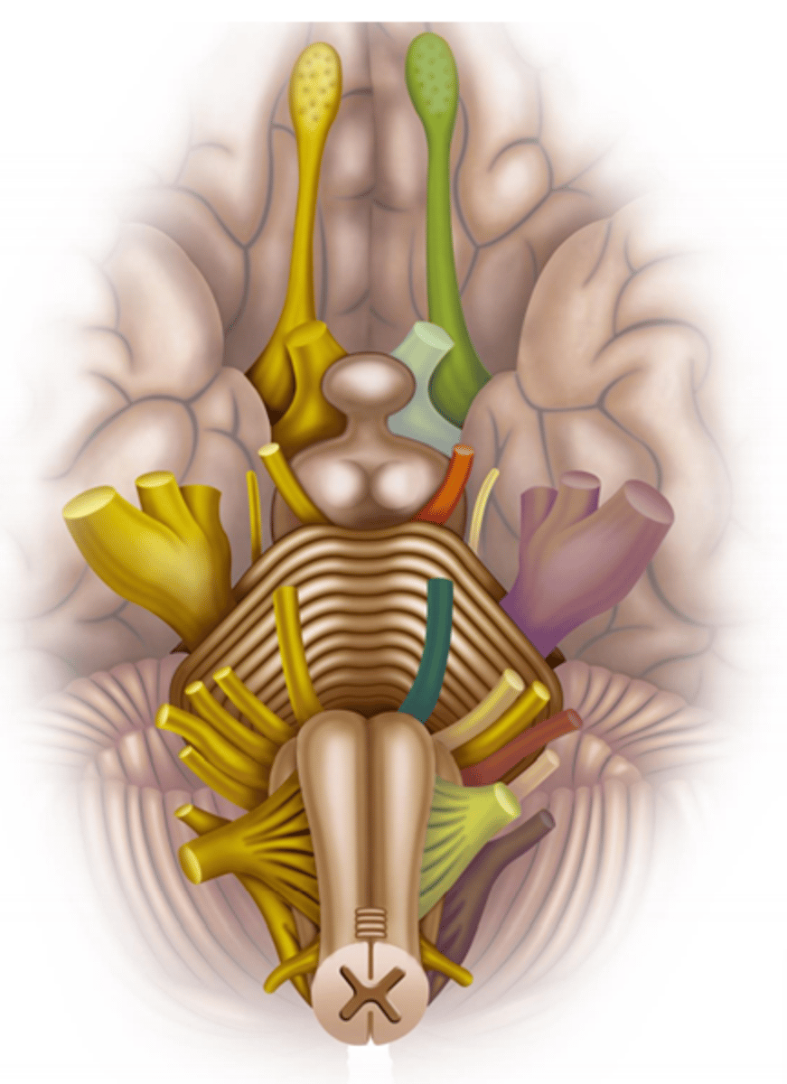
Cranial Nerves
On Occasion Our Trusty Truck Acts Funny Very Good Vehicle Any How
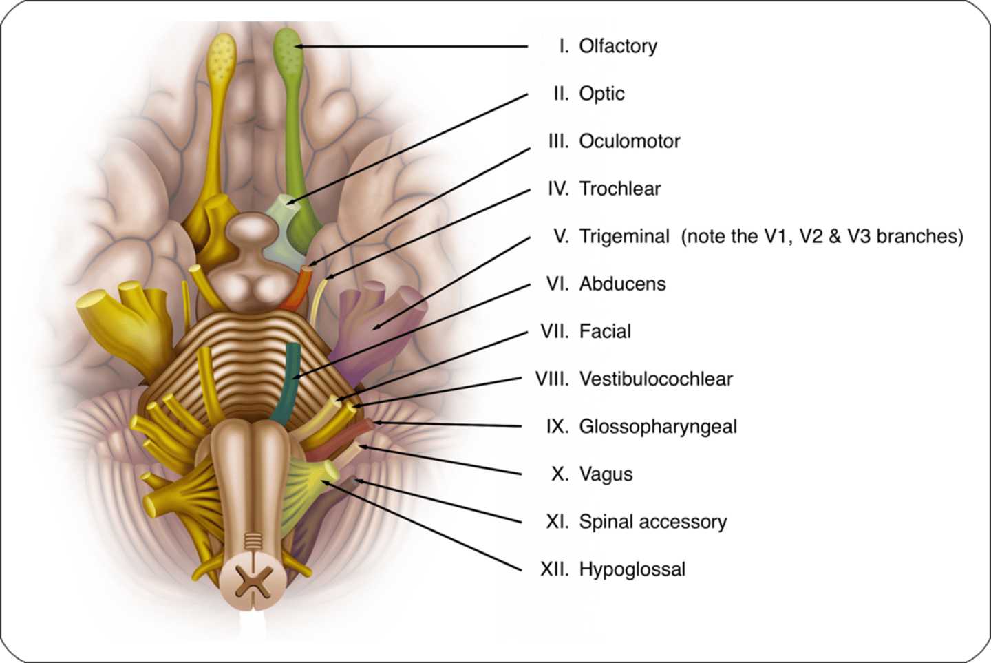
What are the functions of each cranial nerve?
Some Say Marry Money But My Brother Says Big Brains Matter Most
Sensory, Motor, Both
Cranial Nerve I
-Nerve name
-Function
-Bedside test
Olfactory
Sensory
Smell
Cranial Nerve II
-Nerve name
-Function
-Bedside test
Optic
Sensory
Vision
Cranial Nerve III
-Nerve name
-Function
-Bedside test
Oculomotor
Motor
Eye movement and pupil constriction
3 - eye lashes so all movement
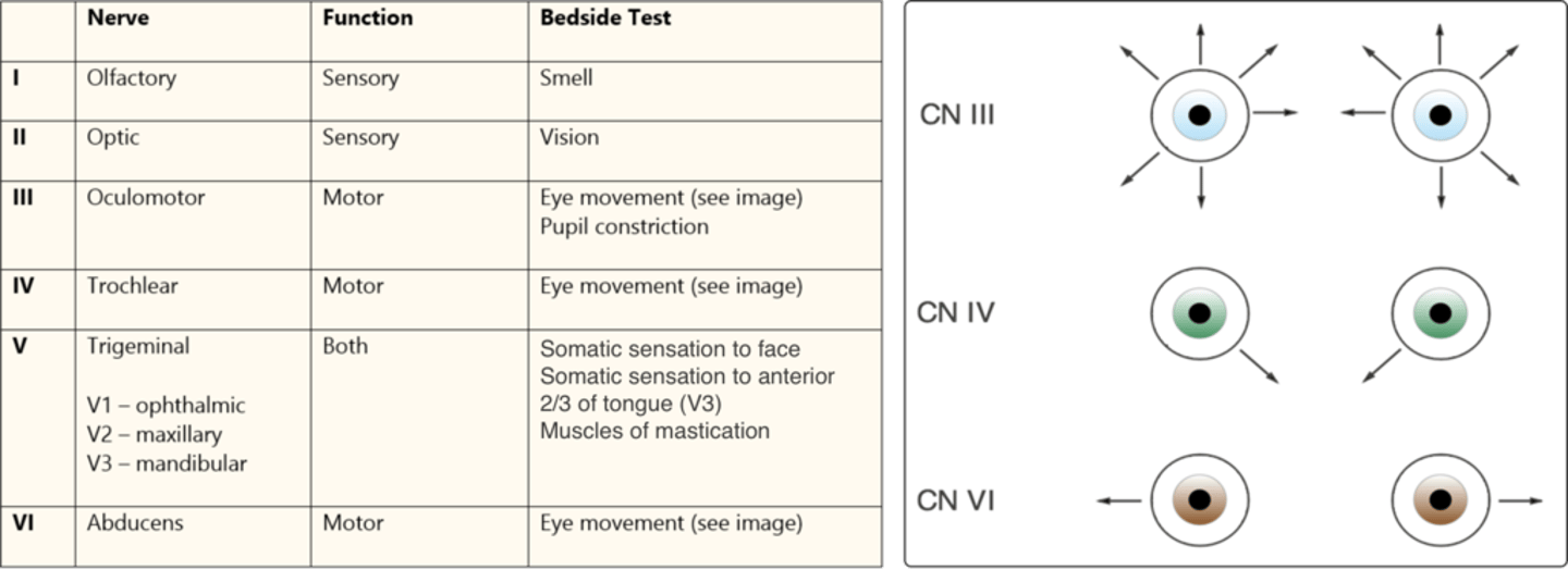
Cranial Nerve IV
-Nerve name
-Function
-Bedside test
Trochlear
Motor
Eye Movement down and center
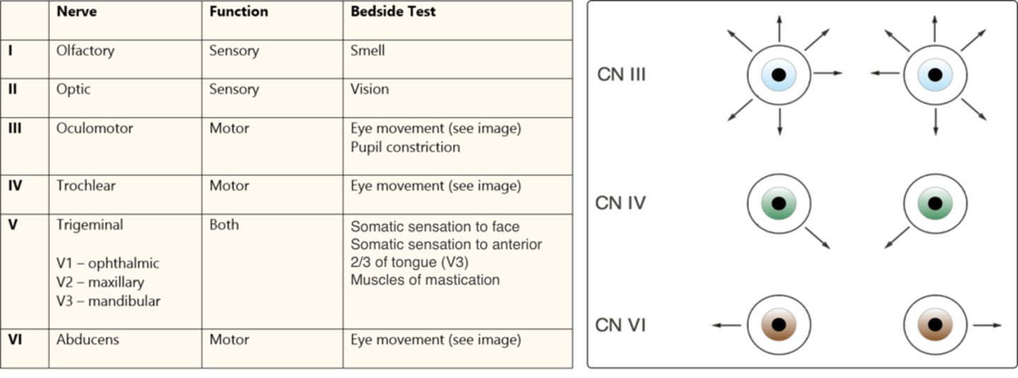
Cranial Nerve V
-Nerve name
-Function
-Bedside test
Trigeminal
-VI: ophthalmic
-V2: Maxillary
-V3: Mandibular
Both
Somatic sensation to face
Somatic sensation to anterior 2/3 tongue
Muscles of mastication
Cranial Nerve VI
-Nerve name
-Function
-Bedside test
Abducens
Motor
Eye Movement out to sides
Cranial Nerve VII
-Nerve name (5 branches)
-Function
-Bedside test
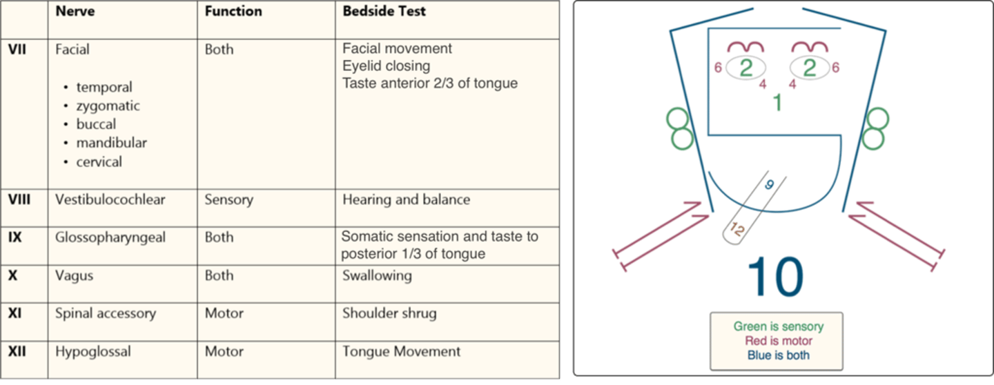
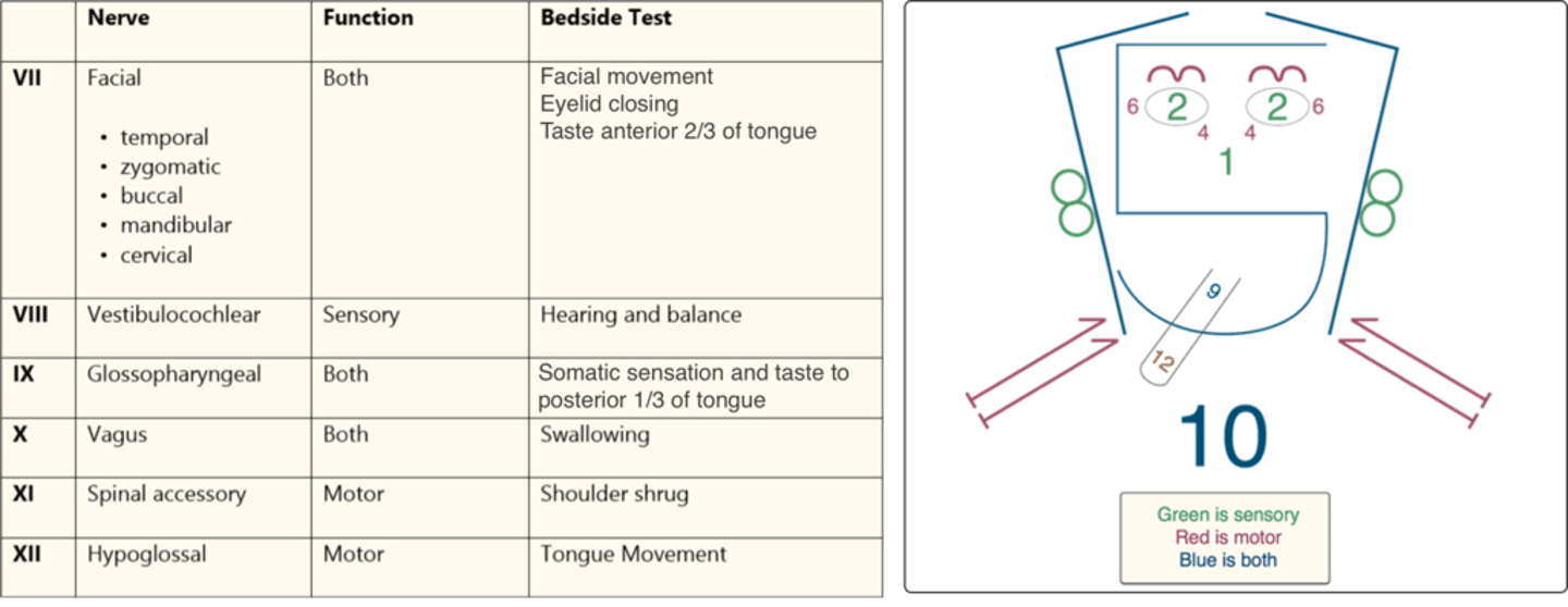
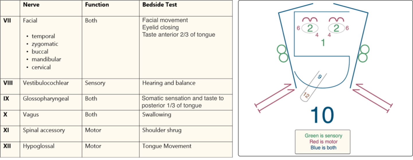
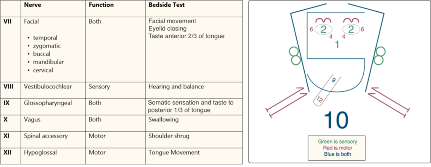
Facial (temporal, zygomatic, buccal, mandibular, cervical)
Both
Facial movement
Eyelid closing
Taste anterior 2/3 of tongue
*Two zebras bit my carrot
*Two zits by my clavicle
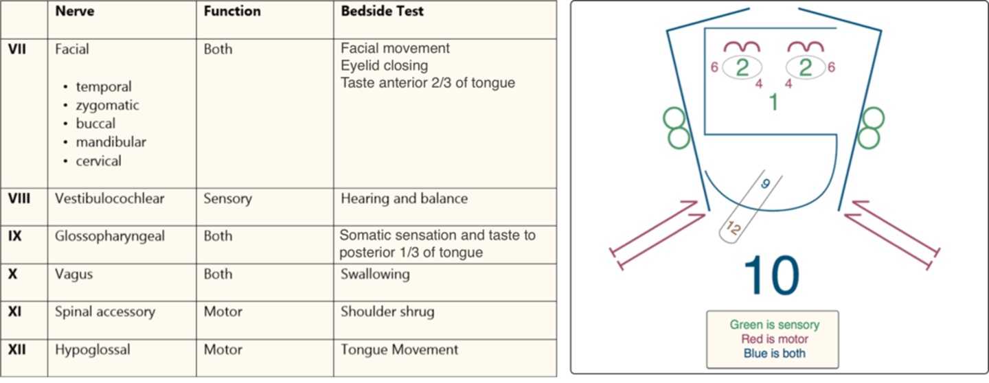
Cranial Nerve VIII
-Nerve name
-Function
-Bedside test
Vestibulocochlear
Sensory
Hearing and balance
Cranial Nerve IX
-Nerve name
-Function
-Bedside test
Glossopharyngeal
Both
Somatic sensation and taste to posterior 1/3 tongue
Cranial Nerve X
-Nerve name
-Function
-Bedside test
Vagus
Both
Swallowing
Cranial Nerve XI
-Nerve name
-Function
-Bedside test
Spinal accessory
Motor
Shoulder shrug
Cranial Nerve XII
-Nerve name
-Function
-Bedside test
Hypoglossal
Motor
Tongue movement
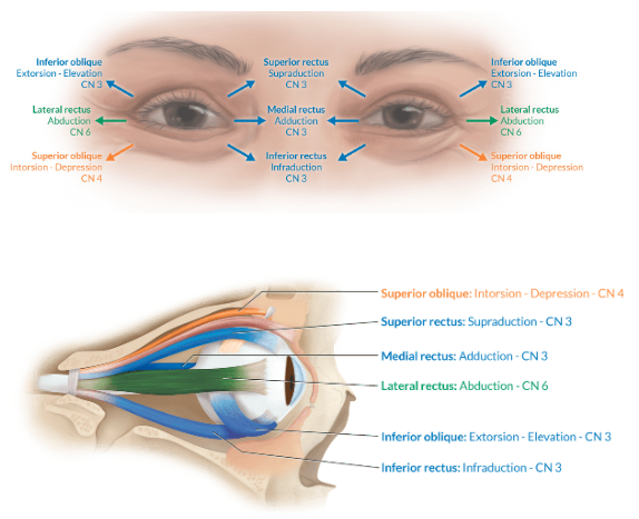
Cranial Nerve Eye functions
Where do cranial nerves arise from?
Brain and brainstem
-12 cranial nerves
Cranial nerves are part of the central or peripheral nervous system?
-Which one is the exception?
-Peripheral nervous system
-Optic Nerve (II) is part of the central nervous system and is surrounded by dura
What is tic doulureaux?
What nerve is affected?
Trigeminal neuralgia CN 5
Excruciating neuropathic pain in the face
What nerves control eye movement
CN 3, 4, 6
Mnemonic for five branches of the facial nerve (CN7)
Two Zebras Bit My Carrot
Bell's palsy results from injury to...
the facial nerve (7)
-causes ipsilateral facial paralysis
Parasympathetic output is carried by which cranial nerves?
III, VII, IX, and X
Which nerve is responsible for 75% of all parasympathetic activity?
Vagus
What does the blood brain barrier separate?
What restricts passage of large molecules and ions within the BBB?
-Separates CSF from plasma
-Tight junctions restrict passage of large molecules and ions (does not have carrier proteins)
When is the BBB dysfunctional?
Sites of tumor, injury, infection, or ischemia
& poorly developed in the neonate
Cerebrospinal fluid functions (3)
-Cushions the brain
-Provides buoyancy
-Delivers optimal conditions for neurologic function
Cerebrospinal fluid locations (3)
-Ventricles (left lateral, right lateral, third, and fourth)
-Cisterns around the brain
-Subarachnoid space in brain and spinal cord
Where is the BBB not present? (5)
Chemoreceptor trigger zone (CTZ)
Posterior pituitary gland
Pineal gland
Choroid plexus
Parts of the hypothalamus
"Diencephalon"
The BBB is poorly developed in...
neonates
CSF volume
150mL
Specific gravity of CSF
1.002-1.009
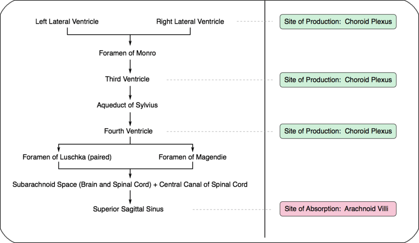
What produces CSF and where is it located?
-Produced by ependymal cells of choroid plexus in cerebral ventricles
-Choroid plexus is located in all 4 cerebral ventricles
Where is CSF reabsorbed
Arachnoid villi in the superior sagittal sinus. Reabsorption is dependent on pressure gradient between CSF and venous circulation
CSF pressure
5-15mmHg
Flow of CSF
Lateral ventricles
Foramen of Monro
3rd ventricle
Aqueduct of Sylvius
4th ventricle
Foramen of Luschka and Magendie
Subarachnoid Space
Superior sagittal sinus/arachnoid villi
"Love My 3 Silly 4 Lorn Magpies"
Production rate of CSF
30mL/hr
Relative tonicity of CSF compared to plasma
Isotonic
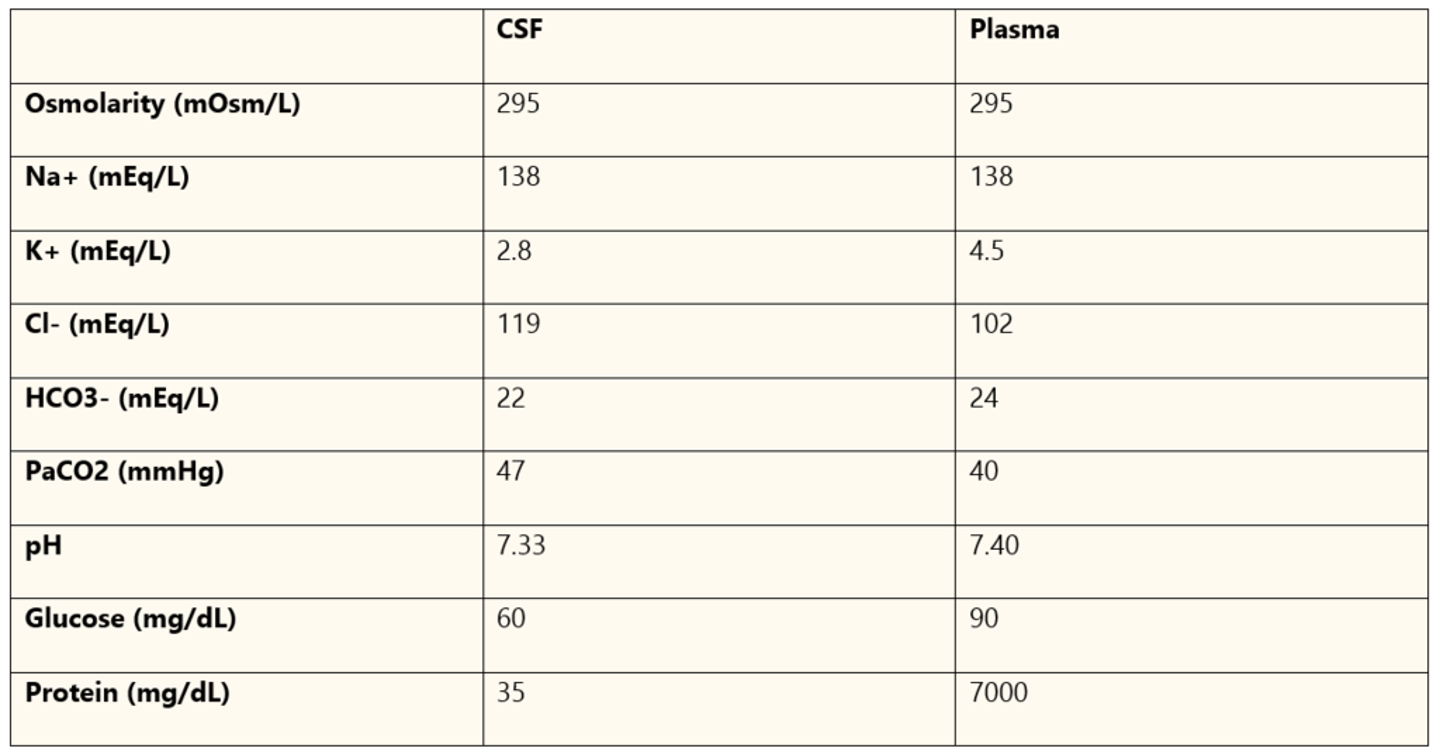
(CSF is isotonic with plasma, it is not an ultrafiltrate of plasma and has its own chemical composition)
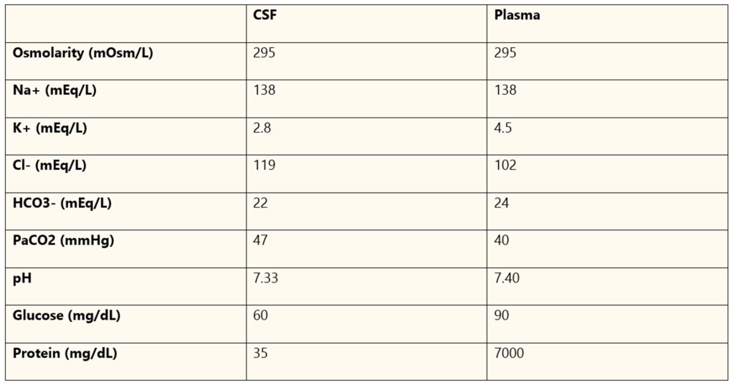
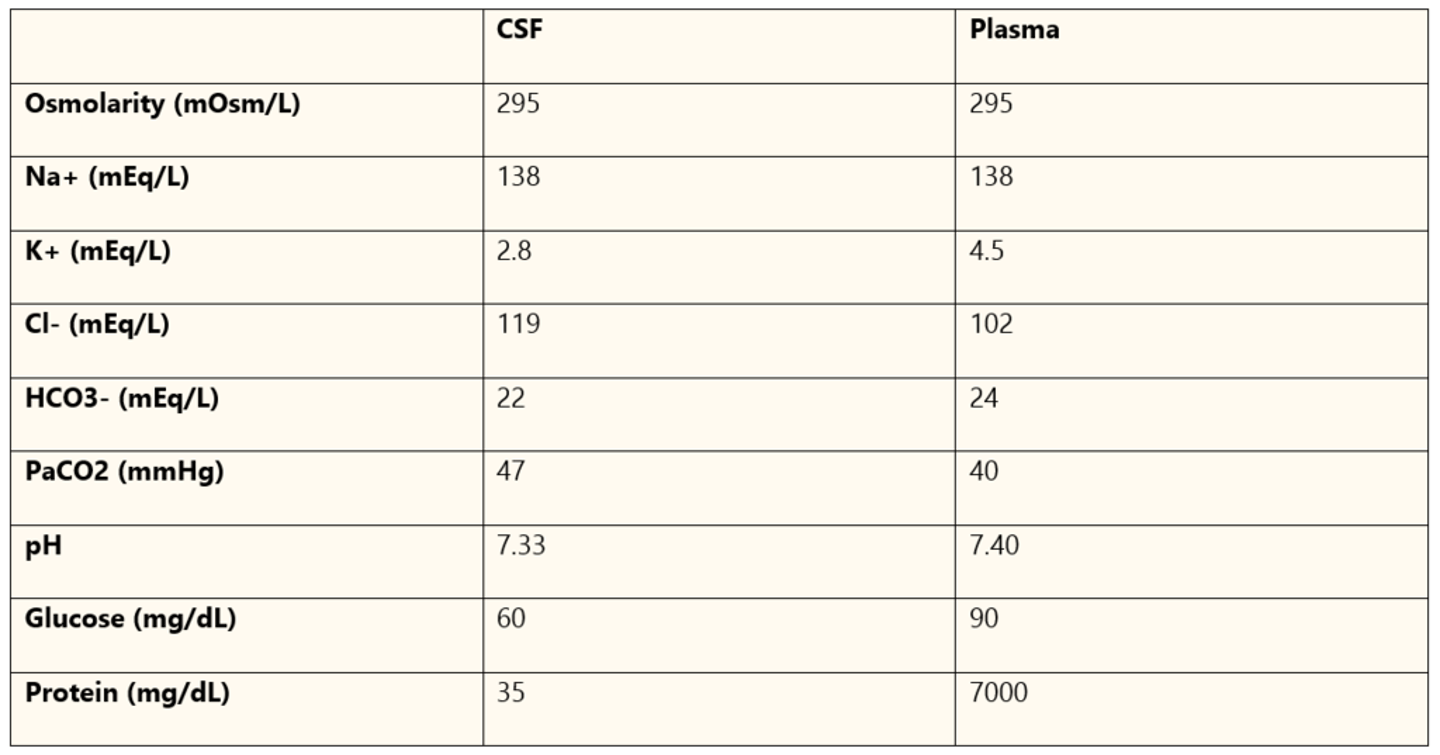
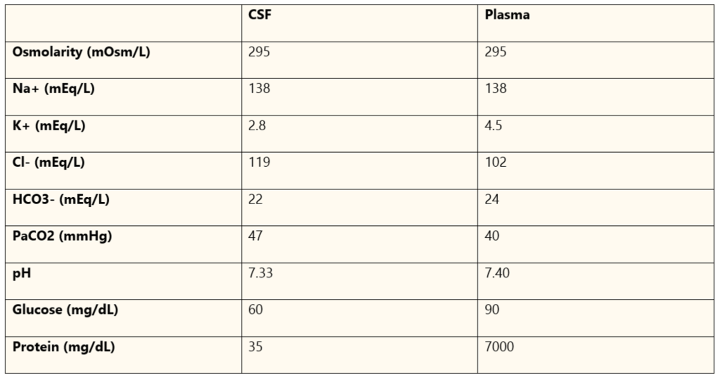
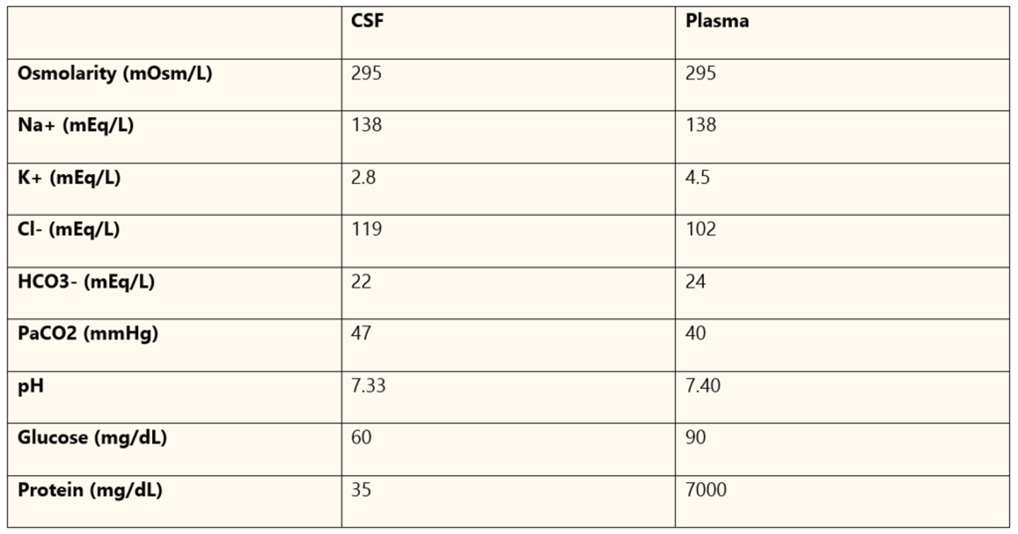
-Osmolarity the same 295 mOsm/L
Site of absorption of CSF
Reabsorbed into venous circulation via arachnoid villi in the superior sagittal sinus
CSF reabsorption is dependent on the pressure gradient between ___ and ___
CSF and venous circulation
Potassium in CSF compared to plasma
2.8 in CSF vs. 4.5 in plasma
"Guess lower - everything is lower except CO2 in the brain"
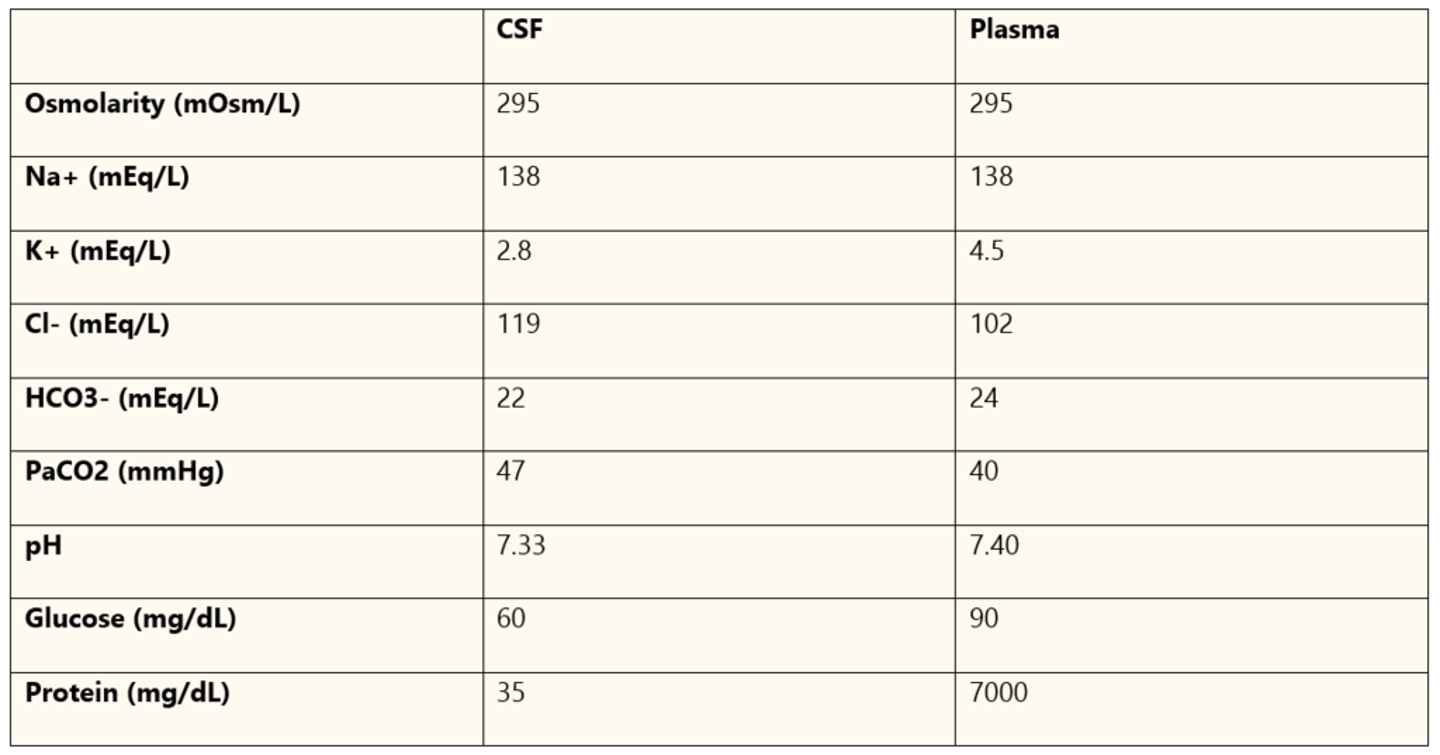
Which electrolytes are similar between CSF and plasma?
Sodium
Bicarb
Chloride (119 in CSF vs 102 in plasma)
PaCO2 (47 in CSF vs 40)
Glucose content in CSF vs. plasma
60 in CSF vs. 90 in plasma
*all used up
Protein in CSF vs. plasma
35 in CSF vs. 7000 in plasma
pH of CSF
7.33
(PaCO2 47 in CSF)
What is hydrocephalus
Excessive accuulation of CSF in the brain which can increase ICP
What are the 2 types of hydrocephalus
Obstructive hydrocephalus- obstruction to CSF flow in the ventricular system (most common)
Communicating hydrocephalus- decreased CSF absorption by arachnoid villi (ICH) or overproduction of CSF
Surgical management for either type of hydrocephalus
Depends on cause
Could either be a catheter in cerebral ventricles to drain CSF or placing a ventriculoperitoneal shunt (brain to heart) or ventriculoperitoneal shunt (brain to belly)
Global cerebral blood flow
45-55mL/100g tissue/min
Percentage of cardiac output going to global cerebral blood flow
15%
Cerebral autoregulation definition
-Formula for cerebral blood flow
The brains ability to maintain a constant cerebral blood flow over a wide range of mean arterial blood pressures
CBF = Cerebral perfusion pressure/Cerebral vascular resistance
-Benefit: ensures steady supply of O2 even with blood pressure fluctuations
Cortical CBF
75-80mL/100g/min
Subcortical CBF
20mL/100g/min
At what level of CBF is there evidence of ischemia?
20mL/100g/min
At what level of CBF is there complete cortical suppression?
15mL/100g/min
At what level of CBF is there membrane failure and cell death?
<15mL/100g/min