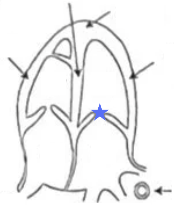
Echocardiographic windows and views
1/159
There's no tags or description
Looks like no tags are added yet.
Name | Mastery | Learn | Test | Matching | Spaced |
|---|
No study sessions yet.
160 Terms
Acoustic Window
the transducer’s position on the thoracic surface enabling imaging of the heart without interference from the lungs and ribs
What are the 5 main windows?
Left parasternal
right parasternal
Apical
Subcostal
Suprasternal notch
windows are
where you put the transducer on the body
Views are
the different images you can obtain at that window by adjusting the probe
Imaging planes in TEE
Long axis
Short axis
4 Chamber

Sagittal View =
LAX
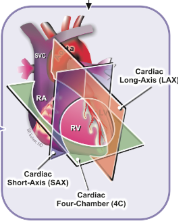
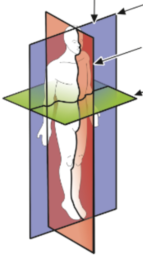
Frontal View =
SAX


Transverse view =
4C

Parasternal Views (P)
PLAX
RVIT
RVOT
PSAX @ Ao
PSAX @ MV
PSAX @ PM
PSAX @ apex
Apical Views (A)
4 Ch
5 Ch
2 Ch
3 Ch / LAX
Subcostal views (SC)
4 Ch
SAX
LAX @ IVC
LAX @ Hep Vn
Suprasternal views (SSN)
LAX @ aortic arch
SAX @ Ao
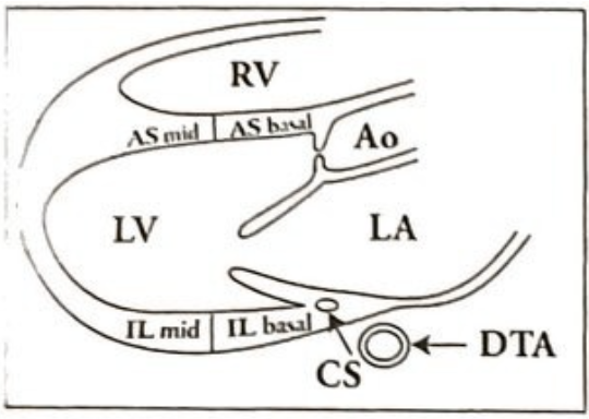
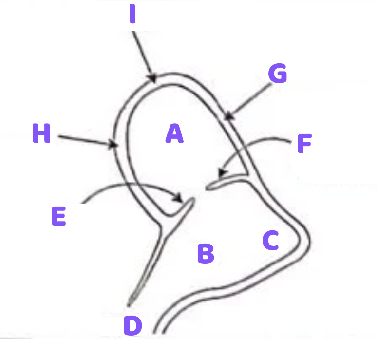
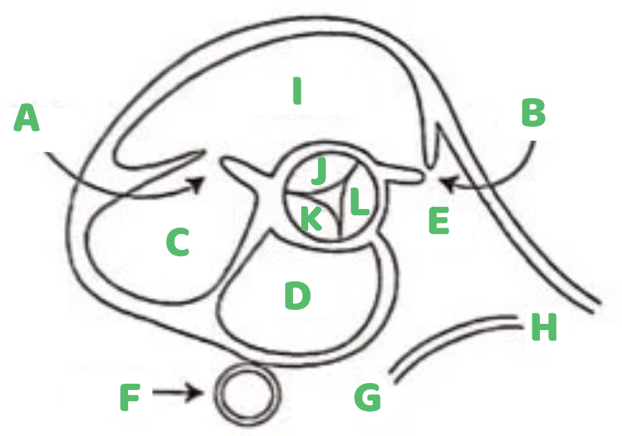
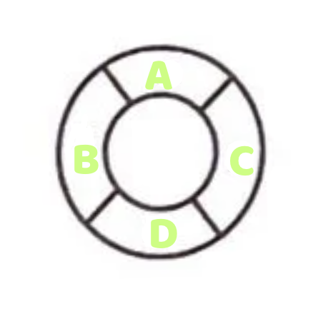
What view is this?
PLAX

A
Right Ventricle

B
Left Ventricle

C
Left Atrium

D
Aorta

E
AnteroSeptal (AS) basal

F
AnteroSeptal (AS) mid

G
descending aorta

H
Inferolateral mid
I
Inferolateral basal
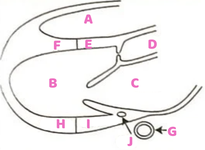
J
Coronary Sinus (CS)
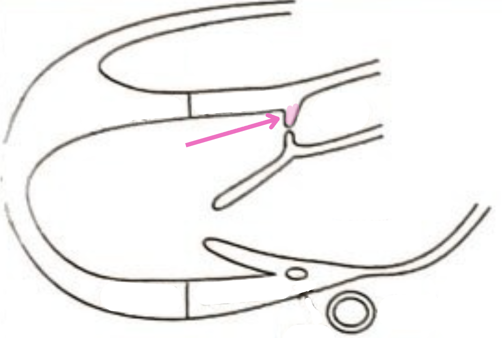
What is this structure?
right coronary cusp (RCC)
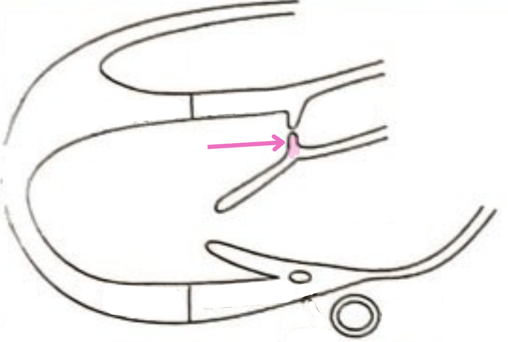
What is this structure?
non coronary cusp (NCC)
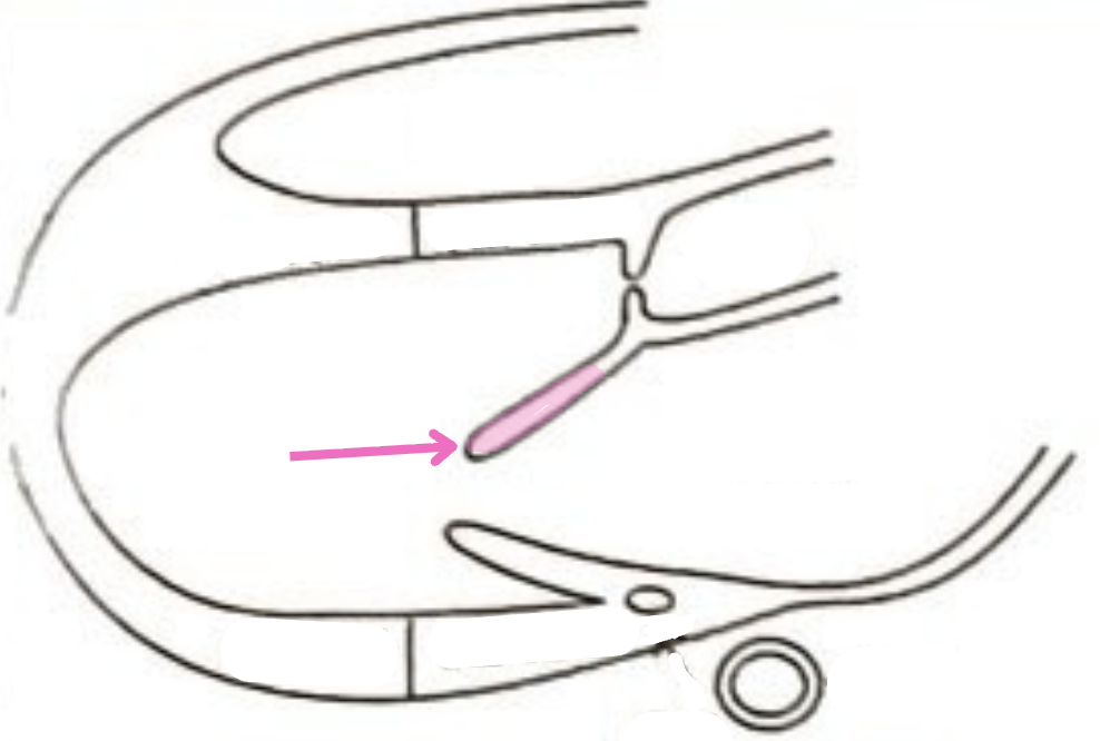
What is this structure?
anterior mitral valve leaflet (AMVL)
What is this structure?
posterior mitral valve leaflet (PMVL)
What is this structure?
Posterior / Inferolateral wall
What is this structure?
IVS
What is this structure?
Right Ventricular Free Wall (RVFW)
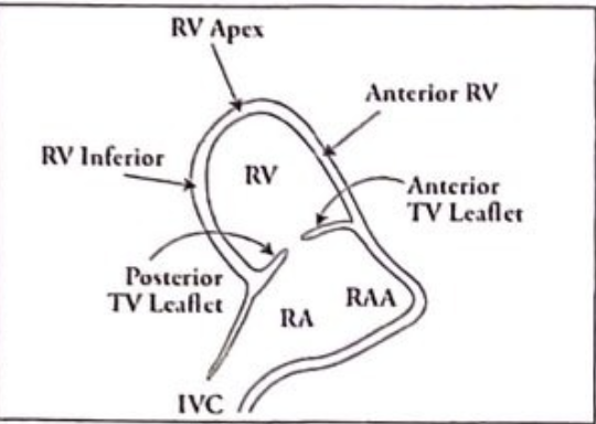
What is this view?
Parasternal RVIT

A
RV

B
RA

C
RAA

D
IVC

E
Posterior TV leaflets

F
Anterior TV leaflets

G
Anterior wall RV

H
RV inferior wall
I
RV apex
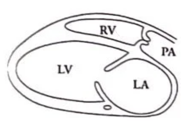
What is view is this?
Parasternal RVOT

A
LV

B
LA
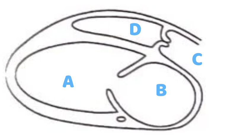
C
Pulmonary Artery (PA)

D
RVOT
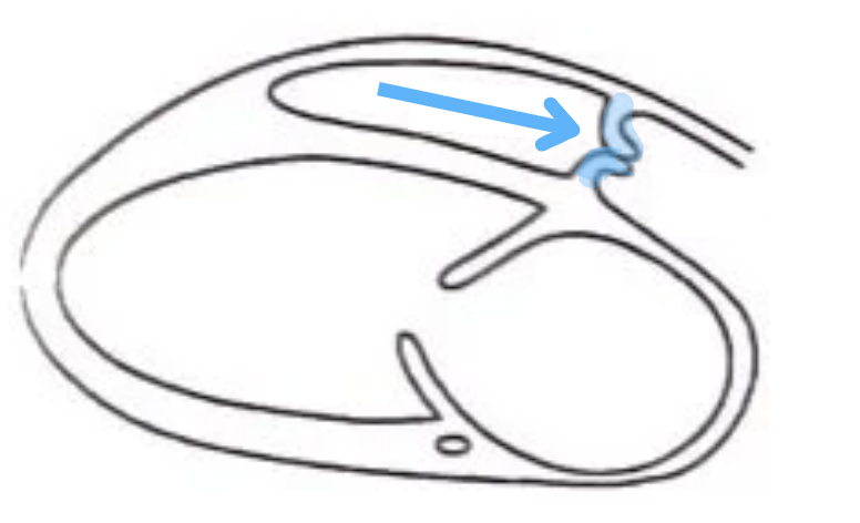
What is this structure?
Pulmonic Valve (PV)
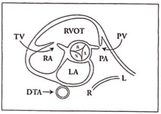
What view is this?
PSAX @ Aorta

A
TV
B
PV

C
RA

D
LA

E
Pulmonary Artery (PA)

F
descending aorta

G
right PA

H
left PA

I
RVOT

J
RCC (right coronary cusp)

K
NCC (non coronary cusp)

L
LCC (left coronary cusp)
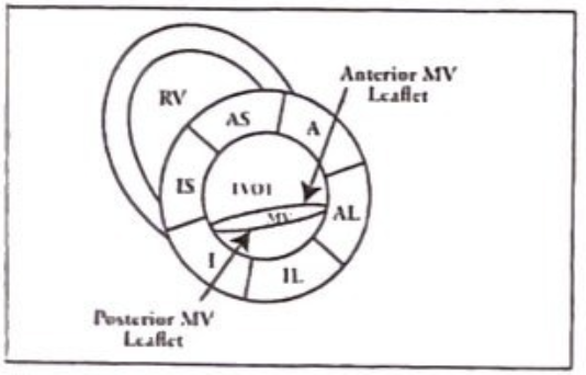
What view is this?
PSAX @ MV

A
RV
B
AS - Anteroseptal

C
A - anterior

D
AL - Anterolateral

E
IL - inferolateral

F
I - inferior

G
IS - inferoseptal

H
Anterior MV leaflet

I
Posterior MV leaflet

J
MV
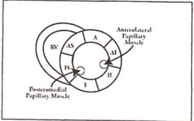
What view is this?
PSAX @ Papillary Muscles
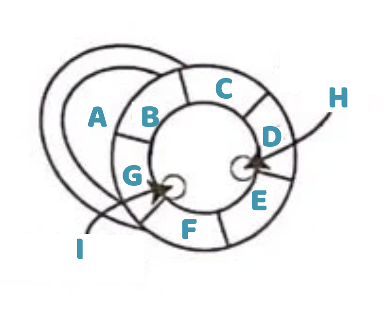
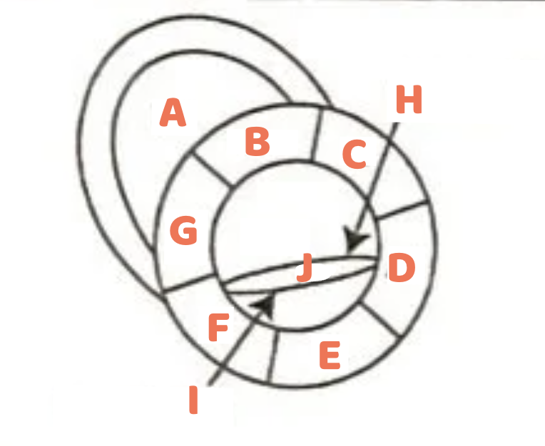
A
RV

B
AS - anteroseptal

C
A - Anterior

D
AL - anterolateral

E
IL - inferolateral

F
I - Inferior

G
IS - Inferoseptal

H
Anterolateral Papillary Muscle

I
Posteromedial Papillary Muscle
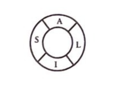
What view is this?
PSAX @ apex
A
A - Anterior

B
S - septum

C
L - lateral

D
I - inferior
What view is this?
Apical 4 Chamber

A
RV

B
LV

C
RA

D
LA

E
IVC

F
Pv (pulmonary vein)

G
Pv

H
Lateral wall
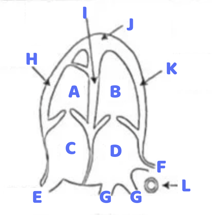
I
Inferior IVS

J
Apex

K
Anterolateral

L
DTA - descending aorta
What structure is this?
TV
What structure is this?
MV