Feline and Canine Integumentary ICVA Diseases
 Call Kai
 Learn
Learn Practice Test
 Spaced Repetition
 Match
Match Flashcards
 Knowt Play
1/118

Earn XP
Description and Tags
Dog: 77%, Feline: 80%
Name | Mastery | Learn | Test | Matching | Spaced | Call with Kai | Chat |
|---|
No analytics yet
Send a link to your students to track their progress
119 Terms
Allergic Dermatitis (Feline)
Atopy is a subtype of Allergic Dermatitis that is year round
Caused by environmental allergens and is inherited
Allergic Dermatitis (Feline): C.S
Pruritus + Alopecia
Erythema + Papules
Crusting and Scaling
Miliary Dermatitis
Ear Infections
Allergic Dermatitis (Feline):D.X
Hx + Exam
Flea control trial
Food trial: 8-12 weeks
Intradermal Skin Testing: Gold Standard
Serologic Testing
Skin Biopsy
Allergic Dermatitis (Feline): T.X
Flea control:FAD
Dietary Managements:Food Allergies
Environmental Management:Atopy
Pharmacologic Therapy:
Corticosteroids: Short term use, Acute flare ups
Antihistamines
Omega-3
Cyclosporine: Long term
Topical Therapy: Medicated shampoos etc
Immunotherapy: Allergen specific immunotherapy (ASIT)
Atopy (Canine):C.S
Pruritus + Erythema
Lichenification + Hyperpigmentation
Alopecia
Otitis externa
Secondary Infections
May present seasonally
Onset:1-3yrs old
Atopy (Canine):D.X
Rule out other pruritic diseases
Allergen Testing: Intraderma, serum IgE
Favrot’s criteria: Meet at least 5 out of 8
Onset of pruritus before 3 years of age
Dog lives mostly indoors
Glucocorticoid Responsive Pruritus
Pruritus w/o primary lesions at onset
Affected front feet
Affected ear pinnae
Non-affected ear margins
Affected ventral abdomen
Atopy (Canine):T.X
Allergen Avoidance
Pharmacotherapy
Corticosteroids: Short term
Cyclosporine: Calcineurin inhibitor
Oclacitinib (Apoquel)*: JAK inhibitor
Short term and long term
Lokivetmab (Cytopoint)*: Monoclonal antibody therapy
Targets the cytokine IL-31
Short term therapy
ASIT
Adjunct therapies
FAD: C.S
Reaction to flea saliva:Type I and IV Hypersensitivity
Intense Pruritus
Lesions:Alopecia + Erythema + papules + excoriations + crusts + moist dermatitis
Chronic: Hyperpigmentation + Lichenification
Secondary Infections
Dogs: Caudo-dorsal distribution
Base of tail
Posterior/Lateral aspect of rear legs
Ventral Abdomen
Cats:Often neck and face
Miliary Dermatitis
Similar distributions as dog
Symmetrical alopecia of the dorsal lateral trunk
FAD (Feline):D.X
Clinical History + Physical Exam: See flea dirt etc
Distribution of clinical lesions
Intradermal Skin Testing: Gold Standard
Serological Testing: IgE tests
Response to treatment
FAD (Feline):T.X
Flea control
Symptomatic Tx
Glucocorticoids
Antihistamines
Fatty Acid Supplements
Treatment of Secondary Infections
Immunomodulatory Therapy
Food Allergy(K9+Feline): C.S
Non-seasonal pruritus
Persistent, Year round
Usually on head, neck, perianal regions
Eosinophilic Granuloma Complex
Indolent ulcers, plaques, linear granuloma
Miliary Dermatitis(Dorsum)
Food Allergy(K9+Feline): D.X
Dietary Elimination Trial using a novel protein or hydrolyzed protein
Gold standard. 8-12 weeks
Allergen Specific IgE testing
Poor sensitivity+ specificity in cats
Intradermal Testing
More commonly used in dogs
Histopath
Food Allergy(K9+Feline): T.X
Dietary Management
Long -term Diet
Avoid Cross contamination
Medications
Corticosteroids
Antihistamines
Fatty Acid Supplements
Management of Secondary Infections
Burns (K9+Feline): Levels of Burns
First-Degree Burns (Superficial)
Second-Degree Burns (Partial Thickness)
Third-Degree Burns (Full Thickness)
Burns covering a large surface can trigger SIRS
Burns (K9+Feline): C.S
Pain
First and Second degree burns
Redness + Swelling
First degree
Blisters
Second degree
Eschar Formation
Third degree, dry blackened or leathery non painful skin
Discharge and Odor
If infected
Systemic Signs
Burns (K9+Feline): D.X
Exam
Assessment of Burn Surface Area (BSA)*
Wound Culture + Sensitivity: if infected*
CBC/CHEM: Look for SIRS, Lyte imbalance, organ function impairment
Lactate Levels: ^ indicates poor perfusion or sepsis in severe burns
Imaging: Deep tissue burns
Fluid Balance
To avoid hypovolemic shock
Burns (K9+Feline):T.X
Immediate Stabilization
Fluid therapy
Pan Management : First + Second Degree
Wound Management
Cooling
Debridement
Dressings: Foam, Silver, Hydrogel
Negative Pressure Wound Therapy
Infection Prevention
Topical antimicrobials: Silvr Sulfadiazine or Honey dressing
Supportive Care
High Calorie +High Protein
Vitamins A,C,Zinc: support collagen formation
Surgical Intervention
Escharotomy
Skin Grafting
Adjunctive Therapies
Hyperbaric Oxygen therapy
Physical Therapy
Cuterebriasis: C.S
Usually late summer and early fall
Well- demarcated,
Soft Subcutaneous swelling with fistula(Breathing Hole)
± upper respiratory or neurologic signs
Larvae migrate to brain, nasal passages, pharynx
Cuterebriasis: D.X
Identification of the botfly larva
Usually 3rd stage larvae
Cuterebriasis: T.X
Careful extraction w/ mosquito forceps followed by wound cleaning.
Ruptured larvae—> Chronic inflammatory reactions/recurrent abscesses or anaphylaxis
Flush area w/ sterile saline, let heal by second intention(no sutures)
IF CNS: Diphenyhydramine and Dexmethasone 1 to 2 hours before giving ivermectin
Dermatophytosis(Feline):C.S
Microsporum canis is the most common
Alopecia + Erythema
Scaling + Crusting
Papules and Pustules
Pruritus
Nail Involvement
Kerion Formation
Many cats maybe asymptomatic Carriers
Dermatophytosis(Feline):D.X
Wood’s Lamp Examination
Trichogram
Fungal Culture: Gold standard
PCR Testing
Skin Biopsy and Histo
Dermatophytosis(Feline):T.X
Systemic Antifungals
Itraconazole*
Terbinafine
Griseofulvin
Topical Therapy
Lime Sulfur Dips
Enilconazole
Miconazole and Chlorhexidine Shampoo
Environmental Decontamination
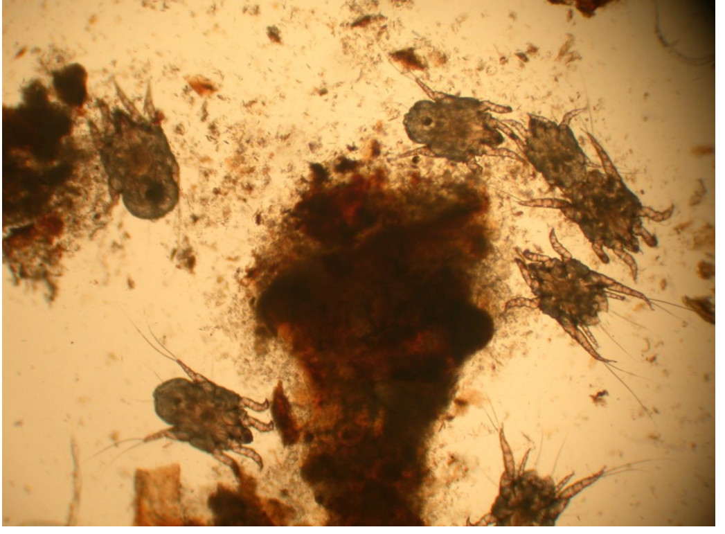
Ear Mites:C.S
Otodectes Cynotis
Black to brown coffee-ground-like debris in the ear canals
Cats produce more than dogs
Drooping Pinnae
More common in dogs
Irritation and Pruritus
Severe: Secondary bacterial/yeast infection, torn eardrums
Pinnal Pedal Reflex
Massage ear causes scratching movement of hindlimbs
Otodectic ascariasis: Generalized/Miliary dermatitis
Ear Mites:D.X
Otoscopic exam + Ear swab cytology
False negatives
Fecal exam may or may not reveal eggs
Ear Mites:T.X
All in-contact animals must be treated for at least 14 days
TX consists of two components
Topical Cerumenolytic
Epi Otic Advanced
Duoxo Micellar
KlearOtic
Not all ear cleansers have Cerumenolytic
Systemic antiparasitic/ascaricde
Revolution (Selamectin)
Advantage Multi (Moxidectin)
Isoxazolines (Fluralaner, Sarolaner)
Ivermectin
OR Topical antiparasitic/ascaricides
Avermectins
Milbemycin
Thiabendazole
Fleas:C.S
Dogs:
Severe itching: Lead to intense scratch, lick, rub, and chew
Papules, crusts, alopecia, erythema, and hyperpigmentation,
primarily on the lower back, tailhead, and thighs.
Chronic FAD:± Extensive alopecia,lichenification), secondary bacterial/yeast infections.
Cats:
Miliary dermatitis, small crusted papules
on the back, neck, and face.
These lesions are not flea bites but a result of a systemic allergic reaction, causing generalized pruritus.
±Alopecia, facial dermatitis, exfoliative dermatitis, and a "racing stripe" pattern of lesions along the back.
Fleas:D.X
Clinical signs
Finding fleas or flea dirt
Intradermal skin testing
Key factors include the frequency of flea exposure, duration of the disease, and the presence of secondary skin conditions.
Fleas: T.X
Eliminating Fleas:
Fast-acting topical or oral flea treatments like
afoxolaner(NexGard),
fluralaner(Bravecto),
lotilaner(Credelio),
sarolaner(Simparica),
spinosad (Comfortis)
Persistent treatment over several months is often necessary to break the flea life cycle.
Environmental Control
Supportive Care
Lymes Disease: Cause
Causes Lyme Disease
Ixodes
Lymes Disease:C.S
Pathognomonic Signs: Shifting leg lameness, polyarthritis Lameness can appear suddenly and may shift from one leg to another over time.
Clinical Signs:
Fever + lethargy + swollen joints.
± Kidney issues if severe: lead to Lyme nephritis, characterized by proteinuria, azotemia, and hypoalbuminemia.
Lyme Disease: D.X
Serological tests:C6 ELISA, SNAP 4Dx, detects antibodies
A positive result + clinical correlation to confirm active infection.
PCR testing: less commonly used in clinical practice.
Lyme Disease: T.X
Antibiotics:
Doxycycline*
Amoxicillin and ceftriaxone are alternatives, particularly for patients who cannot tolerate doxycycline.
Duration: Treatment typically lasts 2-4 weeks, depending on the stage and severity of the disease.
Symptomatic Relief:
NSAIDs: Lyme arthritis.
Pacemakers: Lyme carditis with heart block.
Joint Effusions: Joint aspiration can provide relief from pain and swelling in cases of severe arthritis.
Ehrlichia:C.S
Caused by: Lone star tick (Amblyomma americanum), Brown dog Tick (Rhipicephalus sanguineus)
Pathognomonic Signs: Pancytopenia
Anemia, thrombocytopenia, and leukopenia
Clinical Signs: Presents in three stages
Acute phase,
Fever
Lethargy + weight loss
Lymphadenopathy.
Chronic
Epistaxis + petechiae + ecchymoses
Neurological signs like ataxia or seizures.
Ehrlichia: D.X
Serology: IFA + SNAP 4Dx test (detect antibodies)
CBC: Pancytopenia (Chronic)
BM aspiration might be necessary to confirm the diagnosis.
PCR: Ehrlichia DNA in blood (Acute)
Ehrlichia:T.X
Doxycycline* : For 4 weeks
Supportive Care: In severe cases, particularly those with chronic ehrlichiosis, supportive care such as fluid therapy, blood transfusions, and nutritional support may be necessary.
Rocky Mountain Spotted Fever:C.S
Tick: American Dog tick
Pathogen: Rickettsia rickettsii
Pathognomonic Signs: Petechial or ecchymotic hemorrhages on the mucous membranes
which result from widespread vasculitis caused by the rickettsial infection.
Clinical Signs:
Acute + fever, anorexia, vomiting, diarrhea, lethargy.
± Neurological signs such as ataxia, vestibular deficits, and hyperesthesia.
Severe cases can lead to shock, disseminated intravascular coagulation (DIC), and multi-organ failure.
Rocky Mountain Spotted Fever:D.X
Diagnosis: Diagnosis is primarily based on clinical suspicion and confirmed through serological testing, such as an IFA, which detects antibodies against Rickettsia rickettsii. A fourfold increase in antibody titers between acute and convalescent samples is confirmatory. PCR testing of blood or tissue samples can detect rickettsial DNA, but sensitivity can be variable.
Rocky Mountain Spotted Fever:T.X
Doxycycline: The antibiotic of choice for treating RMSF. Treatment should be initiated based on clinical suspicion, without waiting for test results, as early treatment is critical for preventing severe complications and death. The recommended dosage is 5 mg/kg every 12 hours or 10 mg/kg every 24 hours for 14–21 days.
Supportive Care: Management of dehydration, electrolyte imbalances, and hemorrhagic issues is often necessary. Careful administration of fluids is important due to concerns about vascular integrity.
Neurological Damage: Some dogs may have residual neurological deficits even after successful treatment, but lifelong immunity is typically conferred by natural infection.
Anaplasmosis:C.S
Pathognomonic Signs: Thrombocytopenia
this is not exclusive to the disease.
Clinical Signs: may present with nonspecific signs
Fever+ lethargy, anorexia,polyarthritis,
leading to lameness similar to Lyme disease.
Some dogs may develop splenomegaly or mild hepatomegaly.
Anaplasmosis:D.X
SNAP 4Dx or IFA
PCR
CBC: Thrombocytopenia
Anaplasmosis:T.X
Doxycycline
Supportive Care: May be necessary in severe cases, particularly if there are significant bleeding issues or neurological involvement.
Canine Mites: Sarcoptes C.S
Clinical Signs
Intense pruritus, often out of proportion to the lesions
Erythematous papules and crusting, especially on the ears, elbows, hocks, and abdomen
Alopecia, thickened skin, and secondary bacterial infections may develop
Canine Mites: Sarcoptes D.X
Diagnostics
Gold Standard: Superficial skin scraping to identify mites, though they can be difficult to find.
Additional Tests: Response to treatment can be diagnostic if mites are not found.
Canine Mites: Sarcoptes T.X
Treatment
Gold Standard: Isoxazolines (e.g., fluralaner, afoxolaner, sarolaner) are effective and commonly used.
Other Options: Lime sulfur dips, ivermectin, or selamectin can also be effective. All in-contact animals should be treated due to the high contagion risk.
Canine Mites:Demodex Canis
Clinical Signs
Localized Demodicosis: Alopecia, erythema, and scaling often around the face, paws, and forelimbs.
Generalized Demodicosis: Widespread hair loss, erythema, hyperpigmentation, and potential secondary pyoderma.
Risk Factors
Genetic Predisposition: Certain breeds (e.g., Bulldogs, German Shepherds) are predisposed.
Immune Suppression: Young age, immunosuppressive treatments, or underlying diseases.
Canine Mites:Demodex Canis D.X
Diagnostics
Gold Standard: Deep skin scraping or trichogram (hair pluck) to identify mites within hair follicles.
Additional Tests: A biopsy may be used in challenging cases.
Canine Mites:Demodex Canis T.X
Treatment
Gold Standard: Isoxazolines (e.g., fluralaner, afoxolaner, sarolaner) are highly effective.
Other Options: Amitraz dips, ivermectin, and milbemycin oxime are also used, but treatment duration may be longer.
Canine Mites:Otodectes cynotis C.S
Clinical Signs
Intense ear scratching, head shaking
Dark, coffee-ground-like discharge in the ear canal
Potential secondary otitis externa with erythema and swelling
Risk Factors
Age: Puppies and young dogs are more commonly affected.
Environment: Contact with infected animals (e.g., other dogs or cats).
Canine Mites:Otodectes cynotis D.X
Diagnostics
Gold Standard: Otoscopic examination and microscopic evaluation of ear swabs to identify mites.
Additional Tests: Response to treatment may help confirm diagnosis if mites are not visualized.
Canine Mites:Otodectes cynotis T.X
Treatment
Gold Standard: Topical or systemic isoxazolines (e.g., fluralaner or selamectin).
Other Options: Topical ear medications containing pyrethrins or milbemycin may also be effective.
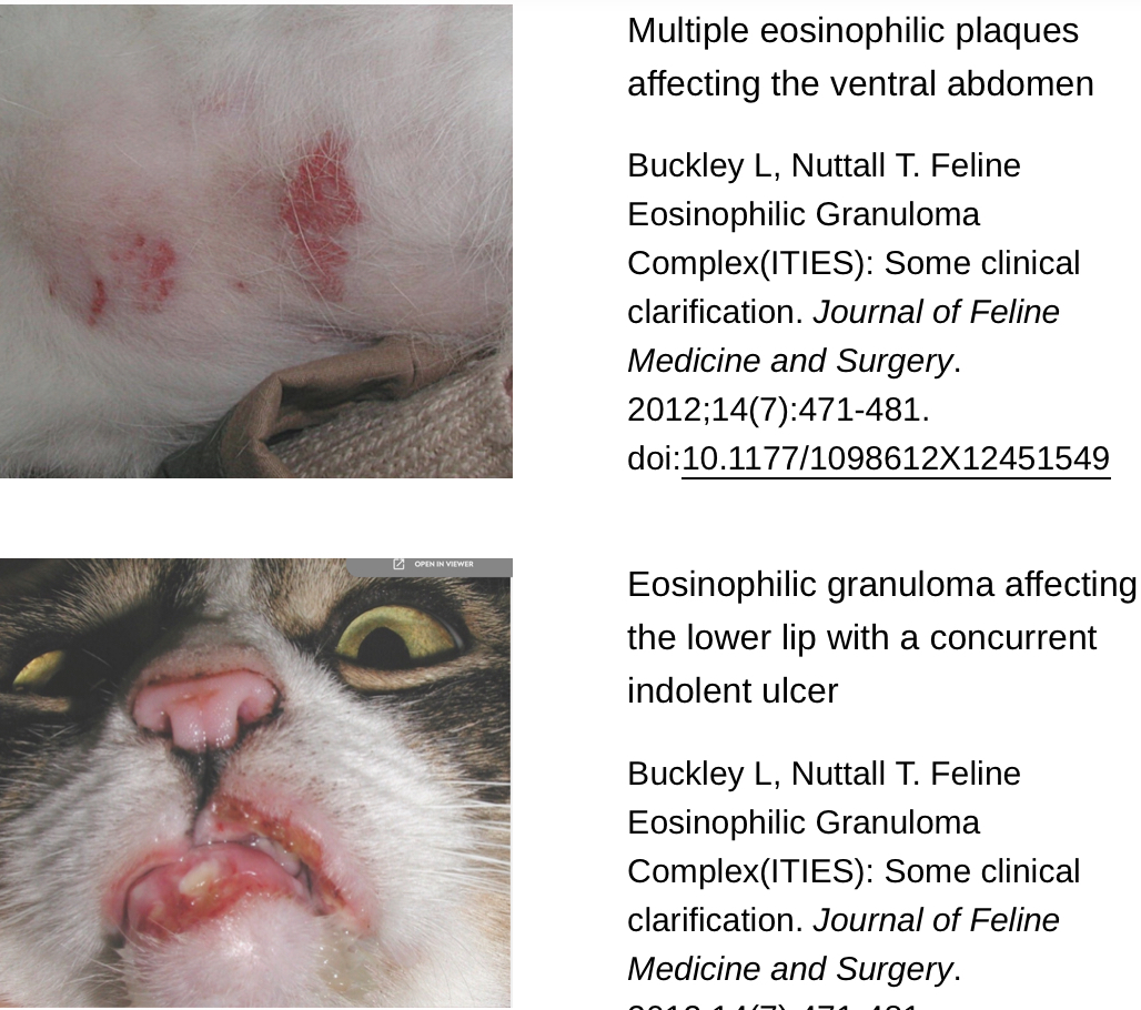
Feline Eosinophilic Granuloma Complex:C.S
Commonly associated w/ hypersensitivity reactions to fleas and food allergens
3 Types of Clinical Presentations
Indolent/rodent/eosinophilic ulcer =
Well-circumscribed erythematous ulcerative or erosive lesion
usually on the upper lip that is non-painful and non-pruritic.
These ulcers are part of the Feline Eosinophilic Granuloma Complex (EGC)
Do not cause discomfort or itching for the cat.
Eosinophilic plaque =
Raised erythematous lesions that can occur anywhere on the body
Intense pruritus;
One manifestation of pyoderma in cats
Commonly seen as medial linear lesions on the thighs and abdomen
Eosinophilic granuloma =
Firm pink, yellow, or orange nodules or other lesion found anywhere on the body,
but most commonly seen in the oral cavity, on the lips, or as linear pencil-like lesions on the caudal thighs
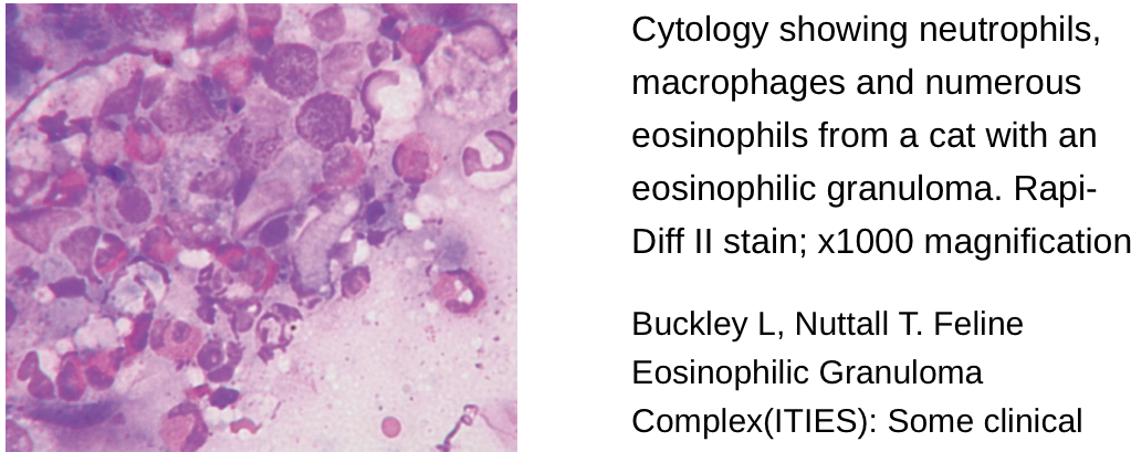
Feline Eosinophilic Granuloma Complex:D.X
Exam:
See lesions
Response to flea control/hypoallergenic diet trial
CBC: Eosinophilia
Impression cytology may reveal a high number of eosinophils +/- neutrophils, basophils.
If there is no response to empiric treatment or the lesions are especially acute and destructive, biopsy + histopathology is recommended to rule out squamous cell carcinoma (most common oral neoplasia in cats), cutaneous lymphoma, and other skin conditions.
Feline Eosinophilic Granuloma Complex:T.X
Resolution can take several weeks!
First-line treatment:
Flea control and/or a hypoallergenic diet trial.
If lesions persist, cats with EGC are treated with either
Prednisolone is considered by some to be the best treatment. It is started aggressively and tapered to the lowest effective dose.
For eosinophilic plaques and lesions with secondary bacterial infections, antibiotics are recommended.
If possible, treat the underlying cause (e.g. flea preventatives, avoidance). Rarely, the underlying cause can’t be identified and cyclosporine (immunosuppressant) may be effective.
Surgical excision, radiation therapy, and laser therapy of the lesions have been reported, but are rarely recommended.
Prognosis
Most cats respond well to medical management, especially when the underlying cause is identified (e.g. flea infestation).
Feline Acne: C.S
Comedones primarily on the chin
Secondary Bacterial infections: farunculis, folliculitis
Moderate to severe inflammation
Erythema, Papules, Pustules, Black Crusty Exudate,
± Pruritis
Severe: Cellulitis and abscess formation
Feline Acne: D.X
Clinical presentation
Skin scrapes and fungal culture are recommended to rule out demodicosis and ringworm
Skin cytology may reveal increased neutrophils +/- secondary bacterial infections.
In refractory or severe cases:
bacterial culture and sensitivity (C/S) and biopsy are recommended to rule out secondary bacterial infections, eosinophilic granuloma complex, any underlying allergies, fungal infections, and neoplasia.
The most commonly isolated opportunistic bacteria include Pasteurella, beta-hemolytic Streptococci, and Malassezia yeast.
Histopathology of feline acne reveals follicular plugging, dilation, hyperkeratosis, and glandular hyperplasia.
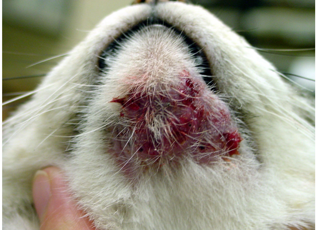
Feline Acne:T.X
No treatment is required for mild cases without secondary infections, but these cats may benefit from:
Shaving the affected areas.
Topical antiseptics — e.g. benzoyl peroxide, salicylic acid, ethyl lactate shampoo, mupirocin
AVOID hydrogen peroxide or alcohol-based products as they can be extremely irritating and worsen the lesions.
Changing plastic food and water bowls to shallow ceramic or stainless steel bowls and cleaning these bowls frequently with soap and water.
Switching to a dry diet.
Cats with moderate or recurrent lesions are treated with topical or systemic steroids.
Severe cases or those with secondary bacterial infections are treated with systemic antibiotics and steroids until the lesions have resolved and an additional two weeks.
For chronic refractory cases:
Isotretinoin (Accutane) has been shown to be effective in ~33% of cats.
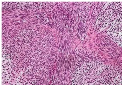
Fibrosarcoma:C.S
In cats, they are particularly significant due to their association with vaccine administration, which has led to the term "vaccine-associated sarcoma" (VAS).
Fibrosarcomas in cats usually present as firm, subcutaneous masses that are often not painful. These masses are typically found on the back, flanks, or limbs and may be adherent to underlying tissues due to their invasive nature. Key clinical signs include:
Swelling or Lump: A firm, palpable mass that may grow slowly over weeks to months.
Ulceration: In some cases, the overlying skin may ulcerate, leading to secondary infections.
Decreased Mobility: Depending on the tumor's location, cats may exhibit lameness or reluctance to move.
Weight Loss: Advanced cases may show signs of cachexia.
Localized Pain: While not common, pain may occur if the tumor invades nerves or bones.
Fibrosarcoma:D.X
Physical Examination: Initial assessment should focus on the size, location, and mobility of the mass, along with a complete physical exam to check for signs of metastasis.
Fine Needle Aspiration (FNA): While FNA can be useful for initial assessment, it often yields non-diagnostic samples due to the dense, fibrous nature of these tumors. Cytology may reveal spindle cells, but histopathology is typically required for a definitive diagnosis.
Biopsy: Incisional or excisional biopsy provides tissue samples for histopathological examination. Biopsy allows for assessment of cellular morphology, mitotic index, and the degree of infiltration into surrounding tissues. Special staining techniques, such as immunohistochemistry, may be employed to differentiate fibrosarcoma from other sarcomas.
Histo
Cellular Composition:
Spindle-Shaped Cells: Fibrosarcomas are primarily composed of spindle-shaped cells, which are elongated and resemble the fibroblasts from which they originate.
Pleomorphism: The tumor cells often exhibit pleomorphism, meaning they vary in size and shape. This variability is a hallmark of malignancy.
Hyperchromatic Nuclei: The nuclei of the tumor cells are often hyperchromatic, meaning they stain more darkly than normal cells due to increased DNA content. The nuclei may also appear large and irregular in shape.
High Mitotic Rate: Fibrosarcomas typically show a high mitotic rate, with frequent mitotic figures (cells in the process of division) visible under the microscope. This indicates the rapid proliferation of tumor cells.
Stromal Features:
Dense Collagenous Stroma: The stroma (connective tissue framework) of fibrosarcoma is often dense and collagen-rich. The tumor cells are embedded in this collagenous matrix, which can make the tumor feel firm or hard upon palpation.
Haphazard Cell Arrangement: The arrangement of tumor cells within the stroma is typically disorganized. Unlike normal tissue, where fibroblasts are aligned in a regular pattern, fibrosarcoma cells are randomly oriented.
Infiltrative Growth Pattern: Fibrosarcomas are characterized by their infiltrative growth pattern. Tumor cells invade surrounding tissues, often extending beyond the visible edges of the mass. This invasiveness contributes to the high recurrence rate of fibrosarcomas after surgical excision.
Tumor Margins:
Poorly Defined Borders: The margins of fibrosarcomas are often poorly defined, making it difficult to distinguish the tumor from surrounding normal tissue. This can complicate surgical removal and increase the likelihood of local recurrence.
Pseudocapsule: Sometimes, a fibrosarcoma may appear to have a pseudocapsule—a layer of compressed tissue surrounding the tumor. However, this pseudocapsule is not a true barrier, and tumor cells can infiltrate through it.
Necrosis and Inflammation:
Areas of Necrosis: Necrosis (cell death) may be present within the tumor, particularly in larger or more aggressive fibrosarcomas. These necrotic areas can appear as pale or eosinophilic (pink-staining) regions under the microscope.
Inflammatory Infiltrate: The presence of an inflammatory infiltrate, consisting of lymphocytes, macrophages, and sometimes neutrophils, is common in fibrosarcomas. This inflammation may result from the body's response to the tumor or secondary infection.
Vascular Invasion:
Invasion of Blood Vessels: In some cases, fibrosarcoma cells may invade nearby blood vessels. This can facilitate metastasis, although fibrosarcomas are generally considered to have a low metastatic potential.
Imaging
Metastasis evaluation
Fibrosarcoma:T.X
The treatment of fibrosarcomas in cats is challenging due to the tumor's aggressive and invasive nature. Treatment typically involves a multimodal approach:
Surgery:
Wide Excision: The primary treatment for fibrosarcomas is surgical excision with wide margins (at least 2-3 cm) to ensure complete removal of the tumor and reduce the risk of recurrence. Incomplete excision is common due to the infiltrative nature of these tumors, leading to high rates of local recurrence.
Amputation: In cases where the tumor involves a limb, amputation may be the most effective option to achieve clean margins.
Marginal Excision with Radiation Therapy: When wide excision is not feasible, marginal excision followed by radiation therapy can be considered to control local disease.
Radiation Therapy:
Preoperative or Postoperative Radiation: Radiation therapy is often employed as an adjunct to surgery, either preoperatively to shrink the tumor or postoperatively to target residual microscopic disease. Radiation can significantly reduce the risk of local recurrence.
Chemotherapy:
Doxorubicin-Based Protocols: Chemotherapy may be considered for cats with non-resectable tumors, metastatic disease, or as an adjunct to surgery and radiation. Doxorubicin is the most commonly used chemotherapeutic agent, though its efficacy is variable. Other agents like cyclophosphamide or carboplatin may also be considered.
Electrochemotherapy: This is an emerging modality that combines chemotherapy with electrical pulses to increase drug uptake by tumor cells. It has shown promise in treating feline fibrosarcomas, particularly in cases where traditional surgery and radiation are not viable.
Immunotherapy:
Immunomodulators: Various immunomodulatory therapies, such as liposome-encapsulated muramyl tripeptide (L-MTP), have been investigated to boost the cat's immune response against the tumor. While still experimental, these treatments may offer an additional avenue for managing fibrosarcomas.
Prognosis
The prognosis for cats with fibrosarcoma depends on several factors, including tumor size, location, completeness of surgical excision, and whether adjunctive therapies are used. Despite aggressive treatment, recurrence rates are high, ranging from 30% to 70%. The median survival time can vary from 6 months to several years, depending on the treatment modality and the tumor's behavior.
Neoplasia/ Neoplastic Disease:Benign Neoplasms
These tumors are typically well-differentiated and non-invasive, often posing little threat to overall health unless they interfere with critical anatomical functions. Common examples include:
Basal Cell Tumors: Often appear as small, firm nodules, typically benign but can occasionally recur after excision.
Papillomas: These are generally exophytic growths associated with viral infections, often resolving spontaneously or remaining stable.
Sebaceous Adenomas: Generally benign and slow-growing, these tumors arise from sebaceous glands and may become irritated or ulcerated.
Lipomas: Soft, movable masses under the skin, composed of adipose tissue, typically benign and only require removal if they cause discomfort or impede movement.
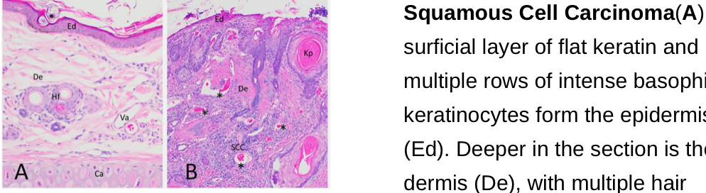
Neoplasia/ Neoplastic Disease:Malignant Neoplasms PICTURES
Malignant tumors are more aggressive and can invade surrounding tissues or metastasize to distant organs. They often present significant diagnostic and therapeutic challenges. Notable examples include:
Squamous Cell Carcinoma (SCC): A common malignant tumor in cats, particularly those with white or lightly pigmented skin. SCC is often associated with chronic UV radiation exposure and typically presents as a non-healing ulcer on the nose, ears, or eyelids.
Mast Cell Tumors (MCT): These tumors can vary in behavior from benign to highly malignant. They are characterized by the presence of mast cells and may cause pruritus and inflammation due to histamine release.
Fibrosarcomas: Often associated with chronic inflammation or injections, such as vaccine-associated sarcomas. These tumors are locally invasive with a high rate of recurrence after surgical excision.
Melanomas: These can occur in various locations, including the skin, oral cavity, and nail beds. Malignant melanomas are highly aggressive and often metastasize to regional lymph nodes and distant organs.
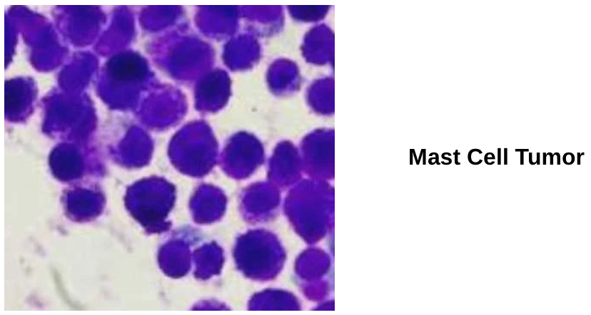
Neoplasia/ Neoplastic Disease:C.S
Masses or Nodules: The most common presentation is a solitary or multiple nodules or masses that can be palpated under the skin. These masses may be firm or soft, movable or fixed, and can vary in size.
Ulceration: Malignant tumors, particularly SCC, often ulcerate, leading to non-healing sores that may bleed or become infected.
Pruritus and Inflammation: Some dermal tumors, especially MCTs, may cause significant pruritus, erythema, and swelling due to the release of histamine and other inflammatory mediators.
Systemic Signs: In cases of malignant neoplasia with metastasis, systemic signs such as weight loss, lethargy, and anorexia may be observed.
Neoplasia/ Neoplastic Disease:D.X
Physical Examination:
Cytology: FNAcan provide preliminary information about the nature of the mass. Cytology is particularly useful in diagnosing MCTs, where characteristic granulated cells can be observed. However, cytology alone may not be sufficient for definitive diagnosis.
Histopathology: Biopsy and subsequent histopathological examination remain the gold standard for diagnosing dermal neoplasia. Histopathology not only confirms the type of tumor but also provides information on the degree of malignancy, cellular differentiation, and invasion of surrounding tissues.
Imaging: Advanced imaging techniques, such as ultrasound, radiography, or CT scans, may be used to assess the extent of the tumor and check for metastasis, particularly in malignant cases.
Immunohistochemistry: In cases where the tumor type is ambiguous, immunohistochemical staining can be used to identify specific tumor markers, aiding in the diagnosis and classification of neoplastic diseases.
Neoplasia/ Neoplastic Disease:T.X
he treatment of dermal neoplasia in cats depends on the type of tumor, its size, location, and whether it has metastasized:
Surgical Excision: Surgery is the treatment of choice for most dermal neoplasms. Wide surgical margins are particularly important in malignant tumors to reduce the risk of recurrence. For benign tumors, less aggressive excision may be adequate.
Radiation Therapy: Radiation therapy is often used as an adjunct to surgery in cases where complete excision is not possible or when dealing with highly invasive tumors like fibrosarcomas. It can also be used as a palliative treatment to reduce the size of the tumor and alleviate clinical signs.
Chemotherapy: Systemic chemotherapy may be indicated for malignant tumors with a high risk of metastasis, such as MCTs and fibrosarcomas. The choice of chemotherapeutic agents depends on the tumor type and the cat's overall health.
Cryotherapy: For small, superficial neoplasms, cryotherapy can be an effective treatment option. It involves freezing the tumor cells, leading to their destruction. This method is most suitable for benign or low-grade malignant tumors.
Immunotherapy: Novel treatments, such as immunotherapy, are being explored for certain types of neoplasms, particularly vaccine-associated sarcomas. These treatments aim to stimulate the cat’s immune system to recognize and destroy cancer cells.
Canine: Squamous Cell Carcinoma
Epithelial Tumors
Pathophysiology:
SCC arises from the malignant transformation of epidermal keratinocytes often triggered by chronic UV exposure.
Clinical Signs:
Ulcerated, crusted plaques or nodules—commonly on the nasal planum, eyelids, limbs, or abdomen.
Risk Factors: Light-colored coats and prolonged sun exposure.
Diagnostics: Biopsy with histopathology reveals keratin pearls and dysplastic keratinocytes.
Treatment:
Wide surgical excision is first-line. Consider radiation or photodynamic therapy for incompletely resected or non-resectable tumors.
Prognosis: Depends on location and degree of invasion.
Canine: Basal cell tumor
Basal Cell Tumors (Trichoblastomas) epithelial tumor
Pathophysiology: Previously termed basal cell tumors, many are now reclassified as trichoblastomas. These arise from basal epithelial cells and are typically benign.
Clinical Signs: Solitary, well-defined nodules, often on the head or neck.
Diagnostics: FNA may be suggestive, but histopathology post-excision confirms the diagnosis.
Treatment: Complete surgical excision is usually curative.
Canine Fibrosarcoma
Mesenchymal (Soft Tissue) Tumor
Pathophysiology: Malignant tumor arising from dermal fibroblasts. Locally aggressive but less likely to metastasize.
Clinical Signs: Firm, fixed masses often found on the trunk or limbs. May ulcerate with progression.
Risk Factors: Large breed and older dogs.
Diagnostics: Biopsy is required; imaging such as CT or MRI assists in surgical planning.
Treatment: Wide surgical excision is key. Radiation is often used post-operatively due to high recurrence risk.
Canine Mast Cell Tumor
Round Cell Tumors
Pathophysiology: Arise from dermal mast cells and are classified as round cell tumors cytologically. They can release histamine and other mediators, causing systemic effects. c-KIT mutations are frequently involved.
Clinical Signs: Firm, variably sized nodules that may wax and wane in appearance, often pruritic.
Risk Factors: Breeds such as Boxers, Pugs, Boston Terriers, and Labrador Retrievers are predisposed.
Diagnostics: FNA often reveals granulated round cells. Histopathologic grading (Patnaik or Kiupel) informs prognosis.
Treatment: Wide surgical excision is standard. Adjunctive options include radiation, chemotherapy, and tyrosine kinase inhibitors (e.g., toceranib) for high-grade or metastatic cases.
Notable Feature: Darier’s sign—erythema and swelling after palpation.
Canine Histiocytoma
Round Cell Tumor
Pathophysiology: Benign tumor of Langerhans cells, most common in young dogs.
Clinical Signs: Rapidly growing, dome-shaped red nodules, typically on the head or limbs.
Risk Factors: Dogs under 3 years of age.
Diagnostics: FNA shows round cells with pale cytoplasm and kidney-shaped nuclei.
Treatment: Often regress spontaneously. Surgery is indicated if they ulcerate, persist, or cause irritation.
Canine Cutaneous Lymphoma
Round cell tumor
Pathophysiology: Neoplastic proliferation of lymphocytes. Can be epitheliotropic (T-cell) or non-epitheliotropic (B-cell).
Clinical Signs: Multifocal plaques, nodules, or diffuse erythema, often accompanied by pruritus.
Risk Factors: Middle-aged to older dogs; Boxers may be predisposed.
Diagnostics: Biopsy with immunohistochemistry is required for phenotyping.
Treatment: Chemotherapy is the mainstay; agents like lomustine and retinoids are commonly used for T-cell variants.
Gold Standard: Immunophenotyping guides treatment selection.
Canine Melanoma
Pathophysiology: Tumors of melanocytes that may be benign or malignant. Location strongly influences behavior—oral and digital melanomas are typically malignant regardless of histology.
Clinical Signs: Darkly pigmented or amelanotic nodules; oral tumors may bleed or ulcerate.
Risk Factors: Breeds with dark pigmentation (e.g., Scottish Terriers, Schnauzers).
Diagnostics: Cytology may suggest diagnosis, but histopathology is needed for malignancy assessment.
Treatment: Surgery is the first-line approach. Malignant cases may benefit from adjunctive immunotherapy (canine melanoma vaccine), radiation, or chemotherapy.
Canine Lipoma
Pathophysiology: Benign neoplasm of adipocytes.
Clinical Signs: Soft, well-encapsulated, subcutaneous masses, often freely movable and non-painful.
Risk Factors: Common in older, overweight dogs.
Diagnostics: FNA shows mature adipose cells.
Treatment: Surgery is elective unless the mass impairs movement or causes discomfort.
Canine Liposarcoma
Pathophysiology: Malignant counterpart to lipoma, locally invasive with potential for recurrence.
Clinical Signs: Firm, fixed masses that may enlarge rapidly.
Risk Factors: Older, large breed dogs.
Diagnostics: Biopsy is required to confirm malignancy.
Treatment: Wide surgical excision is the mainstay. Radiation and chemotherapy may be considered in aggressive or recurrent cases
Otitis Externa Feline:Factors
The inflammatory process begins with irritation of the ear canal lining, leading to increased vascular permeability and immune cell infiltration. This causes swelling, erythema, and increased cerumen production. Secondary infections, particularly with Staphylococcus spp., Pseudomonas aeruginosa, and Malassezia pachydermatis, further complicate the condition, leading to chronicity and potential damage to the tympanic membrane.
Otitis Externa Feline
Pruritus: Scratching at the ears or shaking the head is a common sign of discomfort.
Otorrhea: The presence of discharge, which may be ceruminous, purulent, or mixed, often with a foul odor.
Erythema and Swelling: Redness and swelling of the ear canal and pinna.
Pain: Cats may exhibit signs of pain, such as vocalizing when the ears are touched or head tilt.
Alopecia: Hair loss around the ear due to self-trauma.
Hearing Loss: In cases of severe or chronic otitis externa, hearing impairment may occur.
Hyperplasia and Stenosis: Chronic cases may lead to thickening of the ear canal walls and narrowing of the canal, complicating treatment.
Otitis Externa Feline
Diagnosing otitis externa in cats requires a thorough approach, including history, physical examination, and diagnostic tests.
History and Physical Examination:
Cytology: A cytologic examination of ear discharge is crucial. Samples should be collected from both ears and stained with Diff-Quik or Gram stain. The presence of bacteria, yeast, or inflammatory cells can guide the choice of treatment.
Culture and Sensitivity: In recurrent or refractory cases
Imaging: Advanced imaging, such as CT or MRI, may be indicated in chronic cases to assess the extent of the disease, particularly if there is suspicion of otitis media or extension to the inner ear structures.
Allergy Testing: In cases where hypersensitivity is suspected, intradermal testing or serum allergen-specific IgE testing can help identify the allergens involved.
Biopsy: If neoplasia or autoimmune disease is suspected, a biopsy of the ear canal may be required.
Otitis Externa Feline: T.X
Cleaning the Ear Canal: Before any topical treatments, the ear canal should be cleaned to remove debris and discharge. This can be done with ear cleaners specifically designed for cats, which should be pH balanced and non-irritating. In severe cases, sedation may be required.
Topical Therapy:
Antibiotics: Topical antibiotics, such as gentamicin, neomycin, or polymyxin B, are commonly used to treat bacterial infections.
Antifungals: For yeast infections, topical antifungals such as miconazole or clotrimazole may be used.
Anti-inflammatories: Topical corticosteroids (e.g., dexamethasone or betamethasone) help reduce inflammation and pruritus. Care must be taken with long-term use to avoid systemic absorption and side effects.
Antiparasitics: If ear mites are present, topical acaricides like selamectin or moxidectin are effective.
Systemic Therapy:
Antibiotics or Antifungals: Systemic therapy may be required in cases of severe or chronic infections, particularly if there is otitis media.
Corticosteroids: In cases of severe inflammation, oral corticosteroids may be indicated to reduce swelling and discomfort.
Immunotherapy: In cases where allergies are involved, immunotherapy or long-term management strategies for allergic dermatitis may be necessary.
Surgical Intervention: In cases of chronic otitis externa with irreversible changes, surgical options such as total ear canal ablation (TECA) may be considered. This is often the last resort and is indicated when there is extensive hyperplasia, fibrosis, or neoplasia.
Otitis Externa/Media Canine:C.S
Head Shaking and Ear Scratching: Due to discomfort, dogs often shake their heads and scratch at the affected ear(s).
Erythema and Swelling: Redness, heat, and swelling are common around the external ear canal.
Odor: A foul smell often indicates a bacterial or yeast infection.
Ear Discharge: The type of discharge can vary from waxy and brown to purulent, yellow, or malodorous.
Pain and Sensitivity: Dogs with otitis may be sensitive to touch around the ears and may display signs of pain or agitation.
Behavioral Changes: Due to pain, dogs may become irritable or head shy.
Decreased Hearing: Accumulation of discharge and swelling can impede hearing, especially in cases of chronic otitis.
Otitis Externa/Media Canine:D.X
Otoscopy: An otoscopic exam helps visualize the ear canal, identify any foreign bodies, assess the tympanic membrane, and evaluate the extent of inflammation or discharge. Dogs with painful ears may require sedation for a thorough examination.
Cytology:
Ear Swab Cytology: Samples from the ear canal are examined under a microscope to identify the presence of bacteria, yeast, or inflammatory cells. The presence of rods may indicate a bacterial infection, while round cells may suggest Staphylococcus species or yeast.
Gram Staining: Differentiates between Gram-positive and Gram-negative bacteria, aiding in antibiotic selection.
Culture and Sensitivity Testing:
Bacterial Culture: For cases of recurrent otitis or severe infections, bacterial culture and sensitivity testing help guide appropriate antibiotic therapy.
Fungal Culture: If cytology indicates yeast but fails to respond to treatment, fungal culture may be warranted.
Imaging Studies:
Radiographs, CT, or MRI: Advanced imaging may be needed in chronic cases to assess middle ear involvement or detect structural changes in the ear canal.
Additional Diagnostics:
Endocrine Testing: If an underlying condition like hypothyroidism or hyperadrenocorticism is suspected, additional blood tests should be conducted.
Allergy Testing: In cases with suspected allergies, intradermal testing or serum IgE testing can help identify allergens for long-term management.
Otitis Externa/Media Canine:T.X
Cleaning the Ear Canal:
Ceruminolytic Agents: For cases with excessive cerumen, ceruminolytic solutions help dissolve ear wax. Regular cleaning with these agents can prevent buildup and reduce infection risk.
Antimicrobial Flushes: Infected ears may benefit from antimicrobial solutions that cleanse and disinfect the ear canal.
Manual Cleaning: In severe cases, the ear canal may need to be cleaned manually under anesthesia to remove debris and discharge.
Topical Therapy:
Antibacterial and Antifungal Agents: Topical ear medications are chosen based on cytology and/or culture. Aminoglycosides, fluoroquinolones, or enrofloxacin may be used for bacterial infections, while antifungals like miconazole and ketoconazole target yeast infections.
Corticosteroids: Topical steroids reduce inflammation, alleviate pain, and reduce edema. Steroid drops or ointments are especially useful for addressing the thickened ear canal and inflammation in chronic cases.
Systemic Therapy:
Antibiotics and Antifungals: In severe or chronic cases where the infection extends beyond the ear canal or in cases with marked tissue swelling, systemic antibiotics or antifungals may be required.
Systemic Corticosteroids: In cases of severe inflammation, systemic corticosteroids (e.g., prednisone) may help reduce swelling and discomfort.
Management of Underlying Causes:
Allergen Control: In cases with allergic otitis, managing underlying allergies through dietary modifications or allergen-specific immunotherapy is essential.
Endocrine Therapy: For dogs with conditions like hypothyroidism, appropriate treatment of the endocrine disorder can help reduce recurrence of otitis.
Surgical Intervention:
Lateral Ear Resection: For cases with chronic, recurrent otitis externa, a lateral ear resection can improve airflow and drainage, reducing recurrence.
Total Ear Canal Ablation (TECA): In end-stage otitis, where there is irreversible change or mineralization, a TECA combined with a bulla osteotomy may be required. This procedure removes the entire ear canal and is usually a last-resort option for managing severe cases.
Long-Term Maintenance and Prevention:
Routine Ear Cleaning: Regular ear cleaning with a gentle ear cleanser can help prevent wax buildup and reduce infection risk.
Moisture Control: Avoiding excessive moisture in the ears is critical, particularly for dogs that swim frequently. Owners should be educated on drying the ears after exposure to water.
Feline Pemphigus Foliaceus
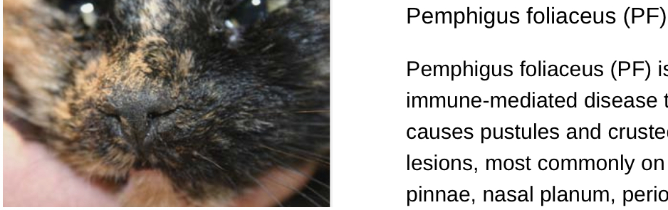
Pemphigus Foliaceus (PF)
Overview: PF is the most common form of pemphigus in cats, primarily affecting the superficial layers of the skin. Autoantibodies target desmoglein 1, a protein crucial for epidermal integrity. Desmoglein 1 is a type of protein that is a key component of desmosomes, which are structures that help cells stick together in the superficial layers of the skin (epidermis). Desmosomes are essential for maintaining the integrity and cohesion of the skin's outermost layers, ensuring that the skin remains intact and resistant to physical stress.
When the body produces autoantibodies against desmoglein 1, as seen in certain autoimmune diseases like Pemphigus Foliaceus, these antibodies mistakenly target and attack this protein. This disruption weakens the connections between skin cells, leading to the breakdown of the superficial epidermis. As a result, the skin forms superficial blisters, pustules, and erosions because the cells are no longer properly held together. These blisters and erosions quickly rupture, leading to crusting and further damage to the skin.
Hallmark Clinical Signs and Lesion Locations:
Superficial Pustules and Crusts: Lesions typically appear on the face (particularly the nasal planum, ears, and around the eyes), paw pads, and around the nail beds. These pustules quickly rupture, forming yellowish crusts.
Erythema: Redness of the skin is often observed around the lesions.
Alopecia: Hair loss occurs around the affected areas and may become more extensive over time.
Pruritus: Itching may or may not be present; secondary bacterial infections can increase pruritus.
Diagnosis:
Cytology: Reveals acantholytic keratinocytes and neutrophils.
Biopsy: The gold standard for diagnosing PF, showing subcorneal pustules with acantholysis.
Immunofluorescence: Detects autoantibodies deposited in the epidermis.
Feline Pemphigus Erythematosus
Pemphigus Erythematosus (PE)
Overview: PE is a milder variant of PF, sharing features with both PF and discoid lupus erythematosus (DLE). It typically affects sun-exposed areas. Pemphigus erythematosus presents with facial lesions that are photosensitive, and histopathology reveals subcorneal pustules with acantholytic keratinocytes. The exacerbation with sunlight is a distinguishing feature of this variant.
Hallmark Clinical Signs and Lesion Locations:
Localized Erythema and Crusting: Lesions are typically confined to the face, particularly the nasal planum, and ears. These areas may exhibit erythema, scaling, and crusting.
Photosensitivity: Lesions often worsen with sun exposure, leading to more pronounced erythema and crusting in affected areas.
Alopecia: Hair loss around the affected regions is common.
Diagnosis:
Biopsy: Reveals features similar to PF but with milder changes, and possible basal layer involvement.
Immunofluorescence: May show antibody deposition along the basement membrane zone and in the epidermis.
Pemphigus erythematosus shares characteristics with both pemphigus foliaceus and lupus erythematosus, often presenting as a more benign form of pemphigus foliaceus. Histopathologically, PE is characterized by several distinct features. Acantholysis, the loss of cohesion between keratinocytes, leads to the formation of intraepidermal blisters, typically seen as suprabasal or subcorneal blisters. These blisters often contain subcorneal pustules filled with neutrophils and eosinophils. Another hallmark of PE is basal cell vacuolization, where the basal layer of the epidermis shows vacuole formation, indicative of early cell death and associated with interface dermatitis, a feature shared with lupus erythematosus. This interface dermatitis is marked by a lichenoid infiltrate, a band-like accumulation of inflammatory cells, predominantly lymphocytes, located just beneath the epidermis. Additionally, direct immunofluorescence (DIF) often reveals the deposition of immunoreactants, such as IgG, IgM, and complement components like C3, within the intercellular spaces of the epidermis and occasionally at the dermoepidermal junction. These combined histopathological features are essential for diagnosing pemphigus erythematosus and distinguishing it from other autoimmune blistering diseases.
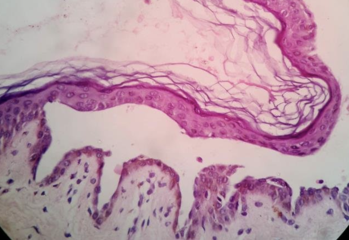
Feline Pemphigus Vulgaris
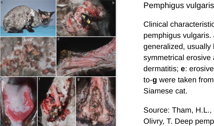
Pemphigus Vulgaris (PV)- VERY RARE
Overview: PV is the rarest and most severe form of pemphigus in cats. Autoantibodies target desmoglein 3, affecting deeper layers of the epidermis, leading to more profound and often life-threatening lesions.
Hallmark Clinical Signs and Lesion Locations:
Deep Ulceration: The primary lesions in PV are deep erosions and ulcers, often starting as vesicles or bullae that rupture. These are most commonly found in the oral mucosa, at mucocutaneous junctions (e.g., lips, nose), and on the skin in the groin area.
Oral Involvement: Oral mucosa involvement is a key feature, leading to significant pain, drooling, and reluctance to eat.
Systemic Illness: Cats with PV may also exhibit signs of systemic illness, including lethargy, anorexia, and fever.
Diagnosis:
Biopsy: Critical for diagnosing PV, typically showing suprabasilar clefting with acantholysis, creating a "tombstone" appearance of basal cells.
Immunofluorescence: Confirms the presence of autoantibodies targeting desmoglein 3 in the epidermis.
Pemphigus:DX
Diagnostics
Clinical Examination:
A thorough dermatologic examination is crucial, focusing on symmetric distribution of lesions, crusts, and erosions, particularly on the face, ears, and paws.
Cytology:
Aspiration or impression smears from intact pustules reveal acantholytic keratinocytes, a hallmark finding in PF.
Skin Biopsy:
Histopathology: Essential for definitive diagnosis. Lesions typically show subcorneal pustules with acantholytic keratinocytes in PF, and suprabasilar clefting in PV.
Immunohistochemistry or Direct Immunofluorescence (DIF): May reveal intercellular deposition of IgG and complement within the epidermis.
Blood Tests:
Routine blood work may show nonspecific changes such as leukocytosis or anemia if there is chronic inflammation or secondary infection.
Autoantibody Testing:
Although not routinely available in all veterinary practices, indirect immunofluorescence can be used to detect circulating autoantibodies.
Differential Diagnosis:
Includes bacterial folliculitis, dermatophytosis, demodicosis, and other immune-mediated diseases like lupus erythematosus.
Pemphigus: TX
Immunosuppressive Therapy:
Corticosteroids: Prednisolone is often the first line of treatment, with high doses typically required initially, followed by gradual tapering.
Adjunct Immunosuppressives: For refractory cases or to reduce steroid doses, drugs like chlorambucil or cyclosporine may be used.
Topical Therapies:
Tacrolimus ointment can be useful for localized lesions, especially in facial areas, to minimize systemic side effects. Tacrolimus ointment is a topical immunosuppressive medication used primarily to treat inflammatory skin conditions like atopic dermatitis and certain autoimmune skin diseases. It works by inhibiting calcineurin, which reduces the activity of T-cells and decreases inflammation and immune response in the affected area. It is often used when other treatments, like corticosteroids, are ineffective or not suitable
Antibiotics:
Secondary bacterial infections are common and may require systemic antibiotics based on culture and sensitivity.
Supportive Care:
Pain management, nutritional support, and addressing concurrent conditions are essential for comprehensive care.
Long-term Management:
Pemphigus is a chronic condition often requiring lifelong immunosuppressive therapy, with regular monitoring and dose adjustments necessary.
Monitoring:
Regular follow-ups are crucial to assess treatment efficacy and adjust dosages. Blood work should be monitored for potential side effects of long-term immunosuppressive therapy.
Plasma Cell Pododermititis: C.S
Swollen, puffy footpads
Though not always symmetrically.
Ulceration:
In more severe cases.
Bleeding
Lameness
Pain on palpation
Secondary infections
Lethargy and anorexia
Plasma Cell Pododermititis: D.X
Clinical examination:
Cytology: Fine needle aspiration (FNA) of the affected pads may reveal a high number of plasma cells. However, this method is not definitive.
Histopathology: A biopsy of the affected tissue is the gold standard for diagnosis. Histopathological examination typically reveals dense infiltrates of plasma cells within the dermis and subcutis. There may also be associated lymphocytes and macrophages, and the epidermis may show hyperplasia or ulceration.
Immunohistochemistry: Staining for immunoglobulins can confirm the plasma cell origin of the infiltrate.
Viral testing: Testing for FIV and FeLV is recommended, as these viruses are associated with the condition.
Blood work: A complete blood count (CBC) and serum biochemistry may be unremarkable but are useful to rule out other systemic conditions.
Imaging: Radiographs are not typically diagnostic but may be used to rule out other causes of lameness or swelling.
Plasma Cell Pododermititis:T.X
Immunosuppressive therapy: Initial dosing may be high, with gradual tapering based on the cat’s response. Long-term therapy may be necessary in some cases.
Doxycycline: for its antibiotic properties, its anti-inflammatory and immunomodulatory effects.
Cyclosporine: alternative immunosuppressant.
Surgical intervention: In cases of severe ulceration or chronic non-healing wounds, surgical debridement may be necessary. However, surgery is generally considered a last resort.
Antibiotics: If secondary bacterial infection is present
Pain management
Supportive care
Canine: Acral Lick Granuloma:C.S
Staphylococcus pseudintermedius or Staphylococcus aureus
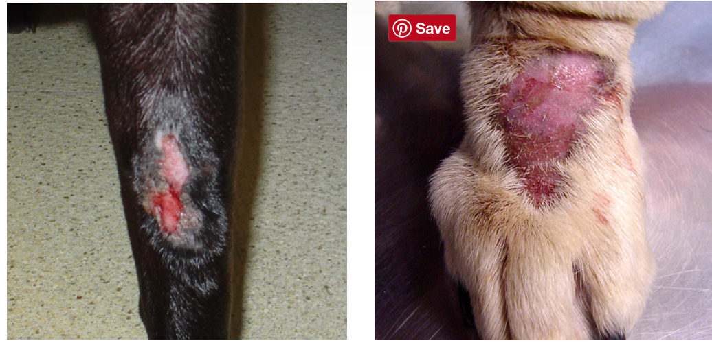
The hallmark lesion is a well-demarcated, raised, and firm granuloma, often on the anterior or lateral aspect of the distal limbs.
Chronic, erythematous, ulcerated lesions on distal limbs.
Firm, thickened, and often alopecic skin over the affected area.
Hyperpigmentation in chronic cases
Serous or serosanguinous discharge if infection is present.
Behavioral evidence of licking, with saliva staining and irritation around the affected region.
Lesions may vary in size but are often solitary and sharply demarcated.
In some cases, a dog may compulsively lick and chew at the area even after ulceration, and signs of frustration, stress, or anxiety may also be noted.
Canine: Acral Lick Granuloma:DX
Exam + HX
Cytology: secondary bacterial infections, typically with Staphy.Cytology may show neutrophilic inflammation and bacterial presence.
Skin Scraping and Fungal Culture: r/o primary dermatologic disease
Bacterial Culture and Sensitivity Testing
Biopsy: For chronic or non-healing lesions, a biopsy can confirm acral lick granuloma by revealing histologic changes: such as dermal fibrosis, acanthosis, and a lymphoplasmacytic infiltrate. This can also help exclude neoplastic processes.
Allergy Testing: Intradermal testing or serum IgE testing may be indicated if allergic dermatitis is suspected.
Canine: Acral Lick Granuloma TX
Requires a multimodal approach to target behavioral, infectious, and inflammatory components.
Behavioral Modification and Environmental Enrichment:
Exercise and Mental Stimulation:
Anti-Anxiety Medications:
Topical and Systemic Antibiotics:
Topical Therapy: Mupirocin or chlorhexidine for secondary infection.
Systemic Antibiotics: If infection is confirmed, systemic antibiotics (e.g., cephalexin, clindamycin) are administered for 4-6 weeks based on culture and sensitivity results.
Anti-Inflammatory and Immunosuppressive Therapy:
Glucocorticoids: Typically used as a short-term measure.
Apoquel (Oclacitinib) or Cytopoint (Lokivetmab):cases associated with atopic dermatitis.
Tacrolimus
Pain Control:
Gabapentin or Pregabalin: These can reduce neuropathic pain, breaking the cycle of licking in cases where pain is a driving factor.
Physical Barriers:
Bandaging or E-collars
Laser Therapy:
Cold Laser Therapy
Surgical Options:
In refractory cases, surgical removal of the lesion may be considered. However, this is generally a last resort due to the risk of recurrence and complications.
Gold Standard Diagnosis and Treatment
While no single treatment is universally successful, the gold standard approach is a combination of:
Comprehensive behavioral modification with environmental enrichment and, if necessary, anti-anxiety medication.
Systemic antibiotics guided by culture and sensitivity for confirmed infections.
Anti-inflammatory therapy with Apoquel, Cytopoint, or glucocorticoids.
Topical and systemic therapies to reduce pruritus and promote healing, tailored to the individual dog's needs.
Decubitus Ulcers:
Decubital ulcers develop due to sustained pressure exceeding capillary closing pressure (approximately 32 mmHg in humans, and similar in animals), leading to ischemia and subsequent tissue necrosis
Delayed Wound Healing
Diseases of Pads
Several factors predispose dogs to pad diseases:
Breed Predisposition: Certain breeds, such as Labrador Retrievers and Border Collies, are prone to zinc-responsive dermatosis. German Shepherds and other large breeds are more prone to trauma and lacerations.
Environmental Factors: Exposure to rough terrain, hot asphalt, or snow can predispose dogs to pad damage and trauma.
Immune Status: Dogs with compromised immune systems, such as those on immunosuppressive therapy or with autoimmune diseases, are at increased risk.
Diet: Diets deficient in zinc or fatty acids, or those based on limited or single-source proteins, may lead to pad abnormalities.
Systemic Disease: Dogs with liver disease, especially those with hepatocutaneous syndrome, are predisposed to paw pad hyperkeratosis and erosions.
Diseases of Pads: Pemphigus Foliaceus
Pemphigus Foliaceus
Pathophysiology: An autoimmune disease causing destruction of keratinocyte cell adhesion.
Clinical Signs: Crusting, ulceration, and erythema of pads, often accompanied by systemic signs.
Diagnostics: Skin biopsy shows acantholytic keratinocytes, a hallmark of pemphigus foliaceus.
Treatment: Immunosuppressive therapy with corticosteroids or cyclosporine. Antibiotics if secondary infection is present.
Diseases of Pads : Zinc-Responsive Dermatosis
Pathophysiology: Zinc deficiency or malabsorption leads to epidermal hyperplasia.
Clinical Signs: Hyperkeratotic, cracked, and thickened pads; scaling and crusting around the face.
Diagnostics: Clinical signs and response to zinc supplementation.
Treatment: Zinc supplementation; essential fatty acids may also help.
Diseases of Pads: Hepatocutaneous Syndrome
Pathophysiology: Associated with liver dysfunction, causing nutrient deficiencies that affect keratinization.
Clinical Signs: Hyperkeratotic, erosive lesions on pads; often described as “frond-like.”
Diagnostics: Biopsy shows a distinctive "red, white, and blue" pattern; liver function tests often abnormal.
Treatment: Intravenous amino acids, essential fatty acids, and diet modification.
Diseases of Pads: Vasculitis
Vasculitis
Pathophysiology: Inflammation of blood vessels leads to ischemic necrosis in affected areas, including pads.
Clinical Signs: Ulcerated, necrotic lesions; pads may be painful and swollen.
Diagnostics: Biopsy with histopathology showing vascular damage.
Treatment: Immunosuppressive drugs, pentoxifylline to improve blood flow, and antibiotics for secondary infections.
Diseases of Pads: Bacterial and Fungal Infections
Bacterial and Fungal Infections
Pathophysiology: Secondary to pad trauma or systemic disease, bacteria (e.g., Staphylococcus spp.) and fungi invade, causing pain and swelling.
Clinical Signs: Red, swollen, purulent lesions; pads may have draining tracts in severe cases.
Diagnostics: Cytology and culture to identify pathogens.
Treatment: Appropriate antibiotic or antifungal therapy, based on culture results.