303: Hepatobiliary System
 Call Kai
 Learn
Learn Practice Test
 Spaced Repetition
 Match
Match Flashcards
 Knowt Play
1/143

There's no tags or description
Looks like no tags are added yet.
Name | Mastery | Learn | Test | Matching | Spaced | Call with Kai | Chat |
|---|
No analytics yet
Send a link to your students to track their progress
144 Terms
Liver
What is the largest gland in the body?
after joining circulation
when are lipids processed by the liver?
Liver
What processes all digested materials & venous blood from spleen & pacnreas
Secretes bile for emulsification of lipids, waste products
What does the exocrine gland of the liver do?
Serum albumin, lipoproteins, thyroid hormone (active form) etc.
What does the endocrine gland of the liver do?
IVC
diaphragm
what two structures suspend the liver?
R. triangular ligament
Identify the structure at #1?
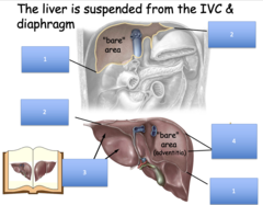
L. triangular ligament
Identify the structure at #2?
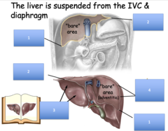
Visceral peritoneum
Identify the structure at #3?
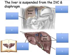
Coronary ligament
Identify the structure at #4?
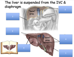
coronary ligament
this ligament helps suspend the liver to the posterior abdominal wall at the "bare area" (adventitia)
right lobe
the bare area is located on the posterior surface of which liver lobe?
liver
what structure is palpable right under the xiphoid process?
Foregut
Are the liver and gall bladder part of the foregut, midgut, or hindgut?
2nd part
The liver, gall bladder & pancreas develop from an outgrowth from what part of the duodenum?
Celiac artery
Which artery is primarily associated with the foregut, supplying blood to the liver, gall bladder, and pancreas?
Superior mesenteric artery
Which artery primarily supplies blood to the midgut, including the small intestine and part of the large intestine?
Inferior mesenteric artery
Which artery primarily supplies blood to the hindgut, including the distal part of the colon and rectum?
2nd part (this explaining the common duct system opening at this location in the adult)
The liver, gall bladder, and pancreas share a common duct system that opens into which part of the duodenum?
Major duodenal papilla (opening for bile duct & main pancreatic duct)
Identify the structure the larger arrow points to
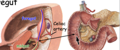
celiac trunk
what main artery supplies the liver?
mesentery
double layer of peritoneum between body wall (usually posterior) and organ
In the mesentery (between two peritoneal layers)
How do neurovascular structures pass to organs?
ligament
double layer of peritoneum between organs or between an organ and the abdominal wall
omentum
double layer of peritoneum between the stomach and another organ
ventral mesentery
The developing liver forms within a...
lesser omentum
the double layer of peritoneum between the stomach and liver:
ventral mesentery
the adult lesser omentum develops from the:
ventral mesentery
the falciform ligament develops from the:
hepatogastric ligament
hepatoduodenal ligament
what two ligaments does the lesser omentum comprise of:
hepatoduodenal ligament
the portal triad travels through what part of the lesser omentum?
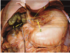
Probe in epiploic foramen (of Winslow) = opening into lesser sac
What is shown at #1?
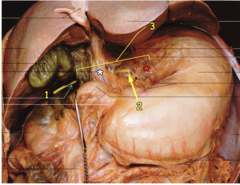
Tip of probe in lesser sac (omental bursa)
What is shown at #2?
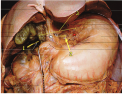
Lesser omentum
What is shown at #3?
Hepatogasatric ligaments
What is shown at the red *
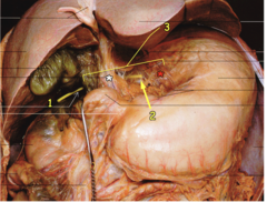
Hepatoduodenal ligaments
What is shown at the white *
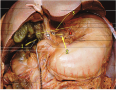
Portal triad
What is the ring around?
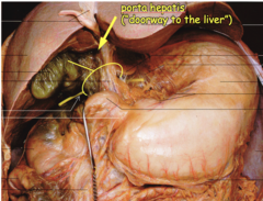
portal vein
hepatic artery
common bile duct
list the components of the portal triad:
posterior to stomach
where is the lesser sac located?
lesser sac
patient has an ulcer on the posterior side of the stomach that has resulted in fluid accumulation. While standing, where would this fluid accumulate?
hepatorenal recess (pouch of Morrison)
patient has an ulcer on the posterior side of the stomach that has resulted in fluid accumulation. While laying supine, where would this fluid accumulate?
Epiploic foramen (foramen of winslow)
what is the name of the opening to the lesser sac?
umbilical vein
during fetal life, what structure ran on the inferior border of the falciform ligament?
ductus venosus
what fetal structure allowed blood to pass through the liver straight into the IVC?
ligamentum venosum
the ductus venosus obliterates and becomes the:
ductus venosus
what is now the ligamentum venosum was the former:
ligamentum teres hepatis (round ligament)
the umbilical vein obliterates and becomes the:
umbilical vein
what is now the ligamentum teres hepatis was the former:
Ductus venosus
What structure is at #1?
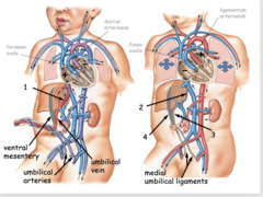
Ligamentum venosum
What structure is at #2?
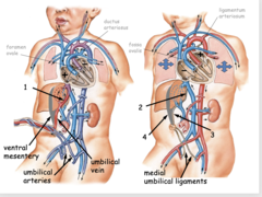
Ligamentum teres hepatis
What structure is at #3?
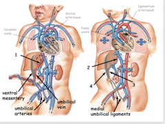
Falciform ligament
What structure is at #4?
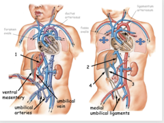
falciform ligament
what structure divides the liver into right and left lobes?
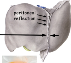
Falciform ligament (from ventral mesentery)
What are the arrows all pointing to?
caudate lobe
which lobe on the posterior surface of the liver sits to the left of the IVC?
quadrate lobe
which lobe on the posterior surface of the liver sits to the left of the gallbladder?
Ligamentum venosum
Ligamentum teres hepatis (round ligament)
What makes up the umbilical fissure?
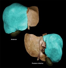
Right lobe of the liver
Identify the structure
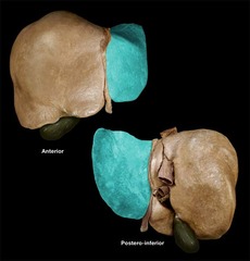
Left lobe of the liver
Identify the structure
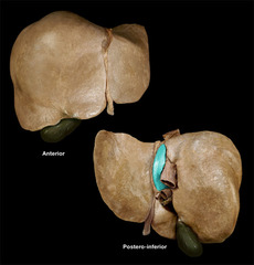
Caudate lobe of the liver
Identify the structure
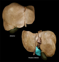
Quadrate lobe of the liver
Identify the structure
True - functional lobes are defined by their dual blood supply
T/F: Functional liver lobes diver from these morphological lobes
1/2 left hepatic a.
1/2 right hepatic a.
the caudate lobe receives blood from what artery:
alcohol abuse
hepatitis infection
autoimmune disease
industrial solvents
list 4 causes of irreversible cirrhosis
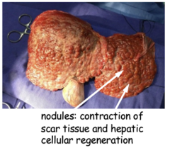
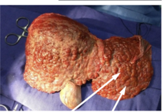
irreversible cirrhosis
patient presents with splenomegaly (due to portal hypertension) and ascites. Liver has "hobnail" appearance. Patient is a long time factory worker and has hep B. what is the diagnosis?
hepatic portal v.
patient presents with esophageal varices. These veins are compensating for what structure?
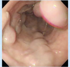
Esophageal varices
This esophagoscope shows what disease?
ascending and descending colon
retroperitoneal varices will present on what structure?
Rectal v.
A patient presents with anorectal varices. Which vein is most likely involved in this condition?
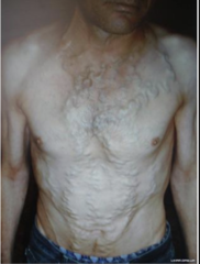
Paraumbilical v.
A patient presents with dilation of periumbilical veins (caput medusa). Which vein is most likely involved in this condition?
hepatic portal v.
venous supply to the liver travels via the:
venous
the majority (75%) of blood that travels to the liver is:
arterial
the minority (25%) of blood that travels to the liver is:
left hepatic a.
the quadrate lobe receives arterial blood from the:
right hepatic a. (Cystic artery)
the gallbladder receives arterial blood from the:
caudate lobe
which liver lobe receives arterial blood from both (50:50) left and right hepatic artery?
right
anatomically, the caudate lobe is a part of the ______ liver
right
anatomically, the quadrate lobe is a part of the ______ liver
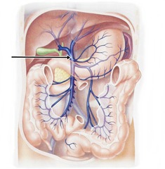
Hepatic portal vein
Identify the structure
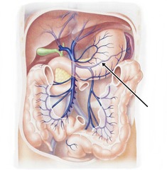
Splenic vein
Identify the structure
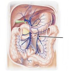
Inferior mesenteric vein
Identify the structure
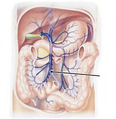
Superior mesenteric vein
Identify the structure
False - all are valveless
T/F - All veins have valves to the liver
Hepatic artery proper
Identify the structure at #1
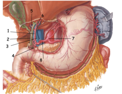
Bile duct
Identify the structure at #2
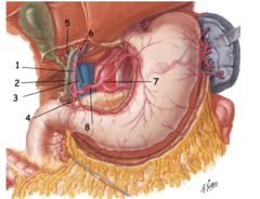
Portal vein
Identify the structure at #3
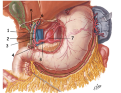
Gastroduodenal artery
Identify the structure at #4
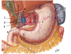
Right hepatic artery
Identify the structure at #5
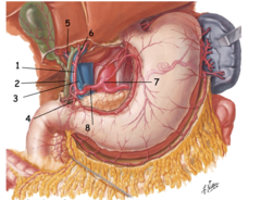
Left hepatic artery
Identify the structure at #6
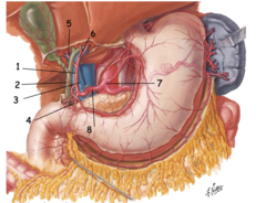
Celiac trunk
Identify the structure at #7
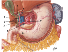
Common hepatic artery
Identify the structure at #8
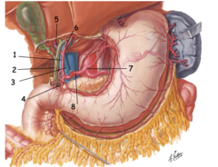
The liver is divided by hepatic arteries, portal
veins (& bile ducts) horizontally and hepatic veins vertically, giving rise to 8 segments
How many segments does the liver have?
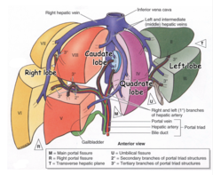
True
T/F Left or right lobe may be used for lobectomy or segmentectomy
70 - regrowth to original size occurs in months
As much as ________% of donor liver can be removed without complications
ligamentum teres hepatis (round ligament)
paraumbilical veins run along what structure?
esophageal v.
rectal v.
retroperitoneal v.
paraumbilical v.
- Chronic shunting may lead to additional pathology
list 4 protocaval anastamoses that can be used during portal hypertension (thus reduced blood flow):
gallbladder
what structure lies between the right and quadrate lobe of the liver?
Gall bladder
Where is the storage and concentration of bile?
IVC
what structure lies between the right and caudate lobe of the liver?
bile canaliculi
what bile structures run in between hepatocytes?