HSCI chapter 7 pt 2, chapter 8
1/44
There's no tags or description
Looks like no tags are added yet.
Name | Mastery | Learn | Test | Matching | Spaced | Call with Kai |
|---|
No analytics yet
Send a link to your students to track their progress
45 Terms
syntharosis
no movement joint
most stable
amphiarthrosis
some joint movement
diathrosis
freely moveable joint
cartilaginous joint
joined by cartilage
fibrous joint
joined by dense regular CT
synovial joints
joint cavity filled with fluid between bones
allows for wide range of motion
flexion
decrease angle
ex. moving elbow at a 90 degree
extension
increase angle
ex. moving elbow joint flat
hyperextension
increase angle beyond
ex. over extending elbow joints
abduction
away from midline
adduction
motion towards midline
circumduction
cone shaped motion
combo of all angular motion
medial rotation
rotation towards midline
lateral rotation
rotation away from midline
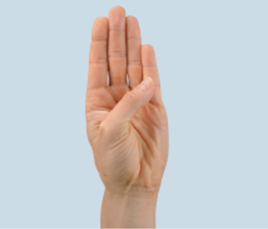
pronation
medial rotation of forearm
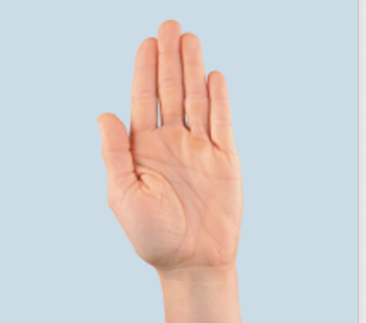
supination
lateral rotation of forearm
opposition
thumb moves toward index finger
reposition
thumb returns to anatomical position
depression
body part moves inferior
elevation
body part moves superior
protraction
body part moves anterior
retraction
body part moves posterior
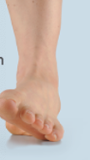
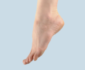
inversion
sole of foot turns medially
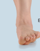
eversion
sole of foot turns laterally
dorsiflexion
flexion of foot
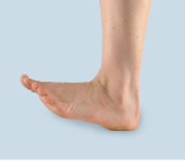
plantarflexion
extension of foot
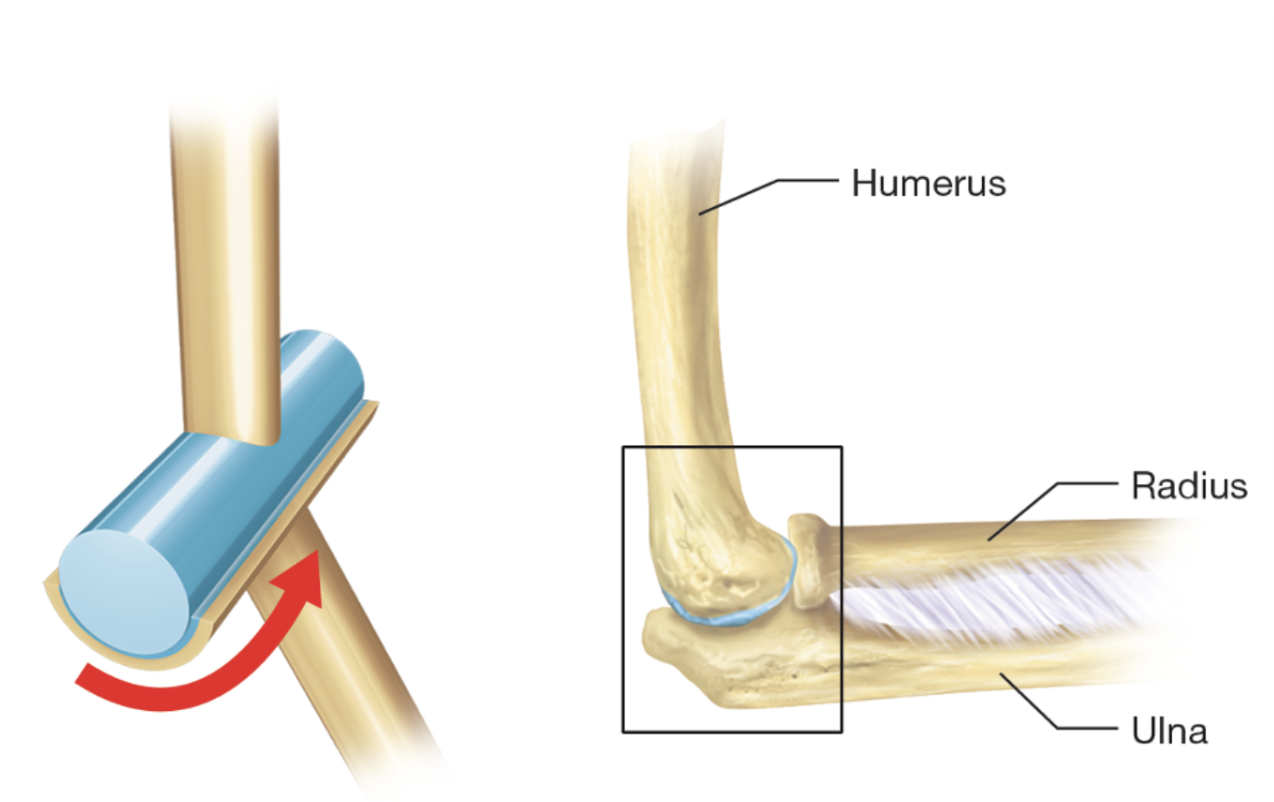
Hinge joint
Convex surface articulate with concave depression
Movements:
Flexion
Extension
Example: elbow,knee, ankle joint
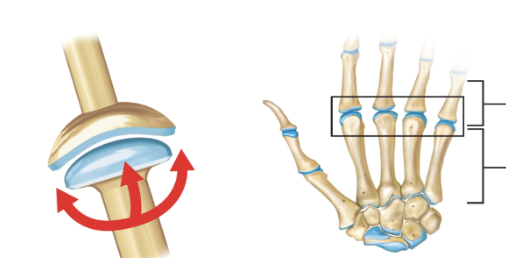
condylar joint
More oval shaped convex surface fits into more shallow concave surface
Movements:
Flexion and extension
Abduction adduction
Circumduction
Example: wrist joint
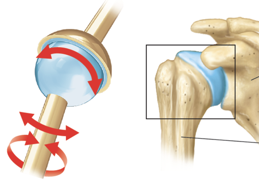
ball and socket joint
Spherical articulating surface fits into cup-shaped depression
Movement:
Flexion and extension
Abduction and adduction
Circumduction
Rotation
Example: shoulder and hip joint
pectoral girdle
bone markings: clavicle and scapula
function: attachment (upper limb + muscles) and mobility
humerus
bone markings: head, capitulum, trochlea
glenohumeral/ shoulder joint
bone marking: humeral head, glenoid cavity
radius
bone markings: radial head
articulates with capitulum
ulna
bone marking: olecranon, trochlear notch
elbow joint
bone markings: trochlear notch (ulna), trochlea (humerus)
radiocarpal/wrist joint
bone markings: distal end of radius, scaphoid, and lunate
pelvic girdle
bone marking: ilium, ischium, pubis
femur joint
bone markings: head, neck, medial and lateral condyles
coxal/hip joint
bone markings: femoral head, acetabulum
patella
markings: quadriceps tendon, patellar tendon
sesamoid bone (embedded inside tendon)
Tibia
bone markings: medial condyle, lateral condyle, tibia tuberosity medial malleolus
fibula
bone markings: head, lateral malleolus
knee joint
bone markings:
tibio femoral
medial and lateral femoral condyles
M/L tribial condyles
patellofemoral
tarsals
bone marking: talus, calcaneus
talocrural/ankle joint
bone marking: medial malleolus of tibia, lateral malleolus of fibula