Osteology of the Skull - Dural Venous Sinuses & Cranial Base
 Call Kai
 Learn
Learn Practice Test
 Spaced Repetition
 Match
Match Flashcards
 Knowt Play
1/86

There's no tags or description
Looks like no tags are added yet.
Name | Mastery | Learn | Test | Matching | Spaced | Call with Kai | Chat |
|---|
No analytics yet
Send a link to your students to track their progress
87 Terms
space within the folds containing blood
Venus sinus
- "tough mother"
- two inseparable layers: periosteal & meningeal
- white inner layer
Dura mater
interior surface of the bone, against the bone
periosteal layer
against the arachnoid mater, most interior, deeper of the two layers
meningeal layer
- "spider mother"
- shiny, translucent
- fibrous, gelatinous-like
- boundary of a real space
Arachnoid mater
- "gentle mother"
- microscopic lining along the surface of the brain, dipping in and out of brain areas
Pia mater
inflammation of the meninges due to viral, bacterial, fungal infections or damage
Meningitis
potential spaces
Epidural and subdural (deep to dura mater) are
- space that can be created where there is normally no space
- can be due to a bleed (hemorrhage, hematoma)
- not supposed to be present
Potential spaces
- real space with vessels and CSF
- deep to the arachnoid mater
Subarachnoid
- protect the brain
- support vessels and venous sinuses
- enclose the subarachnoid space
Function of meningeal layers
scalp > bone > dura mater > subdural (potential space) > arachnoid mater > subarachnoid (real space) > pia mater
Order of layers
- where arachnoid mater dips into the venous sinus
- how the CSF gets out of the subarachnoid space and into the blood system
arachnoid granulations
- connection of dural venous sinuses with veins outside the cranium
- not within the dura but pass through the cranium
- service the scalp
- scalp infections in the "L" layer can spread into the cranial cavity and meninges via these
Emissary veins
spread into, emissary veins
scalp infections in the "L" layer can ______ _____ the cranial cavity and meninges via _______ ______
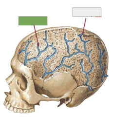
Diploic vv
This is the
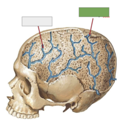
Emissary vv
This is the
connect dural sinuses to scalp veins
The emissary foramina
4 ventricles of the brain itself
CSF is produced in the
brown tissue around the third ventricle continuously producing cerebrospinal fluid
choroid plexus
arachnoid granulations
CSF gets out via
subarachnoid space
The real space that the CSF is in is the
L2 at the end of the spinal cord proper
Subarachnoid space terminates around
- mechanical protection
- pathway for removal of brain metabolites
Function of CSF
- propagations of the arachnoid that protrude through the meningeal layer into the dural venous sinuses
- site of transfer of CSF to venous system
- may cause pits on the internal surface of cranium
CSF to venous sinuses
between hemisphere of cerebrum (sickle shaped)
falx cerebri
in between the hemispheres of the cerebellum
falx cerebelli
between cerebrum and cerebellum is a horizontal fold, creating a sort of tent
tentorium cerebelli
folds around the sides of the sella turcica
diaphragma sella
in falx cerebri:
superior sagittal sinus
in inferior part of the falx cerebri:
inferior sagittal sinus
left, right
There is a ______ and a _______ transverse sinus
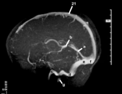
superior sagittal sinus
Radiograph sinuses: 21
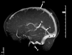
straight sinus
Radiograph sinuses: a
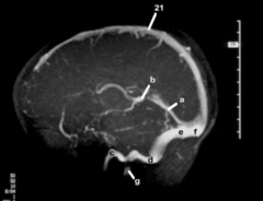
great cerebral vein (Galen)
Radiograph sinuses: b
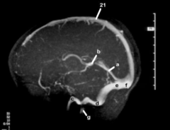
inferior petrosal sinus
Radiograph sinuses: c
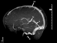
sigmoid sinus
Radiograph sinuses: d
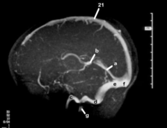
transverse sinus
Radiograph sinuses: e
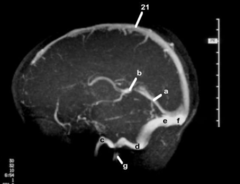
confluence of sinuses
Radiograph sinuses: f
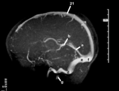
internal jugular vein
Radiograph sinuses: g
- grooves for sinuses
- grooves for the meningeal aa
- foramen for emissary veins
- granular foveolae for arachnoid granulations
Connection to osteology
arachnoid granulation
This is the
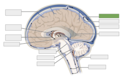
subarachnoid space
This is the
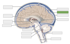
meningeal dural mater
This is the
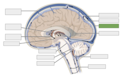
right lateral ventricle
This is the
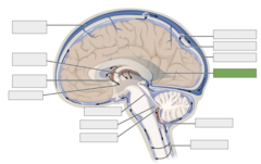
median aperture
This is the
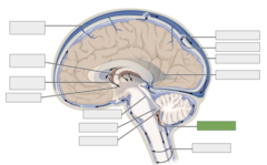
central canal
This is the
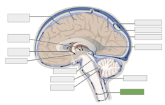
fourth ventricle
This is the
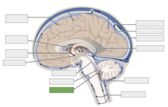
lateral aperture
This is the
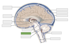
cerebral aqueduct
This is the
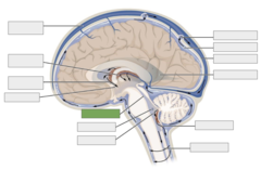
third ventricle
This is the
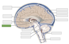
interventricular foramen
This is the
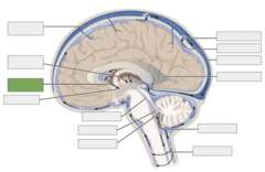
choroid plexus
This is the
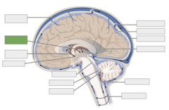
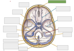
superior sagittal sinus
This is the
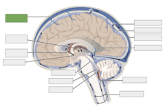
CSF in subarachnoid space
This is the
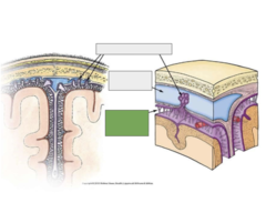
Dural venous sinus
This is the
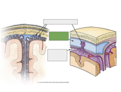
arachnoid granulations
This is the
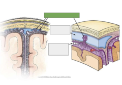
Falx cerebelli
This is the
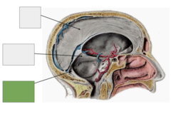
Tenorium cerebelli
This is the
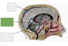
Falx cerebri
This is the
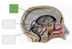
Diaphragma sella
This is the
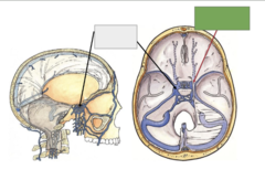
cavernous sinus
This is the
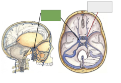
Inferior petrosal sinus
This is the
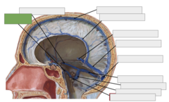
Superior petrosal sinus
This is the
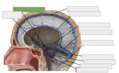
Superior sagittal sinus
This is the
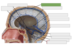
Inferior sagittal sinus
This is the
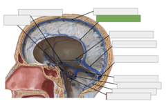
Great cerebral vein
This is the
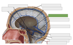
Straight sinus
This is the
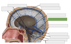
Confluence of sinuses
This is the
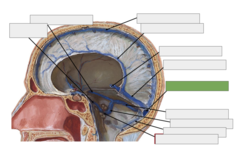
Transverse sinus
This is the
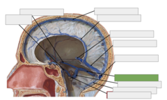
Sigmoid sinus
This is the
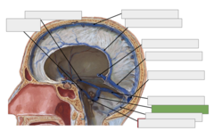
Occipital sinus
This is the
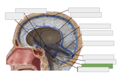
Jugular foramen to IJV
This is the
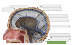
Lt. sphenoparietal sinus
This is the
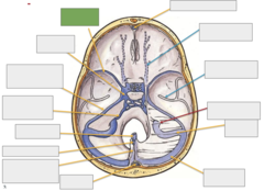
Lt. cavernous sinus
This is the
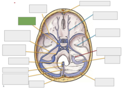
Lt. superior petrosal sinus, draining to sigmoid sinus
This is the
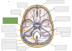
Lt. inferior petrosal sinus, draining to the IJV
This is the
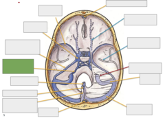
great vein of Galen
This is the
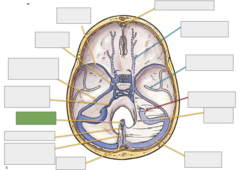
inferior sagittal sinus
This is the
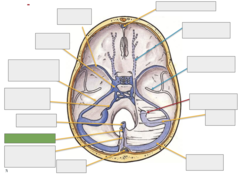
Straight sinus (great vein and inferior sagittal converge)
This is the
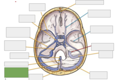
confluence of sinuses
This is the
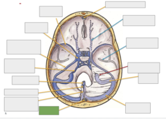
Rt. transverse sinus
This is the
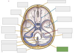
Rt. sigmoid sinus
This is the
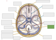
jugular foramen and IJV
This is the
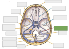
middle meningeal artery
This is the
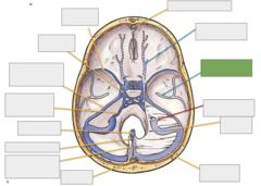
Rt. superior opthalamic v
This is the
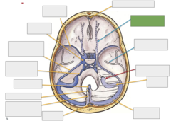
superior sagittal sinus
This is the