Test 1
 Call Kai
 Learn
Learn Practice Test
 Spaced Repetition
 Match
Match Flashcards
 Knowt Play
1/168

There's no tags or description
Looks like no tags are added yet.
Name | Mastery | Learn | Test | Matching | Spaced | Call with Kai | Chat |
|---|
No analytics yet
Send a link to your students to track their progress
169 Terms
Why Do We Need a Standard Reference?
Consistency
Communication
Documentation
Why do we need consistency?
Everyone describes the body from the same reference position
Why do we need communication?
Clinicians can communicate location and movement clearly.
Why do we need documentation?
Findings can be recorded precisely and interpreted by others
Anatomical terminology removes ________.
ambiguity
Anatomical Position
Body upright (or described as if upright)
Head and eyes facing forward
Upper extremities at the sides
Palms facing forward
Lower extremities straight
Feet/toes pointing forward
Anatomical descriptions are based on this standardized position, even…
when the patient is sitting, lying down, or moving
Superior
toward the head / above
Inferior
toward the feet / below
Anterior
toward the front
Posterior
toward the back
Medial
toward the midline
Lateral
away from the midline
PROXIMAL
closer to the trunk or point of attachment
Distal
farther from the trunk or point of attachment
Superficial
closer to the body's surface
Deep
farther from the body's surface
Directional terms describe relationships, not…
isolated structures
IPSILATERAL
on the same side of the body
CONTRALATERAL
on the opposite side of the body
The Three Anatomical Planes
Sagittal
Frontal/Coronal
Transverse
Sagittal Plane
Divides the body into right and left portions.
Common motions: flexion & extension
Frontal/Coronal Plane
Divides the body into anterior and posterior portions.
Common motions: abduction & adduction
Transverse Plane
Divides the body into superior and inferior portions.
Common motions: rotation
Flexion
decreases joint angle
Extension
increases joint angle
ABduction
away from midline
ADduction
toward midline
Internal Rotation
rotation toward midline
External Rotation
rotation away from midline
Pronation
the rotational movement that turns your palm to face downward or backward
Forearm
Supination
rotational movement that turns your palm to face upward or forward
Forearm
Dorsiflexion
the upward movement of the foot at the ankle joint that brings your toes and the top of your foot closer to your shin
Plantarflexion
the movement where you point your foot and toes downward, away from your shin and body
Inverson
the movement where the sole of the foot turns inward toward the midline of the body
Eversion
the anatomical movement of the sole of the foot away from the midline of the body
Elevation
upward movement of the shoulder blades toward the ears
Depression
the downward movement of the shoulder blades (scapula) away from the ears along the rib cage
Protraction
the act of moving a body part forward or extending something in duration
Scapula
Retraction
the posterior and medial movement of a body part back toward the midline or its original resting position
Scapula
Lateral flexion
side-bending
Rotation
a twisting movement where a bone or body part moves around its own central longitudinal axis
VALGUS
Distal segment moves away from the body's midline.
Example: knee valgus
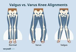
VARUS
Distal segment moves toward the body's midline.
Example: knee varus

Injury initiates a series of responses known as…
inflammation and repair
STANDARD PROCEDURE
The body follows a general process intended to return the injured area toward normal
HEALING PROCESS
The process is consistent and predictable, but it is a changing continuum of events.
PRIMARY INTENTION
Separation of tissue is small.
A bridge of cells binds the tissue together.
Examples: minor wounds; surgery when the ends are sutured together
SECONDARY INTENTION
Wound ends are farther apart.
The body must produce tissue from the bottom and sides of the wound to fill the space.
Example: secondary ligament sprains.
Healing Process | Three Phases
ACUTE INFLAMMATION
Control the area of injury
PROLIFERATION
First stage of tissue repair
MATURATION
Clean up and increase strength
Phase 1 | Acute Inflammation
Sum of the body's tissue reactions to cell injury and death.
Purpose: control the area of injury.
Five cardinal signs are present.
If the duration is excessive, the effects are detrimental
5 CARDINAL SIGNS
Redness
Swelling
Tenderness
Increased temperature
Loss of function
Acute Inflammation | Vascular Response
TRANSIENT VASOCONSTRICTION
Occurs first.
VASODILATION
Follows and increases capillary permeability.
EXUDATE FORMATION
Escaping cells create an osmotic gradient; platelets and serum proteins enter the injured area.
CHEMOTAXIS / CLOTTING
Released products stimulate chemical release and clotting factors
Acute Inflammation | Phagocytosis
5–6 HOURS
Neutrophils and polymorphonuclear leukocytes (PMNs) appear and begin debris removal.
24–48 HOURS
Monocytes and macrophages become the predominant cells.
PHAGOCYTES
PMNs and macrophages act as phagocytes to remove debris.
Phagocytosis =
removal of debris
Acute Inflammation | Clot & Lymphatic Response
CLOT FORMATION
Fibronectin binds with collagen and fibrin.
The clot is temporary, shuts off blood flow to the area, and walls off the injury.
LYMPHATIC DISRUPTION
Small lymph vessels are damaged and can become clogged by fibrin.
This slows removal of fluid from extracellular spaces.
Why does redness occur during inflammation?
Histamine + vasodilation
why does SWELLING / EDEMA occur during inflammation?
Increased substance in the area + blockage of lymph vessels
Why does TENDERNESS / PAIN occur during inflammation?
Chemical irritation of local nerve endings + pressure from edema
Why does INCREASED TEMPERATURE happen during inflammation?
Increased local cellular and chemical activity
Why does LOSS OF FUNCTION occur during inflammation?
Pain + injured tissue
Phase 2 | Proliferation
Removal of debris formed during the inflammatory phase continues.
This is the first stage of tissue repair.
The hallmark is revascularization and formation of granulation tissue.
Scar formation requires vascular production.
Fibroblasts are largely responsible for production of new growth.
Increasing numbers of fibroblasts mark the proliferation phase.
Generally lasts 2–4 weeks
Proliferation | Granulation Tissue
MATRIX
Part of granulation tissue.
CAPILLARY BUDS
Part of granulation tissue.
PLASMINOGEN ACTIVATOR
Breaks down the fibrin network to restore lymph flow.
FIBROUS COMPONENTS
Collagen
Elastin
GROUND SUBSTANCE
GAGs
Proteoglycans
Glycoproteins
Fills gaps between fibrous elements
Proliferation | Collagen Transition
Type III collagen —> Type I collagen
Phase 3 | Maturation
“Cleans up” the area and increases strength of repaired
tissue.
Fibroblasts disappear as they convert to myofibroblasts.
Myofibroblasts shrink or contract the wound size.
Wound contraction is beneficial unless it causes loss of
mobility or function.
The emphasis shifts toward strengthening and organizing the repaired tissue
Maturation | Increasing Tissue Strength
Vascularity is reduced.
Fibroblasts diminish.
Fluid content decreases.
Collagen forms more cross-links.
Tissue tensile strength increases.
Collagen fibers align in a more organized, parallel fashion.
May last up to one year
ACUTE Inflammation
Normally complete in 2 weeks.
SUBACUTE Inflammation
If the reaction continues for 1 month
CHRONIC Inflammation
If the reaction continues for months or years
When Inflammation Persists
Loss of function may be the only sign remaining.
The body is still reacting to the presence of foreign material.
Large quantities of collagen often envelop the affected area
Factors That Modify Inflammation & Repair
PATIENT FACTORS
Age • Health • Nutrition
MEDICATIONS
Corticosteroids may impair the proliferation phase.
IMMOBILIZATION
Prolonged immobilization
TRAUMA
Physical injury or wound produced by an internal or external force
MECHANICAL INJURY
Results from force or mechanical energy that changes the state of rest or uniform motion of matter.
SIGN
Observable
Objectively measurable
Examples:
Swelling • discoloration • deformity • crepitus • redness
SYMPTOM
Subjective complaint
Abnormal sensation described by the patient
Examples:
Pain • nausea • altered sensation • fatigue
ACUTE Injury
Sudden onset
Usually from a single traumatic event
Short in duration
CHRONIC Injury
Gradual onset
Exact mechanism often not known
Patient may not identify a specific date of injury
Prolonged duration
Contusion
“Bruise”
Compression of soft tissue by a direct blow or impact.
Disrupts small capillaries within the tissue.
Results in ecchymosis.
ecchymosis
Bruise
Sprain Injures what?
Ligament or capsular structure
POSSIBLE RESULT: Laxity of the involved ligament or capsule
WHAT DO LIGAMENTS DO?
Ligaments connect bone to bone
Strain is a injury to what?
Muscle or tendon
How does a sprain occur?
Violent, forceful contraction
OR
overstretching of the myotendon unit
Strain V. Sprain
Sprain = ligament/capsule
Strain = muscle/tendon
SUBLUXATION
Incomplete disassociation of two joint surfaces.
More common than a dislocation.
Can be difficult to identify because it may reduce on its own
DISLOCATION
Complete disassociation of two joint surfaces.
Results when forces cause the joint to exceed its normal ROM.
Clinical Evaluation & Diagnosis
A clinical evaluation is more detailed than a basic assessment.
The overall examination outline remains consistent.
Specific special tests vary by body region and suspected
injury.
Evaluation findings help guide a clinical impression, treatment planning, and/or referral
CLINICAL REASONING
Your examination is not a collection of random tests. Each step should help you gather information about the patient's problem
CLINICAL / PHYSICAL DIAGNOSIS
Based on the external examination and clinical findings.
Used to identify the likely pathology or structures involved
MEDICAL DIAGNOSIS
Reserved for physicians in the provided course reference.
May involve additional diagnostic testing
Before You Touch the Patient | Introduce Yourself
1 - WHO YOU ARE
State your name and role.
2 - WHY YOU'RE HERE
Briefly explain the purpose of the evaluation.
3 - WHAT TO EXPECT
Describe what you plan to do before you begin.
Consent Is a Process — Not a Single Question
EXPLAIN
Tell the patient what you want to do and why.
ASK
Obtain permission before beginning the examination or a new hands-on procedure.
CHECK IN
Continue to monitor comfort and willingness as the examination progresses.
PATIENT CONTROL
The patient can ask questions, decline a procedure, or ask you to stop. Respect the response and adapt the examination appropriately
Patient-Centered Orthopedic Examination
LISTEN
Start with the patient's concerns, goals, and description of the problem.
EXPLAIN
Use clear language and tell the patient what you are doing.
RESPECT
Protect privacy, dignity, comfort, and individual preferences.
COLLABORATE
Include the patient in decisions about the pace and progression of the examination.
RESPOND
Modify or stop when symptoms, concerns, or patient preferences require it.
CONFIRM
Check understanding and invite questions throughout
SUBJECTIVE Part tells you what
What does the patient tell you?
OBJECTIVE part tells you what?
What do you observe or measure?
the ASSESSMENT tells you what?
What is your professional impression?
Documentation Matters
Complete and accurate documentation is critical.
Records should be clear, concise, and accurate.
Documentation supports communication among providers.
Good documentation contributes to quality patient care.
The provided course reference also notes its importance for third-party billing.
DOCUMENT THE STORY
Your record should allow another provider to understand what the patient reported, what you found, your impression, and your plan
SOAP Notes | A Documentation Framework
S • SUBJECTIVE
O • OBJECTIVE
A • ASSESSMENT
P • PLAN