W6 ADI
 Call Kai
 Learn
Learn Practice Test
 Spaced Repetition
 Match
Match Flashcards
 Knowt Play
1/42

There's no tags or description
Looks like no tags are added yet.
Name | Mastery | Learn | Test | Matching | Spaced | Call with Kai | Chat |
|---|
No analytics yet
Send a link to your students to track their progress
43 Terms
What 4 other names is Battered Child Syndrome also known as?
Child abuse
Non-accidental injury
Parent- Infant Traumatic Stress Syndrome
Caffey-Kempe Syndrome
What is the general description given to a child suffering from Battered Child Syndrome?
A child with multiple fractures of long tubular bones and subdural haematoma
What are genuine accidental injuries that are NOT findings of Battered Child Syndrome?
Torus fracture (distal radius)
Spiral fracture of tibia (toddler’s fracture)
Clavicle fracture
Skull fracture
What is the hallmark feature of Battered Child Syndrome?
Repeated episodes of multiple fractures
What clinical features make up Battered Child Syndrome?
Children usually less than 6 years old (between 1-4)
Often less than 2 years old when exposed to trauma
After age 4, children can escape abuser or talk about what is happening
Evidence of repeated trauma such as bruising or burns
Repeated episodes of multiple fractures
What radiographic features make up Battered Child Syndrome?
Hallmark feature of multiple fractures (especially ribs) at different stages of healing
Fractures at the corner of metaphyses (especially medial side of distal femoral metaphysis) or exuberant callus in diaphyses or metadiaphyses of long tubular bones
Periosteum loosely attached to shaft of a tubular bone, but more firmly attached to metaphysis
Trauma leads to lifting of periosteum along the shaft (exuberant callus) and avulsion of bone at metaphysis (corner fractures)
Periostitis seen within 1-2 weeks of injury
What sites are often involved in Battered Child Syndrome?
Ribs
Humerus
Tibia
Small bones of hand, foot and skull
Unusual fractures include sternum, lateral clavicle, scapula, spine (spinouses)
Be suss with bilateral acute fractures and lower limb fractures in children who are not walking
Bilateral humeral fractures from severe shaking are seen because the child is held by upper arms
What can severe shaking of children cause?
Brain damage (Cerebral haematoma) (intracranial and subdural haematoma)
Retinal detachment
SP fractures of thoracic and lumbar spine
Rib fractures
Humeral fractures

What is seen in this image? What syndrome could be the cause?
Posterior rib fractures
Battered Child Syndrome

What syndrome is this? What sign can be seen?
Battered Child Syndrome
Metaphyseal corner fractures

What syndrome is this? What sign can be seen?
Battered Child Syndrome
Tibia fractures (can see periosteal response)
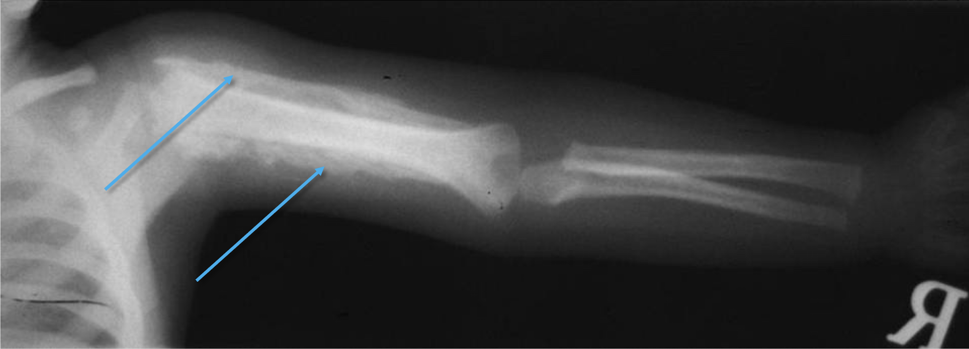
What syndrome is this? What sign can be seen?
Battered Child Syndrome
Massive periosteal reaction in humerus
What is the most common cause of death in Battered Child Syndrome? What signs may you see with it?
Head trauma
Skull fracture
subarachnoid haemorrhage
retinal detachment
cerebral oedema
What is the second leading cause of death in Battered Child Syndrome? What signs may you see with it?
Visceral trauma
Crushing blow to abdomen
Bowel/stomach rupture
Lacerated internal organs
What can be seen on the skin in Battered Child Syndrome?
Burns
Bruises
Lacerations
What are possible internal injuries in Battered Child Syndrome?
Liver fracture (separation)
Pancreas rupture
Bowel contusions
What are typical features of accidental injuries?

What are typical features of non-accidental injuries?

What possible conditions may be mistaken for Battered Child Syndrome?
Infantile cortical hyperostosis
Changes evident in first 5 months, symmetrical, fever, elevated ESR, resolves within few months
Scurvy
Changes after 4 months old, generalised osteopaenia, symmetrical
Congenital syphilis
Symmetrical and diffuse
Osteogenesis imperfecta
Generalised osteoporosis
Congenital insensitivity to pain
Neuropathic joint changes, soft tissue ulcerations, scarred tongue and fingers from burns

What syndrome is this, what sign can be seen?
Battered Child Syndrome
Metaphyseal corner fractures

The image provided is of a 3 month old female. What syndrome could explain the fracture? Provide reasoning.
Battered Child Syndrome
Lower limb fracture in a child who is not walking

What syndrome is depicted in this image?
Battered Child Syndrome

What syndrome is shown here? What hallmark feature helps you to determine this?
Battered Child Syndrome
Hallmark feature of multiple fractures (especially ribs) at different stages of healing

What is shown in this image from a child suffering Battered Child Syndrome?
Left Subdural haematoma

What signs are shown in this image of a child suffering from Battered Child?
Left lateral rib fractures (blue)
Left sided pleural effusion (yellow)

What signs are shown in this image of a child suffering from Battered Child?
Distal radius and ulna fracture

What signs are shown in this image of a child suffering from Battered Child?
Multiple parietal fractures
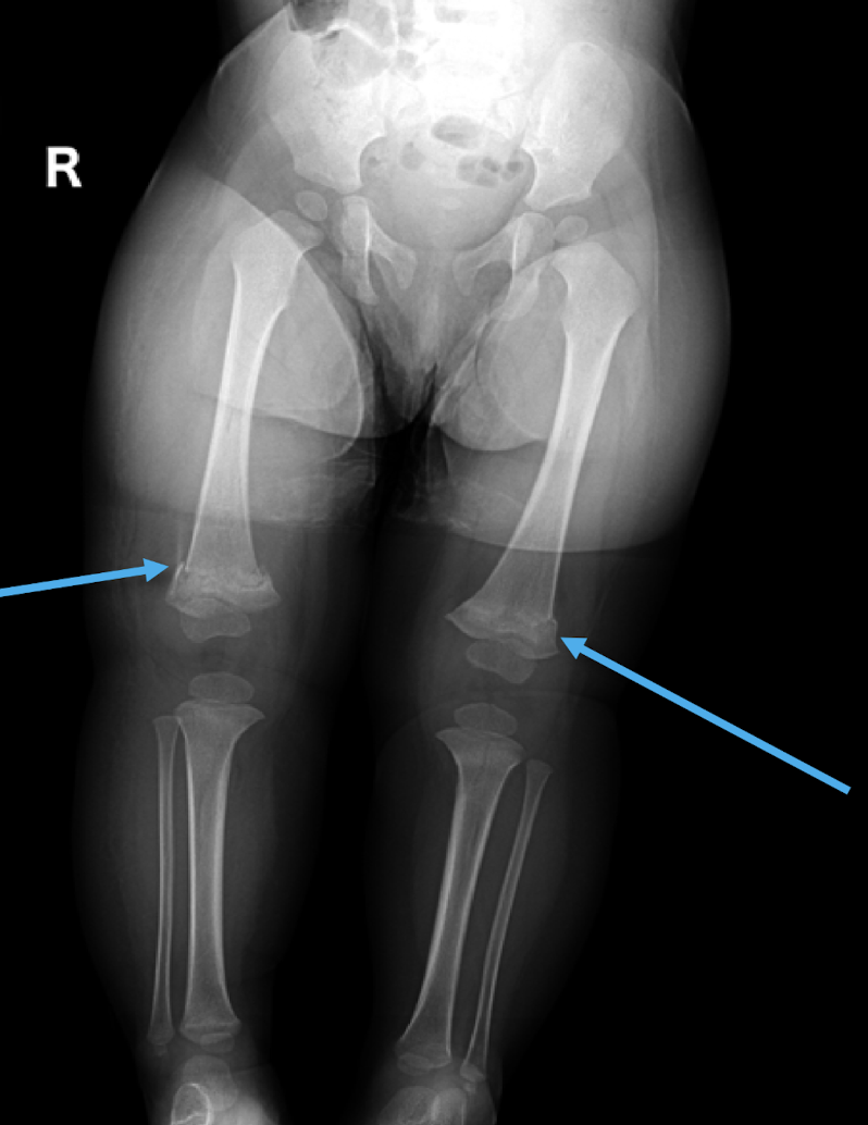
What signs are shown in this image of a child suffering from Battered Child Syndrome?
Bilateral distal femur fractures
I don’t really know what to say for this one so here’s an image describing soft tissue calcification (myositis ossificans).
I think its just saying how prevalent it is.

What is most radiographically visible calcification due to?
Dystrophic calcification
Tissue damage leads to non-specific inflammatory response
Response can include calcification
What is another name for myositis ossificans?
Heterotopic ossification
What is heterotopic ossification?
Bone formation outside it’s usual location
Where does the process of heterotopic ossification occur?
Muscle and connective tissue
What are the most common sites for heterotopic ossification?
Elbow→ Brachialis anterior
Thigh→ Quadriceps femoris and adductor muscles
Knee→ Medial collateral ligament of the knee (Pellegrini-Stieda disease)
Shoulder→ Coracoclavicular ligament rupture
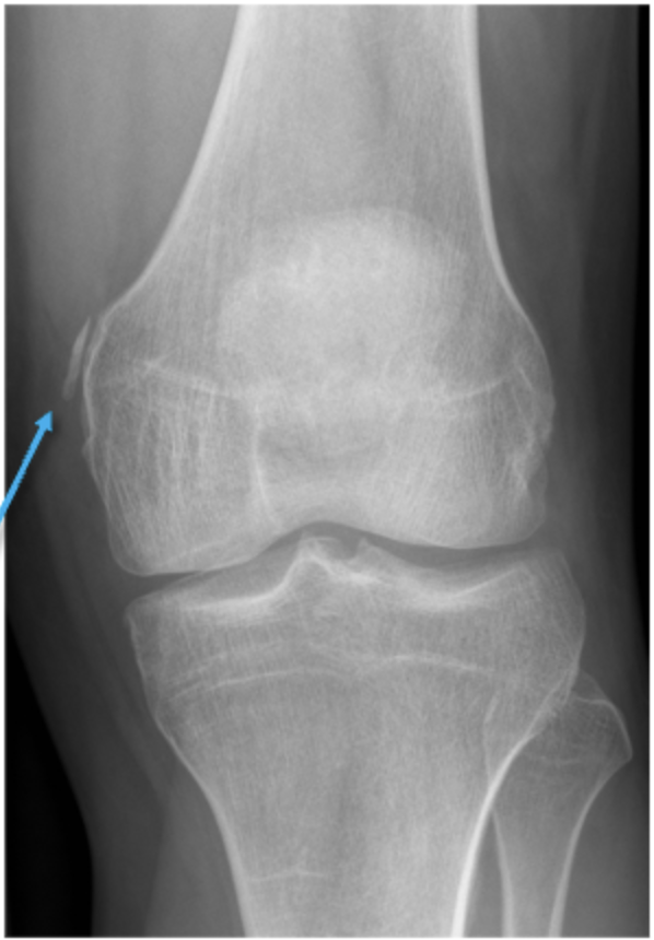
What can be observed in this image and what structure is being affected? What is this indicative of?
Ossification of the medial collateral ligament
Heterotopic ossification

What can be seen in this image and what is this indicative of?
Calcification
Heterotopic calcification
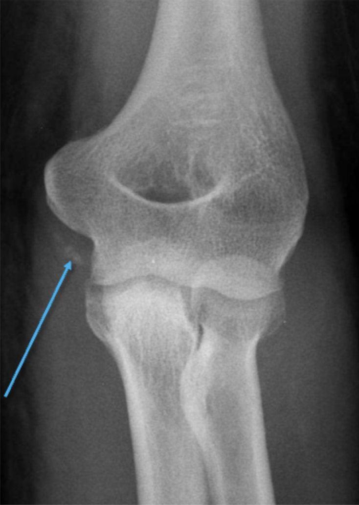
What can be observed in this image and what structure is being affected? What is this indicative of?
Calcification of the ulnar collateral ligament
Indicative of Heterotopic ossification
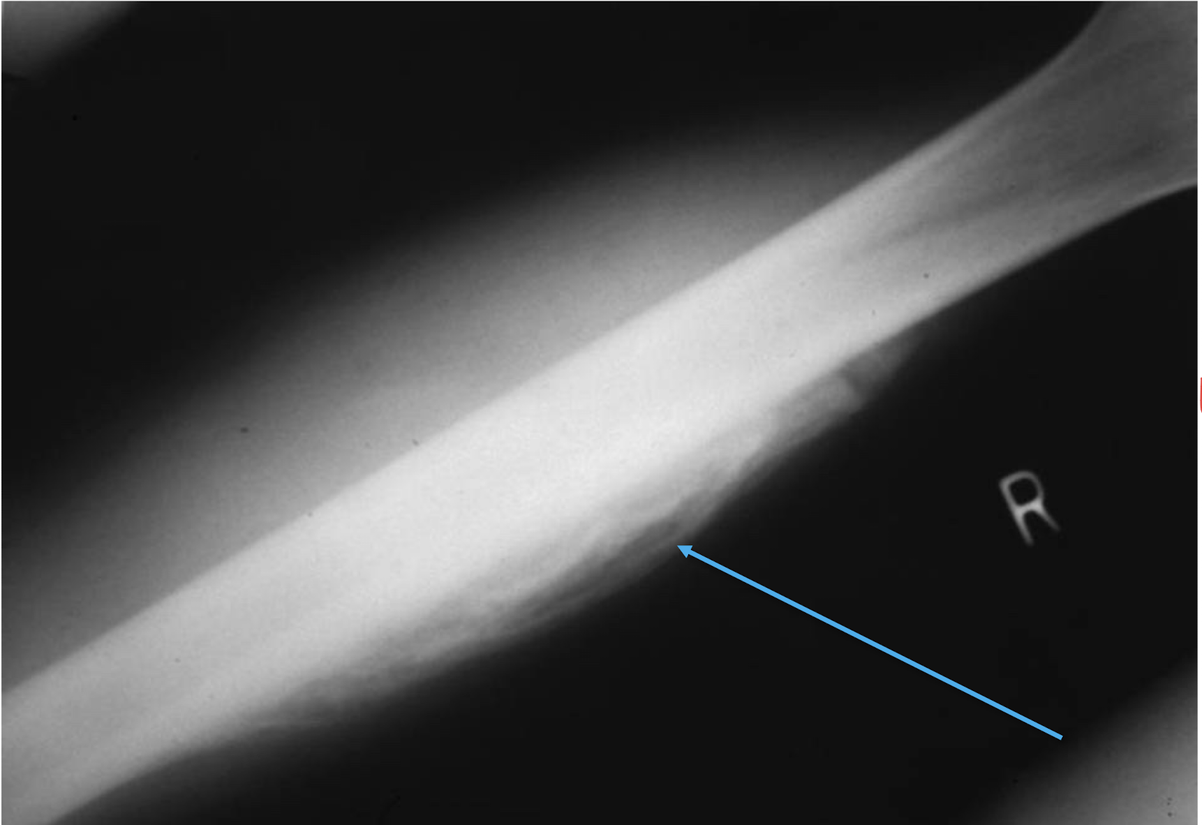
What can be observed in this image? What is this indicative of?
Calcification (almost ossification) of soft tissue structures around the bone
Indicative of Heterotopic ossification

What key sign of Heterotopic ossification can be seen in this image? (Hint: shown by right arrow)
Cleavage sign

What can be seen in this example of Heterotopic ossification and what structure is affected?
Ossification of the gluteal muscle

What structure/s has been affected in this case of Heterotopic ossification?
Distal calf and achilles tendon
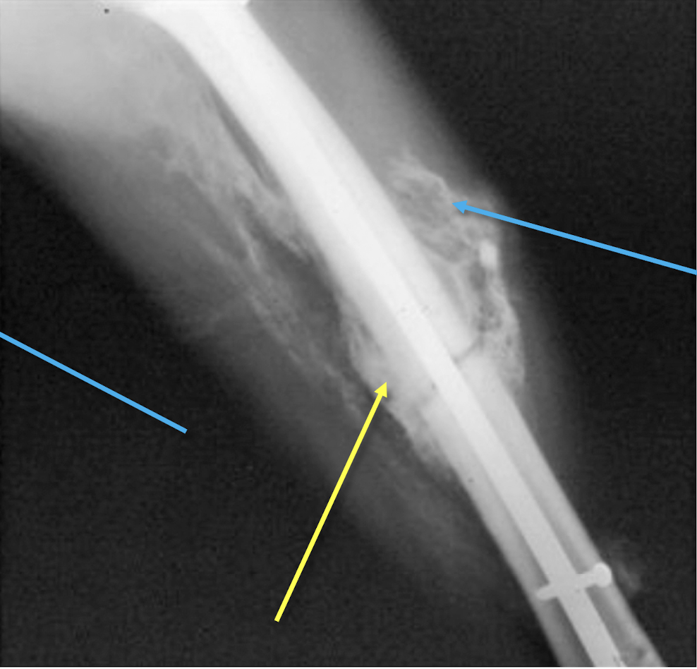
What is shown in this example of a post traumatic femur?
Heterotopic ossification
What are possible differential diagnoses of dystrophic soft tissue calcifications? (Hint: think of examples that may cause calcification using VINDICATE)
Vascular
Venous insufficiency
Infection
Parasitic infestation
Cysticercosis
Dracunculiasis
Armillifer armillatus
Neoplasm
Osteoma
Osteosarcoma
Tumour
Drugs
Vitamin D
Autoimmune
Dermatomyositis
Scleroderma
Trauma
Heterotopic calcification
Injection granulomas
Necrosis