Histology and Tissue Classification
 Call Kai
 Learn
Learn Practice Test
 Spaced Repetition
 Match
Match Flashcards
 Knowt Play
1/30

Earn XP
Description and Tags
A comprehensive set of vocabulary flashcards covering epithelial, connective, muscle, and nervous tissues based on histology lecture material.
Name | Mastery | Learn | Test | Matching | Spaced | Call with Kai | Chat |
|---|
No analytics yet
Send a link to your students to track their progress
31 Terms
Simple Epithelium
Epithelial tissue consisting of one cell layer thick where all cells make direct contact with the basement membrane; functions in absorption, filtration, or secretion.

Stratified Epithelium
Epithelial tissue containing two or more layers of cells where only the deepest layer makes direct contact with the basement membrane; functions in protection or to resist abrasion.

Pseudostratified Epithelium
A single layer of cells that appears stratified because all cell nuclei are not located the same distance from the basal surface; all cells contact the basement membrane, but not all reach the apical surface.

Squamous Cell Shape
Epithelial cells that are flattened and have irregular borders, displaying a flattened nucleus in cross section.

Cuboidal Cell Shape
Epithelial cells that are as tall as they are wide, characterized by a very round, plump nucleus with equal amounts of cytoplasm on all sides.

Columnar Cell Shape
Epithelial cells that are taller than they are wide, with oval or round nuclei that generally line up in a row close to the basal surface.

Transitional Cell Shape
Epithelial cells located on the apical surface that change between flattened and dome-shaped depending on tissue stretch, allowing stretching and relaxing without tearing apart.

Cilia
Small, hairlike structures that extend from the apical surface of epithelial cells and actively move to propel substances along an epithelial sheet.

Microvilli
Extremely small extensions of the plasma membrane on the apical surface of cells that increase surface area and enhance absorption, appearing as a fuzzy brush border.

Keratinization
A homogeneous, acellular-looking portion of stratified squamous epithelium containing keratin, which imparts strength and protection to dead skin epithelial cells.

Goblet Cells
Specialized epithelial cells that are rounded near the apical surface and narrow toward their basal surface (wineglass shape); they produce mucus for protection, transport, or lubrication.

Collagen Fiber
Connective tissue fibers thicker than elastic or reticular fibers that stain pink or blue and resist tensile (stretching) forces along their long axis.
Elastic Fiber
Connective tissue fibers appearing as fine, thin black or wavy fibers that can stretch to greater than 150\text{\null}\% of their resting length and recoil without damage.
Reticular Fiber
Connective tissue fibers composed of a fine, thin type of collagen that appear as an irregular network of short, thin black fibers under silver stain, forming a delicate inner supporting framework for organs.
Mesenchyme
Embryonic connective tissue containing mesenchyme cells, ground substance, collagen fibers, and elastic fibers.
Areolar Connective Tissue
Connective tissue containing predominantly fibroblasts, collagen fibers, and elastic fibers along with some reticular fibers.

Adipose Connective Tissue
Connective tissue characterized by large adipocytes held together with a loose arrangement of collagen fibers.

Elastic Connective Tissue
Connective tissue found in the wall of the aorta consisting of collagen fibers and wavy elastic fibers (staining black) oriented in the same direction.

Dense Regular Connective Tissue
Connective tissue consisting of bundles of collagen fibers all oriented in the same direction, with fibroblasts located between the bundles.

Dense Irregular Connective Tissue
Connective tissue located in the dermis of the skin, consisting of bundles of collagen fibers oriented in many different directions with fibroblasts between the bundles.

Reticular Connective Tissue
Connective tissue consisting of reticular fibers (a fine, thin form of collagen) that appear black when special stain is used, surrounded by cells such as lymphocytes.

Hyaline Cartilage
Cartilage containing prominent lacunae with chondrocytes and no visible fibers in the extracellular matrix, surrounded by a perichondrium with flattened chondroblasts.

Fibrocartilage
Cartilage containing visible bundles of collagen fibers within its matrix, where chondrocytes in lacunae often line up in rows.

Elastic Cartilage
Cartilage containing chondrocytes in lacunae and visible elastic fibers in its extracellular matrix that stain dark blue, purple, or black.

Compact Bone
Tissue composed of multiple osteons, where each osteon consists of a central canal surrounded by concentric layers of bony matrix called lamellae, housing osteocytes in lacunae with canaliculi.

Blood
Fluid connective tissue containing red blood cells (erythrocytes), white blood cells (leukocytes), platelets (thrombocytes), and an extracellular matrix called plasma.

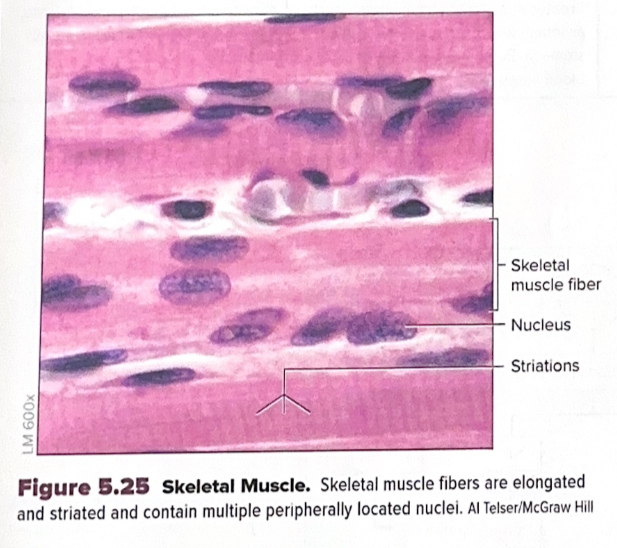
Skeletal Muscle
Muscle tissue consisting of elongated, striated fibers that contain multiple peripherally located nuclei.
Cardiac Muscle
Muscle tissue consisting of short, branched, striated cells containing 1−2 centrally located nuclei and intercalated discs.

Smooth Muscle
Muscle tissue with short, spindle-shaped, non-striated fibers containing a single centrally located nucleus that can coil into a spiral shape upon contraction.

Neurons
Very large cells in nervous tissue with a prominent nucleus, nucleolus, and dark-staining endoplasmic reticulum known as chromatophilic substance.
Glial Cells
Nervous tissue cells that are much smaller and more abundant than neurons.