FINAL TEST
1/149
Earn XP
Description and Tags
Spine and thorax
Name | Mastery | Learn | Test | Matching | Spaced |
|---|
No study sessions yet.
150 Terms
Neck vertebra
7 cervical
Chest vertebra
12 thoracic
Lower back vertebra
5 lumbar
Pelvic vertebra
5 sacral
Coccyx vertebra
4 coccygeal
Function of vertebral column
The vertebral column supports the body, helps move the head for vision, and protects the spinal cord. It also helps transfer weight to the legs and supports upper limb movement by anchoring key muscles.
What are the key components of a typical vertebra?
Vertebral body (weight-bearing), vertebral arch (protection), transverse and spinous processes (muscle/ligament attachment), and vertebral foramen (spinal cord passage).
The body
Anterior, weight-bearing part facing the thorax/abdomen.
Vertebral arch
Central hole formed by arch and body; contains spinal cord.
Pedicle
Short bony stalk connecting the body to the arch (anterior part of arch).
Lamina
Flat plates extending from pedicles to midline (posterior part of arch).
Transverse process
Paired lateral projections for muscle/ligament attachment.
Spinous process
Posterior projection from the arch; palpable on back.
What are distinguishing features of thoracic vertebrae?
Heart-shaped body, long downward-sloping spinous process, costal facets for rib articulation.
What are distinguishing features of lumbar vertebrae?
Large kidney-shaped bodies, short thick spinous processes, and no costal facets.
What are distinguishing features of cervical vertebrae?
Small body, bifid spinous process, transverse foramina for vertebral arteries. C1 (atlas) has no body/spinous process; C2 (axis) has the odontoid process (dens).
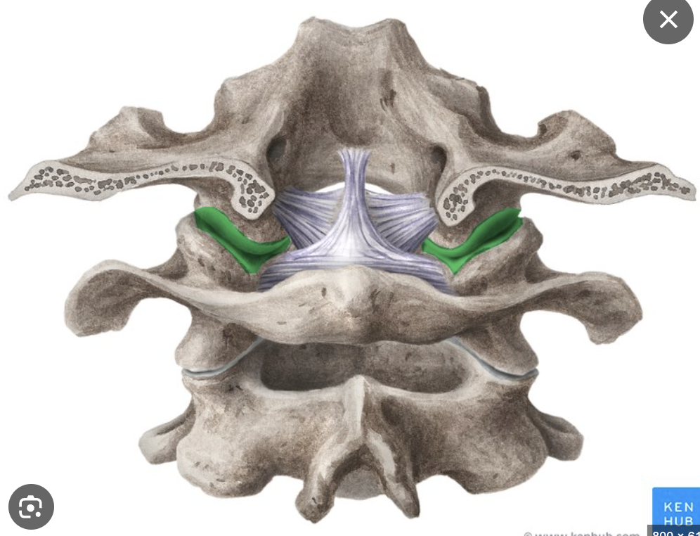
C1 structure
C1 has no body and instead forms a ring. It articulates with the occipital bone of the skull at two points called occipital condyles, forming the atlanto-occipital joint, which allows nodding ("yes") motion.
C2 structure
C2 has a special upward projection called the dens, which fits into C1 and acts like a pivot. This forms the atlanto-axial joint, which allows the head to rotate side to side ("no" motion).
What is the sacrum and coccyx, and how do they differ in females?
The sacrum (5 fused vertebrae) and coccyx (usually 4 fused) form the spine’s base and connect to the pelvis. In females, they are shorter and wider to assist childbirth.
What are the two main types of spinal joints and their functions?
Intervertebral joints (between vertebral bodies): Cartilaginous, absorb shock.
Facet (zygapophyseal) joints (between articular processes): Synovial, allow controlled movement.
What movement occurs at the atlanto-occipital joint?
Flexion and extension of the head ("yes" motion).
What is an intervertebral disc and what is a herniated disc?
Intervertebral discs cushion vertebrae and absorb shock. A herniated disc occurs when the soft center bulges out through a tear in the tough outer layer, pressing on nerves.
What do spinal ligaments do, and what are the key ones?
Spinal ligaments stabilize the spine and limit movement. Key ones are the anterior/posterior longitudinal ligaments, supraspinous, interspinous, and ligamentum nuchae.
Anterior longitudinal
Runs along the front (anterior) side of the vertebral bodies, preventing excessive backward bending (extension).
Posterior longitudinal
Runs along the back (posterior) side of the vertebral bodies inside the vertebral canal, limiting forward bending (flexion).
Supraspinous
Runs along the tips of the spinous processes down the back of the spine, connecting them together.
Interspinous
Located between adjacent spinous processes, helping control flexion and stabilize vertebrae.
Ligamentum Nuchae
A strong ligament in the neck that supports the head and helps muscles keep it upright.
What is the spinal cord and where does it end?
The spinal cord is about 45 cm long and carries sensory (afferent) and motor (efferent) signals. It ends at L1–L2, where nerve roots continue as the cauda equina.
How are spinal nerves named?
Spinal nerves are named for the vertebrae they’re near. Cervical nerves exit above their vertebrae (C1–C7), C8 exits below C7, and all others exit below their vertebrae.
What parts of the spinal cord carry sensory and motor information
The posterior (back) part carries sensory fibers; the anterior (front) carries motor fibers.
What are spinal rami and what do they supply?
Each spinal nerve splits into a large anterior ramus (front body) and a smaller posterior ramus (back and deep back muscles).
What is the sympathetic trunk?
The sympathetic trunk is a vertical chain of nerves that delivers sympathetic signals throughout the body.
Spinal flexion
Bending the spine forward, like when you touch your toes.
Spinal extension
Bending the spine backward, arching your back.
Lateral flexion
Bending the spine sideways, bringing your ear toward your shoulder.
Rotation
Twisting the spine left or right, like shaking your head “no.”
Circumduction
Moving the spine in a circular motion, combining flexion, extension, lateral flexion, and rotation.
What are the main functions of the thorax?
The thorax protects vital organs like the heart and lungs, resists internal pressure changes during breathing, and provides attachment points for upper limb muscles, supporting their weight and movement.
What forms the posterior and anterior thorax?
The posterior thorax is formed by the spine (T1–T12), with each thoracic vertebra articulating with a rib. The anterior thorax includes ribs and the sternum, with the first seven ribs attaching directly to the sternum via costal cartilage joints that allow some movement.
What are the superior and inferior thoracic apertures?
The superior thoracic aperture is the top opening of the thorax, and the inferior thoracic aperture is the bottom opening.
How do ribs articulate with the vertebrae?
Each rib’s head articulates with the vertebral bodies, usually spanning the vertebra above and below or a single vertebra via a demi-facet. The rib’s tubercle articulates with the transverse process of the vertebra. Ribs angle posteriorly around the chest.
What are the parts and joints of the sternum?
The sternum’s superior part is the manubrium, which forms the sternoclavicular joint and connects with the first ribs. The sternal angle joins the manubrium and sternum body, marks the second ribs’ attachment, and is immobile. The sternum body articulates with ribs 2–7 via costal cartilage. The inferior end is the xiphoid process.
What forms the superior thoracic aperture and what passes through it?
The superior thoracic aperture is formed by the first rib, costal cartilage, manubrium, and T1 vertebra. It allows passage of the superior lobes of the lungs and major vessels like the brachiocephalic and subclavian veins returning blood to the heart.
How are the arteries arranged on the left and right sides of the thorax?
On the left, the left ventricle and aorta give off the carotid and subclavian arteries to supply the head and arms. On the right, the brachiocephalic artery branches into the right carotid and subclavian arteries. The arterial and venous systems are asymmetrical.
What is the structure and function of the trachea?
The trachea is a rigid airway supported by D-shaped cartilage rings, located anteriorly and connected to the mouth. It allows air to reach the lungs and lies in front of the muscular esophagus, which changes shape to allow food passage.
What are the sympathetic trunks and what is their function?
The sympathetic trunks are paired vertical nerve chains running alongside the spine outside the vertebral canal, carrying sympathetic fibers that contribute to the autonomic “fight or flight” response by sending signals to peripheral organs.
Where does the phrenic nerve originate and what does it do?
The phrenic nerve originates from spinal nerves C3, C4, and C5 and innervates the diaphragm, controlling breathing by passing through the superior thoracic aperture and mediastinum.
What is the role of the vagus nerve in the thorax and abdomen?
The vagus nerve arises from the brainstem and provides parasympathetic innervation, lowering heart rate and increasing digestive activity, supporting the “rest and digest” state.
What is the thoracic duct and why is it important?
The thoracic duct is the main lymphatic vessel running along the vertebral column, draining lymph fluid into the brachiocephalic vein near the heart, essential for immune function and maintaining fluid balance.
What is the brachial plexus?
A nerve network from the upper spine (neck area) that sends branches down into the shoulder, arm, and hand to control movement and sensation.
What do the nerves from the brachial plexus innervate?
All the muscles and skin of the shoulder, arm, and hand — they control movement and feeling.
What are intercostal nerves?
Nerves from the middle spine that run between the ribs, controlling the muscles that move the rib cage (important for breathing) and some abdominal muscles.
What muscles do intercostal nerves innervate?
The intercostal muscles (between the ribs) and abdominal muscles — helping with breathing and trunk movement.
What nerves supply the deep back muscles?
Small, individual nerve branches that come directly from the spine — they don't form big networks and supply the muscles that move and stabilize the spine.
What is the phrenic nerve?
A nerve that comes from the upper spine (neck region: C3, C4, C5) and controls the diaphragm — the main breathing muscle.
Why is the phrenic nerve important?
It allows the diaphragm to contract and relax, which makes breathing possible.
What is the general pattern of spinal nerves supplying the body?
Upper spine nerves = arms and shoulders (via brachial plexus)
Middle spine nerves = ribcage and abs (via intercostal nerves)
Lower/middle back = deep back muscles
Neck nerve (phrenic) = diaphragm (breathing)
What is the thoraco-abdominal diaphragm and what does it do?
It's a dome-shaped muscle that separates the thorax from the abdomen. When it contracts, it pulls down its central tendon to increase thoracic volume and allow air into the lungs (inhalation). It relaxes to exhale.
What nerve innervates the diaphragm?
The phrenic nerve, which comes from spinal levels C3–C5.
What structures pass through the diaphragm?
The vena cava, descending aorta, esophagus, thoracic duct, azygos vein, and sympathetic trunks.
Why do the thorax and abdomen need to be separated?
So they can change volume independently. This is important for functions like breathing, coughing, sneezing, and childbirth.
What are external intercostal muscles and their function?
Muscles between the ribs that run in the same direction as external obliques. They lift the ribcage during inhalation.
What are internal intercostal muscles and their function?
Deeper rib muscles that run opposite to the externals. They pull the ribs down during forced exhalation.
What nerves innervate the intercostal muscles?
Intercostal nerves (they run between the ribs under each one, in the neurovascular bundle).
What is the difference between quiet and active breathing?
Quiet breathing uses the diaphragm and intercostals. Active breathing adds accessory muscles like sternocleidomastoid and scalenes to lift the ribcage more forcefully.
What does the sternocleidomastoid muscle do?
It turns the head side-to-side (unilaterally), flexes the head forward (bilaterally), and helps elevate the ribcage during deep breathing.
Where does the sternocleidomastoid originate and insert?
Originates at the manubrium and medial clavicle, inserts behind the ear at the mastoid process.
What do the scalene muscles do in breathing?
They help elevate the first and second ribs during deep breathing.
Where do the scalene muscles run?
From the cervical vertebrae to the first and second ribs. The brachial plexus passes between them.
What role do abdominal muscles play in breathing out?
They squeeze the abdominal cavity, pushing the diaphragm up to help force air out during coughing, sneezing, or shouting.
What structures are part of the upper respiratory tract?
Nose and pharynx — which humidify, warm, and filter air. The pharynx is shared with the mouth and splits into nasopharynx, oropharynx, and laryngopharynx.
What stops food from entering the airway when swallowing?
The epiglottis, a flap that covers the trachea during swallowing.
What is the larynx and its function?
Also known as the voice box, it controls airflow and the tension of the vocal folds to produce sound.
What structures are in the lower respiratory tract?
Lungs and mediastinum. The mediastinum includes the heart, trachea, bronchi, vessels, and esophagus.
Where does the trachea split into bronchi?
At the sternal angle, it splits into right and left main bronchi.
Why is the right bronchus steeper than the left?
Because the heart pushes the left bronchus to angle more horizontally around it.
What are alveoli and their function?
Tiny air sacs in the lungs that allow gas exchange with the blood. They provide a massive surface area like a tennis court!
How are the lungs divided?
The right lung has 3 lobes, the left lung has 2 lobes and a cardiac notch for the heart.
What is the hilum of the lung?
A depression on the medial surface where structures like bronchi, arteries, and veins enter and exit the lung.
What is the root of the lung?
The collection of structures (airways, vessels, nerves) that pass into or out of the lung at the hilum.
What are the pleura and their types?
Visceral pleura covers the lungs, parietal pleura lines the thoracic cavity. Fluid between them allows smooth lung movement.
How do lungs move if they have no muscles?
Movement is caused by the ribcage and diaphragm, and the pleural fluid keeps the lungs "stuck" to the inside of the ribcage so they follow its movement.
How is the lung blood supply organised?
Pulmonary artery: brings deoxygenated blood from the heart to the lungs
Pulmonary veins: return oxygenated blood to the heart
Bronchial arteries: from the aorta, bring oxygenated blood to the lung tissue itself
What movement occurs at the atlanto-axial joint?
Rotation of the head ("no" motion).
What are the meninges of the spinal cord?
The spinal cord is protected by three meninges: pia mater (inner), arachnoid mater (middle), and dura mater (outer, tough layer).
What movements can the spine perform, and where are they most prominent?
The spine can flex, extend, rotate, laterally flex, and circumduct. The cervical spine is best at flexion and rotation, the thoracic at rotation, and the lumbar at flexion/extension.
What is the origin of the erector spinae group?
Sacrum, iliac crest, and thoracolumbar fascia.
Where do the erector spinae muscles insert?
Iliocostalis: Ribs and lower cervical transverse processes.
Longissimus: Transverse processes and mastoid process.
Spinalis: Spinous processes.
What are the actions of the erector spinae?
Extension of spine and head, lateral flexion, and rotation.
What nerve innervates the erector spinae?
Posterior rami of spinal nerves.
What is the function of the thoracolumbar fascia?
It provides a strong aponeurotic support structure in the lower back, anchoring muscles like the latissimus dorsi and erector spinae.
Why is rotation limited in the lumbar spine?
Due to the sagittal orientation of lumbar facet joints which favour flexion/extension but restrict rotation.
What is the origin, insertion, action, and innervation of the Rectus Abdominis?
Origin: Pubic crest and pubic symphysis
Insertion: Xiphoid process and costal cartilages of ribs 5-7
Action: Flexes the trunk and compresses the abdomen
Innervation: Thoracoabdominal nerves (anterior rami of T7–T11) and subcostal nerve (T12)
What is the origin, insertion, action, and innervation of the External Oblique?
Origin: External surfaces of ribs 5-12
Insertion: Linea alba, pubic tubercle, and anterior iliac crest
Action: Compresses abdomen, flexes and rotates the trunk to the opposite side
Innervation: Thoracoabdominal nerves (T7–T11) and subcostal nerve (T12)
What is the origin, insertion, action, and innervation of the Internal Oblique?
Origin: Thoracolumbar fascia, anterior iliac crest, and lateral inguinal ligament
Insertion: Inferior borders of ribs 10-12, linea alba, and pubis
Action: Compresses abdomen, flexes and rotates trunk to the same side
Innervation: Thoracoabdominal nerves (T7–T11), subcostal nerve (T12), and first lumbar nerves (L1)
What is the origin, insertion, action, and innervation of the Transversus Abdominis?
Origin: Internal surfaces of ribs 7-12, thoracolumbar fascia, iliac crest, and lateral inguinal ligament
Insertion: Linea alba with aponeurosis of internal oblique, pubic crest
Action: Compresses abdomen
Innervation: Thoracoabdominal nerves (T7–T11), subcostal nerve (T12), and first lumbar nerves (L1)
Where is the heart located?
The heart lies in the anterior, inferior mediastinum, opposite T5–T8, directly behind the sternum. It crosses the midline and sits on the diaphragm.
What are the layers covering the heart?
The heart is covered by the pericardium — a double sac with an outer fibrous layer and an inner serous layer. Between them is pericardial fluid, which reduces friction during movement.
What are the key features of the heart?
The heart is a hollow, muscular organ made of cardiac muscle. It has 4 chambers and a pacemaker that controls its rhythm. It is about 12 cm long, 9 cm wide, and lies obliquely.