BioL121 Quiz
1/13
There's no tags or description
Looks like no tags are added yet.
Name | Mastery | Learn | Test | Matching | Spaced |
|---|
No study sessions yet.
14 Terms
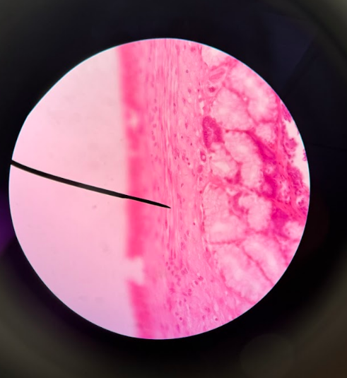
Frog Skin:
stratified squamous epithelium
many layers of epidermis
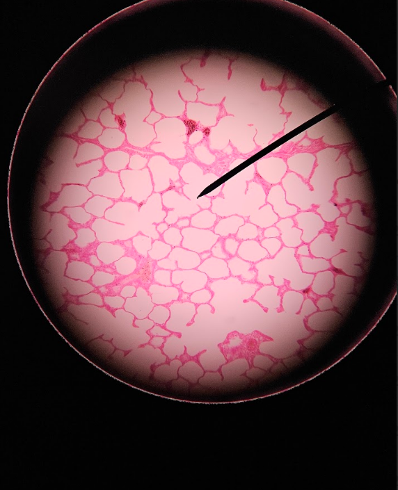
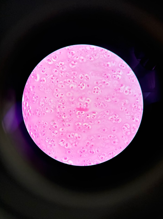
Lung (Human):
simple squamous epithelium
thin layer of epidermis (one singular layer)
has “holes” of empty space
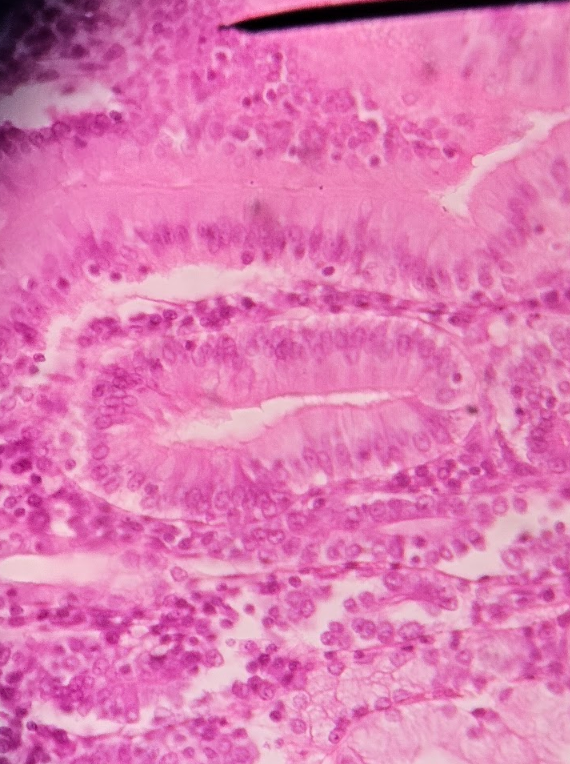
Duodenum:
columnar epithelium
elongated cells
Skin Tissue: Hair Follicle:
follicle and hair bulb
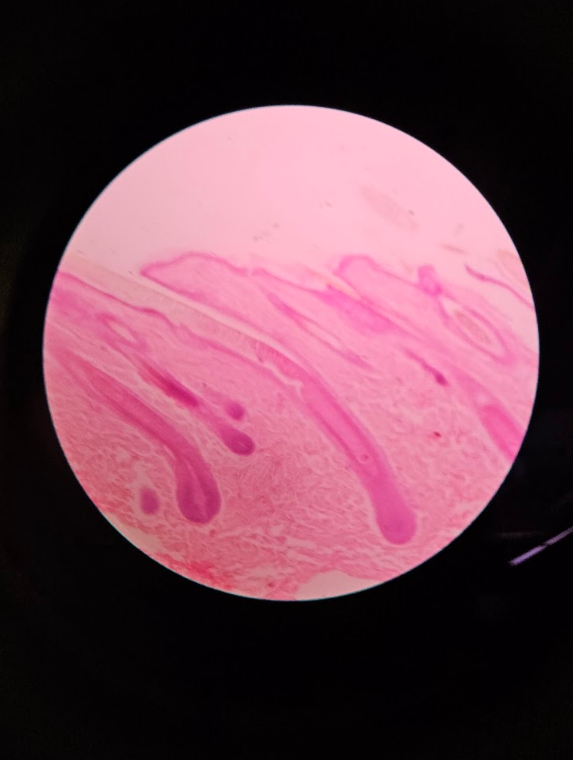
Skin Tissues: Sole of Foot:
thick layer of epidermis (darker than dermis because it is a thick layer of DEAD skin cells)
Skin Tissues: Footpad Squirrel:
thin layer of epidermis and many cells
very similar to the human sole of foot, just fewer dead skin cells → making a thinner epidermis layer
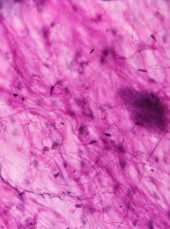
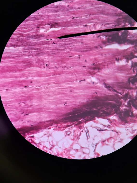
Connective Tissues: Areolar Tissue:
stringy fibers
dark think “strings” = elastic fibers
thick lighter “strings” = collagen fibers
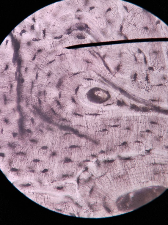
Connective Tissues: Bone Tissue:
black and white; arranged in circled format
osteocyte (in lacuna) = dark dashes around Haversian Canal
Haversian Canal = whiteish circles/blank spaces in the middle of an osteon
Osteon = the entire group of the haversian canal surrounded by all the osteocytes
Canaliculi = hair-like cracks that are near osteocyte
Ossified Matrix = white space between osteons
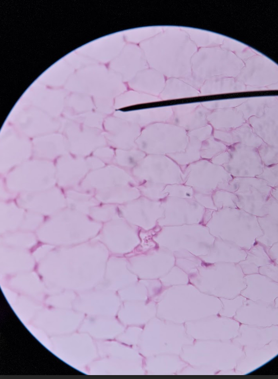
Connective Tissues: Adipose Tissue:
empty spaces → plasma membrane is what stained rather than the actual cell
emptier than the lung tissue
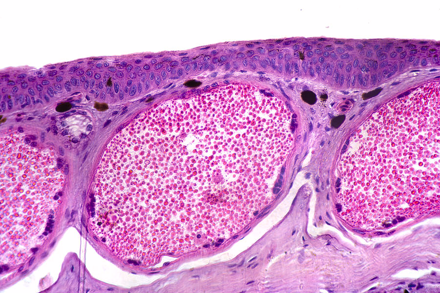
Connective Tissues: Cartilage Tissue:
thin strip with many circles/holes
Chondrocyte = dark “cell-like” shape inside lacuna
Lacuna = small little “ball-like” structures w/dark spots inside
Matrix = empty space surrounding lacuna
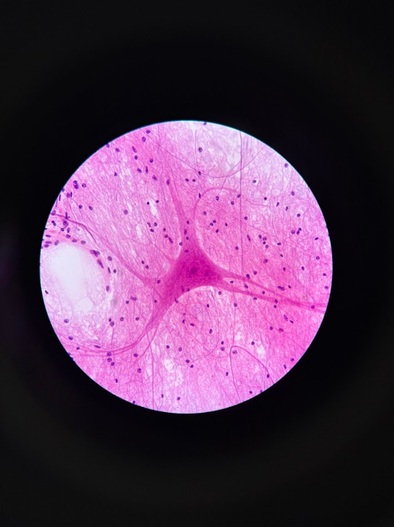
Nervous Tissue: Ox Spinal Cord:
Neurons = bigger blobs w/tentacle-like arms
Glial cells = small little “dots”
Axons = transmits signal away from neuron
Dendrites = receives signals toward the cell
Impossible to tell which of the arms are axons or dendrites without context
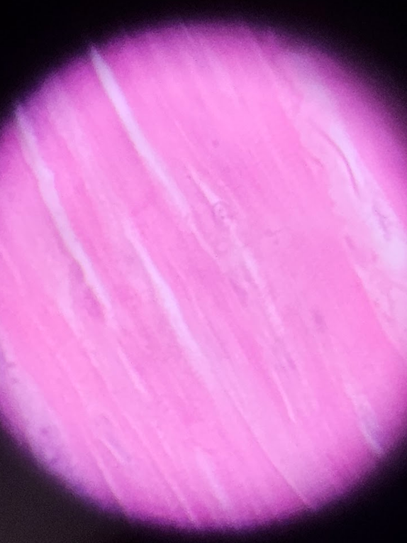
Muscle Tissues: Skeletal Muscle:
VOLUNTARY MUSCLE
many tightly packed rows of squiggly lines of cells
darker in color (purplish)
tube-like format
very coarse and rough looking
Muscle Tissues: Cardiac Tissue:
Branched tubular fibers
Less rows w/more purplish cells
Muscle fibers are more visible (pink in color)
Muscle Tissues: Smooth Muscle:
INVOLUNTARY MUSCLE
Thick layer on edge of skin
Looks similar to cardiac muscle in formatting
Has squiggly lines of purplish color