Exam 2: Knee joint

0.0(0)
Studied by 0 people
call kaiCall Kai
Locked
learnLearn
examPractice Test
spaced repetitionSpaced Repetition
heart puzzleMatch
flashcardsFlashcards
GameKnowt Play
Card Sorting

1/53

encourage image

There's no tags or description

Looks like no tags are added yet.

Last updated 2:25 AM on 7/26/26
Name
Mastery
Learn
Test
Matching
Spaced
Call with Kai
Chat

No analytics yet

Send a link to your students to track their progress

54 Terms

1
New cards

Knee Joint Overview

(bicondylar) 2 Joints in 1 capsule

  • Tibiofemoral

  • Patellofemoral

Contradictory mobility and stability demands

2
New cards

Tibiofemoral Joint

  • Bicondylar (or double condyloid) synovial joint

  • Degrees of freedom

    • 2 rotatory available

    • 1 obvious DoF in function

  • Convex femoral condyles/concave tibial plateau
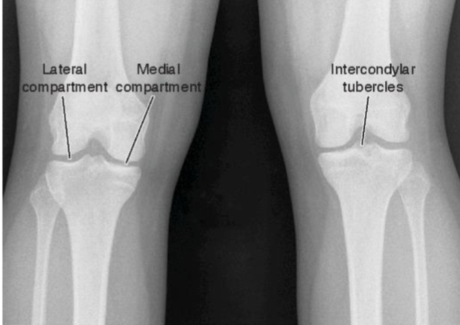
<ul><li><p><span><strong>Bicondylar (or double condyloid) synovial joint</strong></span></p></li></ul><p></p><ul><li><p><span><strong>Degrees of freedom</strong></span></p><ul><li><p><span> 2 rotatory available</span></p></li><li><p><span> 1 obvious DoF in function</span></p></li></ul></li></ul><p></p><ul><li><p><span><strong>Convex femoral condyles/concave tibial plateau</strong></span></p></li></ul><p></p>
3
New cards

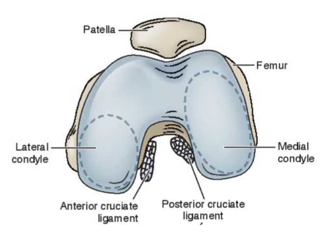
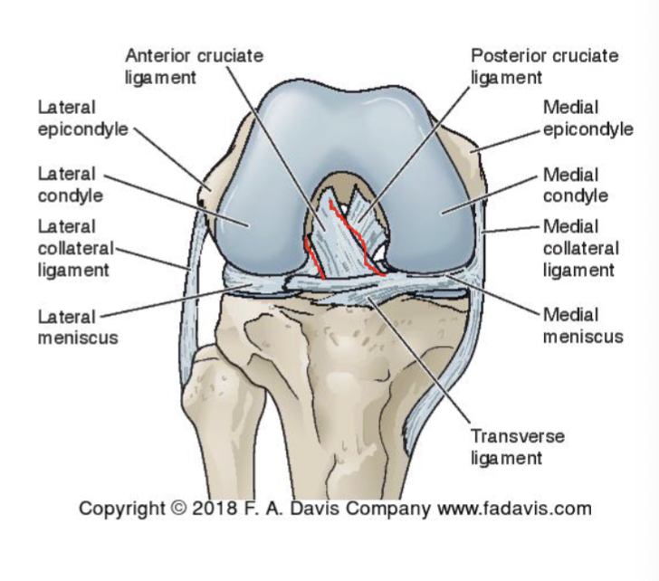
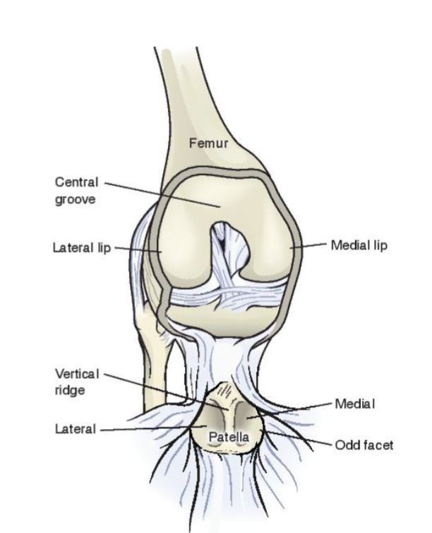
Femur

Medial and lateral condyles form articulating surfaces

  • Larger medial condyle (larger articulating surface)

  • Separated by intercondylar notch

  • Contains articulating surface for PFJ

<p><span>Medial and lateral condyles form articulating surfaces</span></p><ul><li><p><span><strong>Larger medial condyle</strong> (larger articulating surface)</span></p></li><li><p><span>Separated by intercondylar notch</span></p></li><li><p><span>Contains articulating surface for PFJ</span></p></li></ul><p></p>
4
New cards

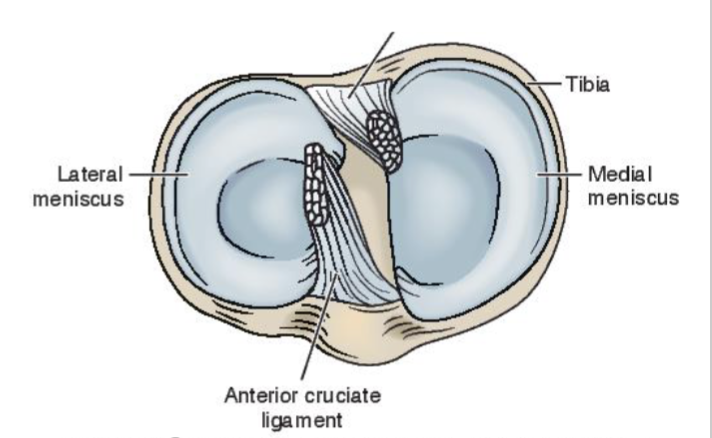
Tibia

Flat tibial condyles/plateaus

  • Longer medially (A/P direction)

  • Slopes posteriorly

  • Separated by bony spines

<p><span>Flat tibial condyles/plateaus</span></p><ul><li><p><span>Longer medially (A/P direction)</span></p></li><li><p><span>Slopes posteriorly</span></p></li><li><p><span>Separated by bony spines</span></p></li></ul><p></p>
5
New cards

Tibiofemoral Articulation

Closed pack = full extension

  • good bony congruency + stability

<p>Closed pack = full extension</p><ul><li><p>good bony congruency + stability </p></li></ul><p></p>
6
New cards

We have convex femoral condyles with a flat tibial
plateau - WHAT DOES THIS JOINT NEED TO BE FUNCTIONALLY
STABLE?!!!!!

  • joint capsule, ligaments → static (No control of it)

  • tendon, muscle → dynamic (motor control of knee)

7
New cards

Deviations from Ideal posture

LoG is anterior → wanting extension

Tension forces give internal moment of flexion to stop excessive extension
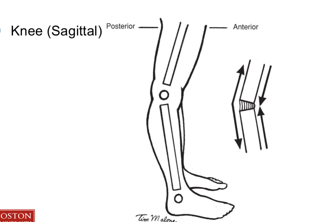
<p>LoG is anterior → wanting extension </p><p></p><p>Tension forces give internal moment of flexion to stop excessive extension</p>
8
New cards

Tibiofemoral Alignment

Knee (Sagittal)

  • knee surgery, injury —> No full extension

  • Quads = Internal moment for extension

  • Soleus = Internal moment for plantarfexion
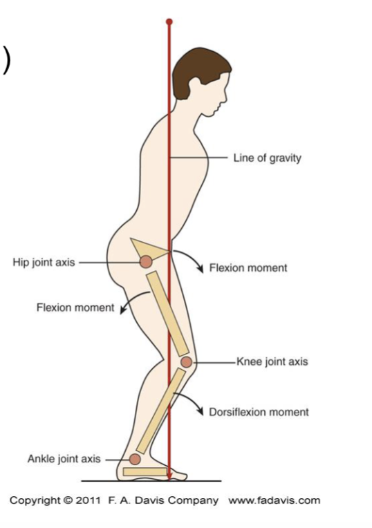
<p><span>Knee (Sagittal)</span></p><ul><li><p>knee surgery, injury  —&gt; No full extension </p></li><li><p>Quads = Internal moment for extension </p></li><li><p>Soleus = Internal moment for plantarfexion</p></li></ul><p></p>
9
New cards
<p>Tibiofemoral angulation </p>

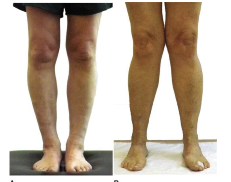
Tibiofemoral angulation

Normal angulation:

  • some vara

  • femur is more oblique for femoral condyles to be in the same plane

Genu valgum:

  • > 180 dg

  • lateral tensile stress

Genu vara:

  • < 180 dg

  • medial compressive stress (hyaline cartilage + meniscus)
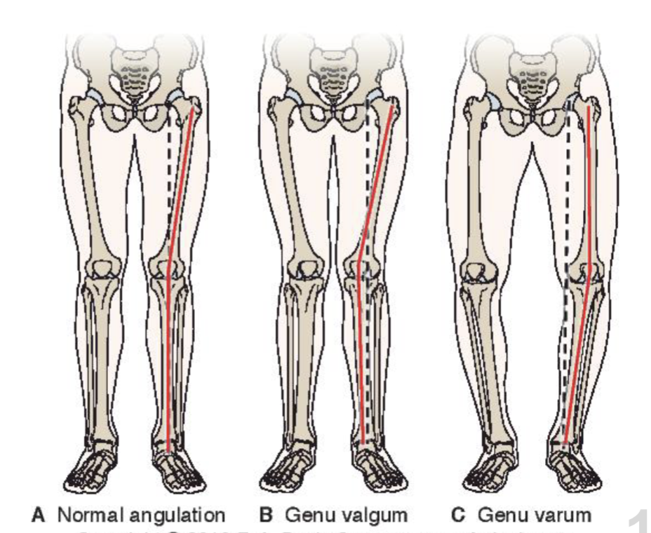
<p><strong>Normal angulation:</strong></p><ul><li><p>some vara </p></li><li><p>femur is more oblique for femoral condyles to be in the same plane</p></li></ul><p></p><p><strong>Genu valgum:</strong></p><ul><li><p>&gt; 180 dg</p></li><li><p>lateral tensile stress</p></li></ul><p></p><p></p><p><strong>Genu vara:</strong></p><ul><li><p>&lt; 180 dg</p></li></ul><ul><li><p>medial compressive stress (hyaline cartilage + meniscus)</p></li></ul><p></p>
10
New cards

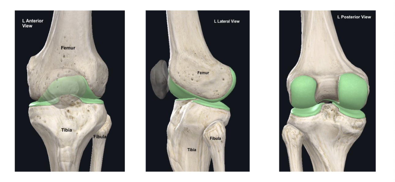
Role of Meniscus

Roles:

  • Increase the concavity of the
    tibial articulating surfaces

    • good for femoral condyles

  • Distribution of WB’ing forces

    • Stress → gives wider surface area → low WBing stress to hyaline cartilage

  • Reducing friction between articulating surfaces (tibia + femur)

  • Shock absorbers to decrease load on articular cartilage (can
    assume over 50% of load)
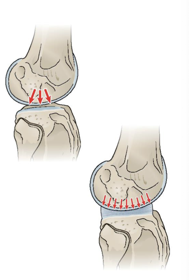
<p><span>Roles:</span></p><ul><li><p><span>Increase the concavity of the<br>tibial articulating surfaces</span></p><ul><li><p>good for femoral condyles </p></li></ul></li><li><p><span>Distribution of WB’ing forces</span></p><ul><li><p><span>Stress   → gives wider surface area → low WBing stress to hyaline cartilage</span></p></li></ul><p></p></li><li><p><span>Reducing friction between articulating surfaces (tibia + femur) </span></p></li></ul><p></p><ul><li><p><span>Shock absorbers to decrease load on articular cartilage (can<br>assume over 50% of load)</span></p></li></ul><p></p>
11
New cards

Meniscus structure


Semicurcular fibrocartilagenous discs
Wedge shaped (thick peripherally, thin centrally). —> inside
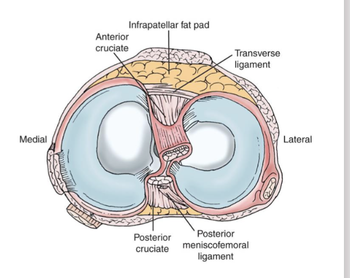
<p><span><br></span><span data-name="black_small_square" data-type="emoji">▪</span><span> Semicurcular fibrocartilagenous discs<br></span><span data-name="black_small_square" data-type="emoji">▪</span><span> Wedge shaped (thick peripherally, thin centrally).    —&gt; inside<br></span></p>
12
New cards

Menisci (Nutrition + Innervation)

Nutrition

  • Synovial fluid primarily

  • Only the outer 1/3 is vascular

  • Capacity to heal/regenerate?

    • outside can be healed

Innervation

  • Nociceptors

  • Mechanoreceptors

    • Proprioceptive function: communicate w/ CNS via sensory messages → dynamic stability

13
New cards

Meniscal Attachments

Medial
Tibia
Patella
Medial capsule
MCL
ACL
PCL
Semimembranosus

Lateral
Tibia
Patella
ACL
PCL
Femur (meniscofemoral ligament)
Popliteus

14
New cards

Joint Capsule

  • Close packed position= full extension

  • Loose packed position= 25-30 degrees flexion

  • Contains both the PFJ and tibiofemoral joints

  • Innervated w/ nociceptors and mechanoreceptors

    • Proprioceptive function

  • 2 Layers

    • Superficial fibrous layer

    • Deep synovial layer

15
New cards
<p>Joint Capsule fibrous layer</p>

Joint Capsule fibrous layer

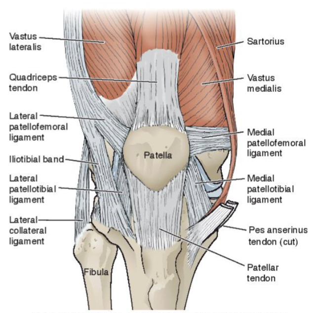
Anterior Portion

  • Spans length of the patella

  • Extensor Retinaculum

    • Medial retinaculum (MPFL)

    • Lateral retinaculum (LPFL)

Posterior Portion

  • femoral condyles to tibial condyles

  • reinforced by arcuate ligament (lateral) and oblique popliteal ligament (medial)
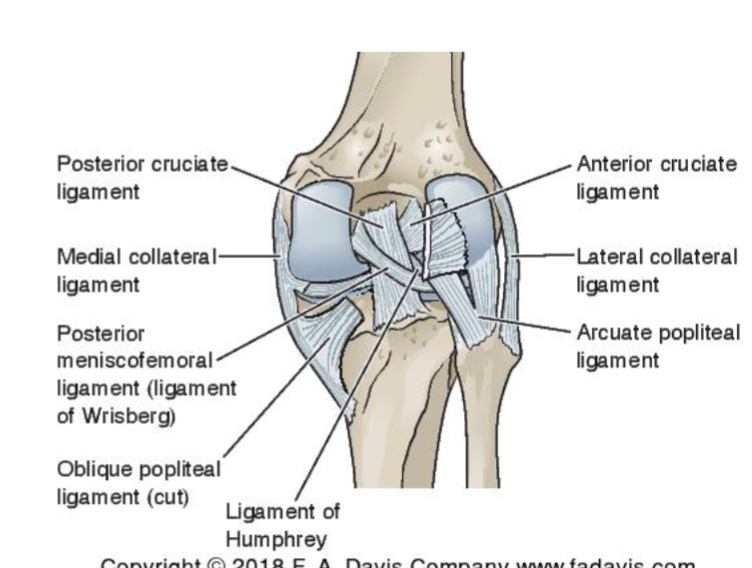
<p><span><strong>Anterior Portion</strong></span></p><ul><li><p><span>Spans length of the patella</span></p></li><li><p><span>Extensor Retinaculum</span></p><ul><li><p><span>Medial retinaculum (MPFL)</span></p></li><li><p><span>Lateral retinaculum (LPFL)<br></span></p></li></ul></li></ul><p><strong>Posterior Portion</strong></p><ul><li><p>femoral condyles to tibial condyles</p></li><li><p>reinforced by arcuate ligament (lateral) and oblique popliteal ligament (medial) </p></li></ul><p></p><p></p><p></p>
16
New cards

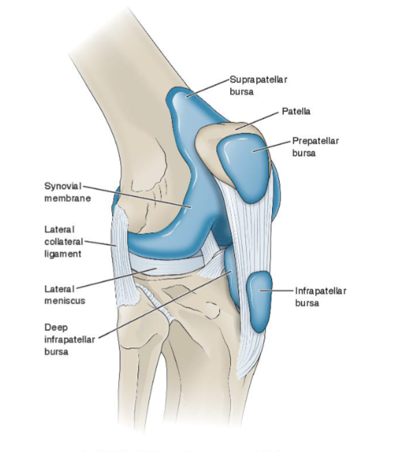
Joint capsule


Intracapsular

  • Everything inside of the fibrous capsule

  • ex: hyaline cartilage and deeper parts of lateral meniscus are intrasynovial getting nutrition from synovial fluid

Extrasynovial

  • Inside the fibrous capsule but outside of the synovial layer
    ACL/PCL
    Fat pads

  • they get nutrition from blood supply

<p><span><br><strong>Intracapsular</strong></span></p><ul><li><p><span>Everything inside of the fibrous capsule</span></p></li><li><p><span>ex: hyaline cartilage and deeper parts of lateral meniscus are intrasynovial getting nutrition from synovial fluid </span></p></li></ul><p></p><p></p><p></p><p><span><strong>Extrasynovial</strong></span></p><ul><li><p><span> Inside the fibrous capsule but outside of the synovial layer<br></span><span data-name="black_small_square" data-type="emoji">▪</span><span> ACL/PCL<br></span><span data-name="black_small_square" data-type="emoji">▪</span><span> Fat pads</span></p></li><li><p><span>they get nutrition from blood supply </span></p></li></ul><p></p>
17
New cards

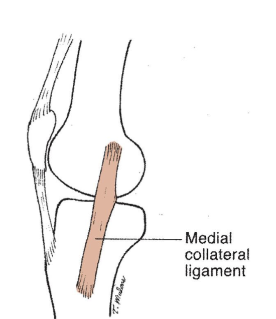
MCL

  • Superficial (origin)

    • Medial epicondyle of femur to pes anserinus

  • Deep (insertion)

    • Continuous w/ joint capsule

    • Firmly attached to medial meniscus


Direct blood supply

<p></p><ul><li><p>Superficial (origin)</p><ul><li><p>Medial epicondyle of femur to pes anserinus</p></li></ul><p></p></li><li><p>Deep (insertion)</p><ul><li><p>Continuous w/ joint capsule</p></li><li><p>Firmly attached to medial meniscus</p></li></ul></li></ul><p><br><span data-name="black_small_square" data-type="emoji">▪</span> Direct blood supply</p><p></p>
18
New cards

MCL Role

Primary:

  • resists genu valgus stress via joint rxn force

  • resists ER of tibia

Secondary:

  • resists anterior shear (implicates ACL inury)

  • works best in full extension and important in slight flexion

Injury of MCL: valgus stress
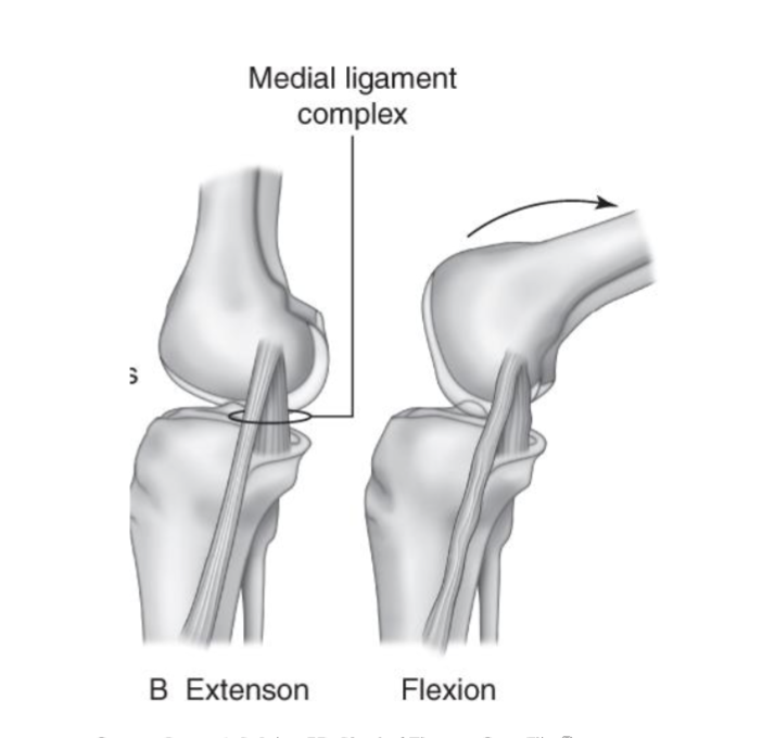
<p><strong>Primary:</strong></p><ul><li><p>resists genu valgus stress via joint rxn force </p></li><li><p>resists ER of tibia</p></li></ul><p></p><p><strong>Secondary:</strong></p><ul><li><p>resists anterior shear (implicates ACL inury)</p></li><li><p>works best in full extension and important in slight flexion</p></li></ul><p></p><p><strong>Injury of MCL: </strong>valgus stress</p>
19
New cards

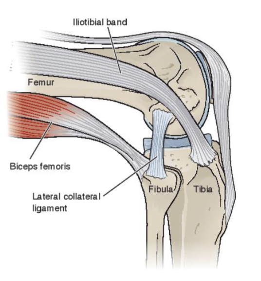
LCL

  • Lateral femoral condyle to fibular head

  • Joins biceps femoris at attachment

  • Separate from the joint capsule

  • Blood supply

<ul><li><p><span>Lateral femoral condyle to fibular head</span></p></li><li><p><span>Joins biceps femoris at attachment</span></p></li><li><p><span>Separate from the joint capsule</span></p></li><li><p><span>Blood supply</span></p></li></ul><p></p>
20
New cards

Role of LCL

  • resists genu varum stress

  • resists ER of tibia (b/c insertion is further from origin and is @ more of angle)

  • Best work in full extension and important in slight flexion

21
New cards

ACL (Anterior Cruciate ligament)


Proximal attachment= posterior medial aspect of lateral femoral condyle

Distal attachment= lateral edge of medial tibial spine


2 Bands

  • Anterior/medial

  • Posterior/lateral

Direct blood supply

<p><span><br><strong>Proximal attachment= </strong>posterior medial aspect of lateral femoral condyle<br></span></p><p><span><strong>Distal attachment=</strong> lateral edge of medial tibial spine</span></p><p></p><p><span><br><strong>2 Bands</strong></span></p><ul><li><p><span>Anterior/medial</span></p></li><li><p><span>Posterior/lateral</span></p></li></ul><p></p><p><span><strong>Direct blood supply</strong></span></p><p></p>
22
New cards

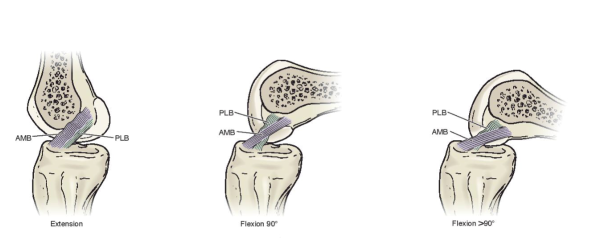
ACL in multiple motions

Extension:

  • PLB is more taut

Flexion 90 dg:

  • both PLB and AMB are NOT taut and in loose pack

Flexion > 90 dg:

  • AMB is more taut

<p><strong>Extension:</strong></p><ul><li><p>PLB is more taut </p></li></ul><p></p><p><strong>Flexion 90 dg:</strong></p><ul><li><p>both PLB and AMB are NOT taut and in loose pack</p></li></ul><p></p><p><strong>Flexion &gt; 90 dg:</strong></p><ul><li><p>AMB is more taut</p></li></ul><p></p>
23
New cards

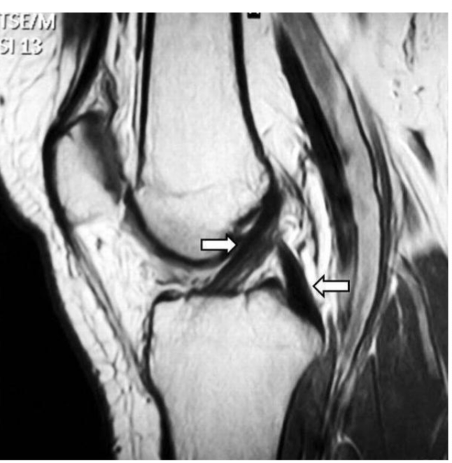
Role of ACL

Primary:

  • resist anterior shear on tibia (b/c insertion is further from origin)

  • resist transverse and frontal combination
<p>Primary:</p><ul><li><p>resist anterior shear on tibia (b/c insertion is further from origin)</p></li><li><p>resist transverse and frontal combination</p></li></ul><p></p>
24
New cards

What leads to ACL tear

  • loose pack (slightly flex) w/ valgus, IR of tibia

25
New cards

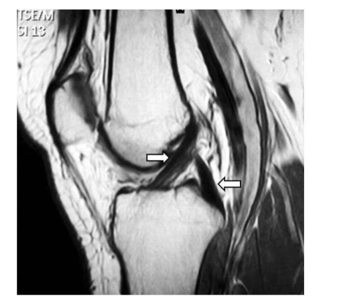
PCL’s role

Primary:

  • resist posterior shear of tibia (b/c Insertion is further from origin)

Secondary:

  • resist ER of tibia

Impact of Hamstring contraction
<p>Primary:</p><ul><li><p>resist posterior shear of tibia (b/c Insertion is further from origin) </p></li></ul><p></p><p>Secondary:</p><ul><li><p>resist ER of tibia </p></li></ul><p></p><p>Impact of Hamstring contraction</p><p></p>
26
New cards

What tears PCL?

direct force anterior

27
New cards

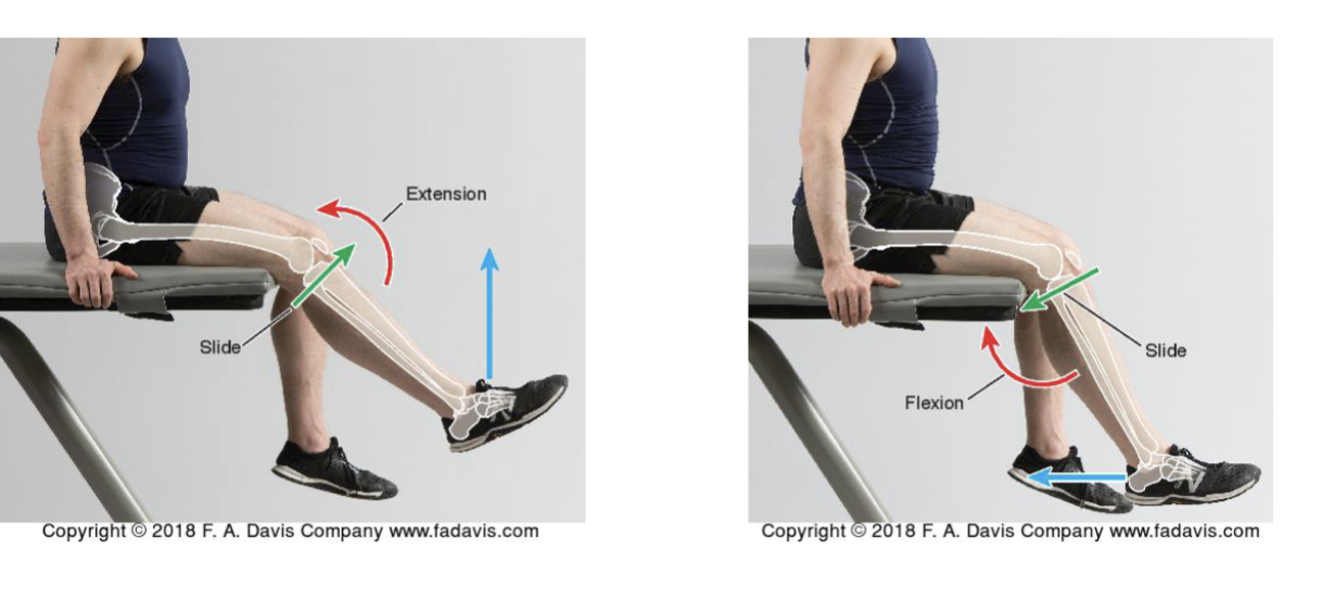
Tibiofemoral Joint Function

2 Rotational DoF
Flex/Ext
ER/IR
Ab/Add (valgus/varus - rot in frontal plane)

  • ABD → distal end of tibia (knee valgus) and NO muscle control of it, so not really DoF

  • ABD → distal end of tibia (knee varus) and NO muscle control of it, so not really DoF


Associated rolling and glide
28
New cards
term image
knowt flashcard image
29
New cards
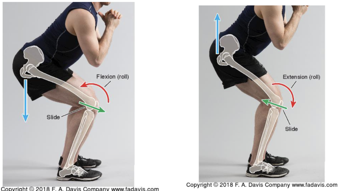
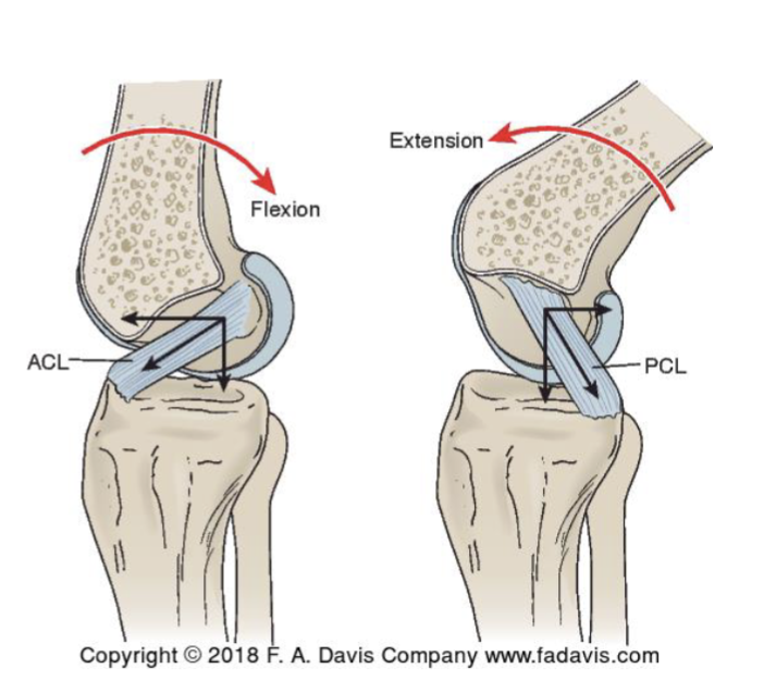
<p>Flex/Ext of Tibiofemoral Joint</p>

Flex/Ext of Tibiofemoral Joint

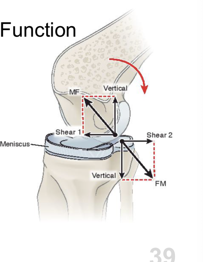
Role of the meniscus
Wedge shaped
Assists in gliding
Deformable
- Post during flexion
- Ant fduring ext.

rxn force from meniscus helps glide of femur

Role of cruciate ligaments

  • ACL helps w/ ant glide of femur during flexion

  • PCL helps w/ post glide of the femur during ext
<p><span>Role of the <strong>meniscus</strong><br></span><span data-name="black_small_square" data-type="emoji">▪</span><span> Wedge shaped<br></span><span data-name="black_small_square" data-type="emoji">▪</span><span> Assists in gliding<br></span><span data-name="black_small_square" data-type="emoji">▪</span> Deformable<br>    - Post during flexion<br>     - Ant f<span>during ext. </span></p><p>rxn force from <strong>meniscus helps glide</strong> of femur </p><p></p><p></p><p></p><p><span>Role of cruciate ligaments</span></p><ul><li><p><span><strong>ACL </strong>helps w/ <strong>ant glide</strong> <strong>of femur</strong> during<strong> flexion</strong></span></p></li><li><p><span><strong>PCL </strong>helps w/ <strong>post glide of the femur </strong>during <strong>ext</strong></span></p></li></ul><p></p><p></p><p></p>
30
New cards

Internal/External and Rotation (Med/Lat) of Tibiofemoral Joint

  • Transverse plane

  • Max rotation available at 90 deg of flex

  • Tibial ER needed for full knee extension (open chain)

    • medial part needs to go more anteriorly b/c it is longer

    • for closed chain

      • tibial IR needed for full extension

ER → posterior glide of lateral parts + anterior glide of medial parts
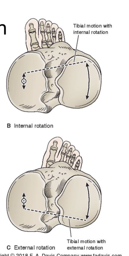
<ul><li><p><span>Transverse plane</span></p></li><li><p><span>Max rotation available at 90 deg of flex</span></p></li><li><p><span>Tibial ER needed for full knee extension (<strong>open chain) </strong></span></p><ul><li><p> medial part needs to go more anteriorly b/c it is longer</p></li></ul><p></p><ul><li><p>for <strong>closed chain</strong></p><ul><li><p>tibial IR needed for full extension</p></li></ul></li></ul></li></ul><p></p><p></p><p>ER → posterior glide of lateral parts + anterior glide of medial parts </p>
31
New cards

Knee flexors

  • hamstrings

  • medial knee flexors → internal rotation (brings medial side back)

  • lateral knee flexors → internal rotation

32
New cards

Active / Passive Insufficiency

Active insufficiency of hamstrings or passive insufficiency of RF
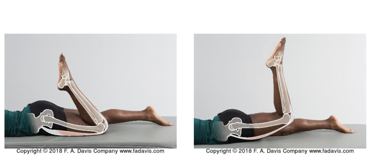
<p>Active insufficiency of hamstrings or passive insufficiency of RF </p>
33
New cards

Flexor group

Fy = rotate tibia and posterior shear

Fx = compression
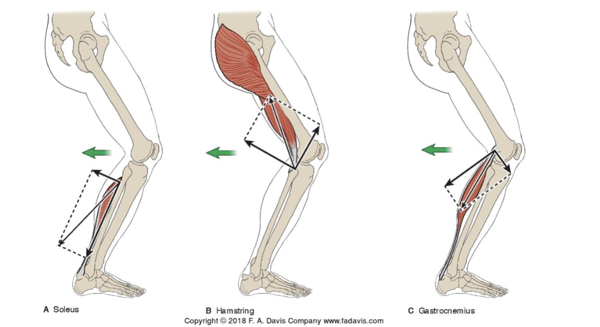
<p>Fy = rotate tibia and posterior shear</p><p></p><p>Fx = compression</p>
34
New cards

Extensor group of knee

Quadriceps

patella goes up and lateral and compress patellofemoral joint
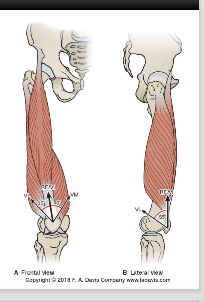
<p>Quadriceps </p><p></p><p>patella goes up and lateral and compress patellofemoral joint </p>
35
New cards

Quads (patella)

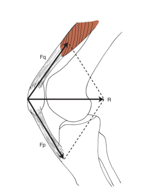
Patella

  • Anatomical pulley to increase MA of Quads

  • Peak MA and peak force (length/tension) occur between 45 and 60 deg of flexion

    • Impact on torque production: 45-60 dg have a lot of force from quads

  • No patella → smaller MA needing more force from quads
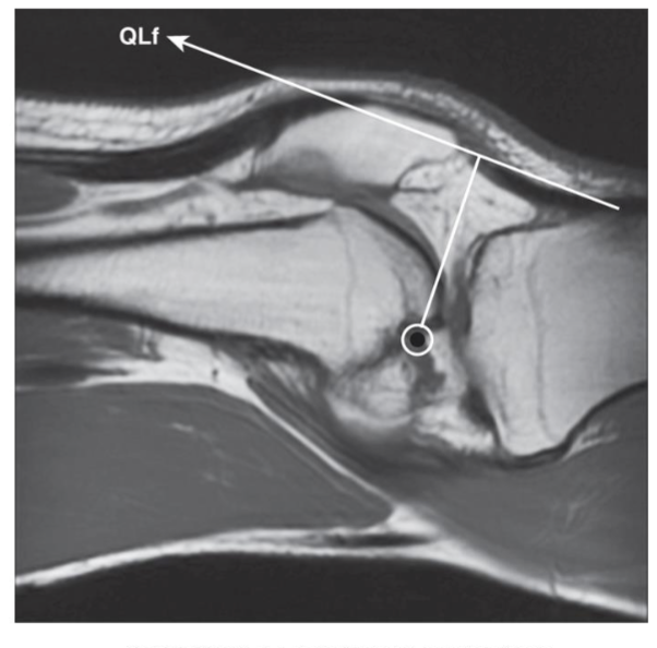
<p><span><strong>Patella</strong></span></p><ul><li><p><span>Anatomical pulley to increase MA of Quads</span></p></li><li><p><span>Peak MA and peak force (length/tension) occur between 45 and 60 deg of flexion</span></p><ul><li><p><span><strong>Impact on torque production: </strong>45-60 dg have a lot of force from quads</span></p></li></ul></li></ul><p></p><ul><li><p>No patella → smaller MA needing more force from quads </p></li></ul><p></p>
36
New cards

Quad in flexion

Fy (anterior shear + flexion)

Fx (parallel → compress tibia into femur)

@ 60 flexion → quads do NOT make anterior shear → No strain to ACL
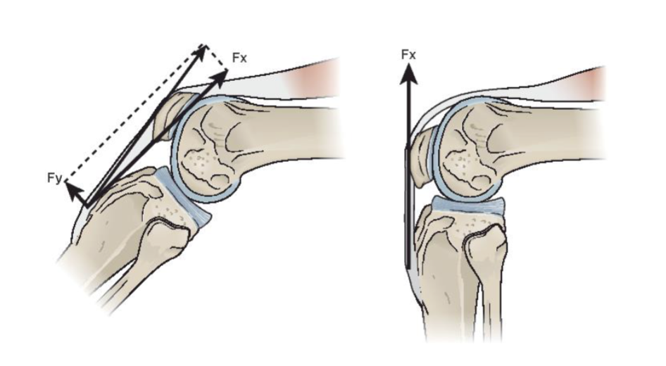
<p>Fy (anterior shear + flexion) </p><p></p><p>Fx (parallel → compress tibia into femur) </p><p></p><p>@ 60 flexion → quads do NOT make anterior shear → No strain to ACL</p>
37
New cards

Patellofemoral Joint

The Patella

  • Triangular shaped bone articulating with the femoral condyles/sulcus

    • Vertical ridge separating into medial and lateral facets

      • helps w/ bony congruency

    • Odd facet

  • Subject to high compressive
    forces (hyaline cartilage @ back of patella

  • Anatomical pulley for the
    quadriceps

  • Very incongruent joint

<p><span>The Patella</span></p><ul><li><p><span>Triangular shaped bone articulating with the femoral condyles/sulcus</span></p><ul><li><p><span>Vertical ridge separating into medial and lateral facets</span></p><ul><li><p>helps w/ bony congruency</p></li></ul></li><li><p><span>Odd facet</span></p></li></ul></li></ul><p></p><ul><li><p><span>Subject to high compressive<br>forces (hyaline cartilage @ back of patella</span></p></li></ul><p></p><ul><li><p><span> Anatomical pulley for the<br>quadriceps</span></p></li></ul><p></p><ul><li><p><span>Very incongruent joint</span></p></li></ul><p></p>
38
New cards

Congruence and Contact Pressures

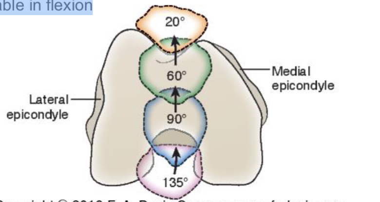
Little to no contact in extension

  • Variations in starting position

  • Implications for stability: increased patella + femur contact press w/ flex

Variable contact pressures in flexion

  • More stable in flexion (60-90 dg)

<p><span><strong>Little to no contact in extension</strong></span></p><ul><li><p><span>Variations in starting position</span></p></li><li><p><span>Implications for stability: <strong>increased </strong>patella + femur contact press w/ <strong>flex</strong><br></span></p></li></ul><p><span><strong>Variable contact pressures in flexion</strong></span></p><ul><li><p><span>More stable in flexion (60-90 dg)</span></p></li></ul><p></p>
39
New cards

Motions of Patella

Knee Flexion
Inferior glide
Medial rotation
Medial glide

Knee Extension
Superior glide
Lateral rotation
Lateral glide

**Apex of the patella follows the tibial tubercle**

40
New cards

Frontal Plane Stability

At rest in extension…
Normal valgus angle
Shallow superior sulcus
Lateral line of pull from quads


During flexion…
Increased stability as patella glides inferior into deeper sulcus

41
New cards

More about frontal plane stability (longitudinal + transverse stabilizers)

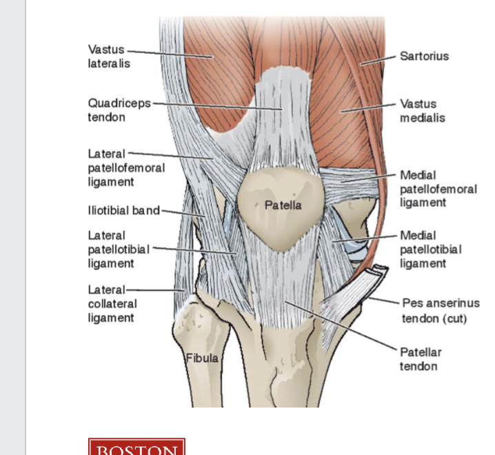
Longitudinal stabilizers
Patella tendon
Quad tendon
Patellotibial ligaments

Transverse stabilizers
Extensor retinaculum
MPFL
Lateral lip of sulcus
Muscle balance

<p><span><strong>Longitudinal stabilizers</strong><br></span><span data-name="black_small_square" data-type="emoji">▪</span><span> Patella tendon<br></span><span data-name="black_small_square" data-type="emoji">▪</span><span> Quad tendon<br></span><span data-name="black_small_square" data-type="emoji">▪</span><span> Patellotibial ligaments<br></span></p><p><span><strong>Transverse stabilizers</strong><br></span><span data-name="black_small_square" data-type="emoji">▪</span><span> Extensor retinaculum<br></span><span data-name="black_small_square" data-type="emoji">▪</span><span> MPFL<br></span><span data-name="black_small_square" data-type="emoji">▪</span><span> Lateral lip of sulcus<br></span><span data-name="black_small_square" data-type="emoji">▪</span><span> Muscle balance</span></p>
42
New cards

PFJ Stresses

Stress= increased Force/unit area

  • influenced by quadriceps activity (force) primarily!!!

    • to find joint rxn force of patella

<p><span>Stress=  increased Force/unit area</span></p><ul><li><p><span>influenced by quadriceps activity (force) primarily!!! </span></p><ul><li><p>to find joint rxn force of patella</p></li></ul></li></ul><p></p>
43
New cards

Knee flexion angle for PF

Knee flexion angle matters somewhat in that it adds passive quadriceps tension
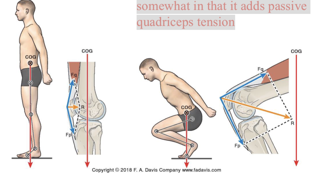
<p><span>Knee flexion angle matters somewhat in that it adds passive quadriceps tension</span></p>
44
New cards

Open chain (do EMA, IMA, Quad force, ACL strain)
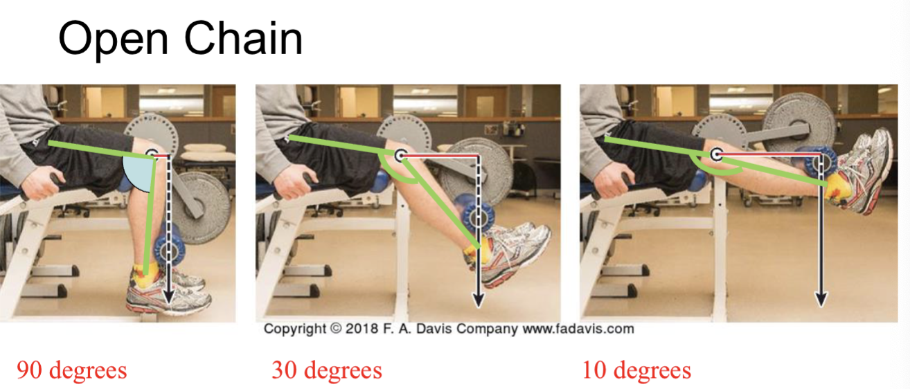
knowt flashcard image
45
New cards

Close to ___ knee flexion, has high ACL strain

0 dg

46
New cards

Closed chain (do EMA, IMA, Quad force, ACL strain)
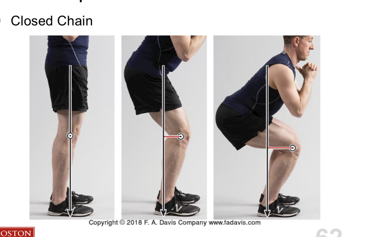
<p></p>
47
New cards

Closed chain pt 2 (do EMA, IMA, Quad force, PF stress)
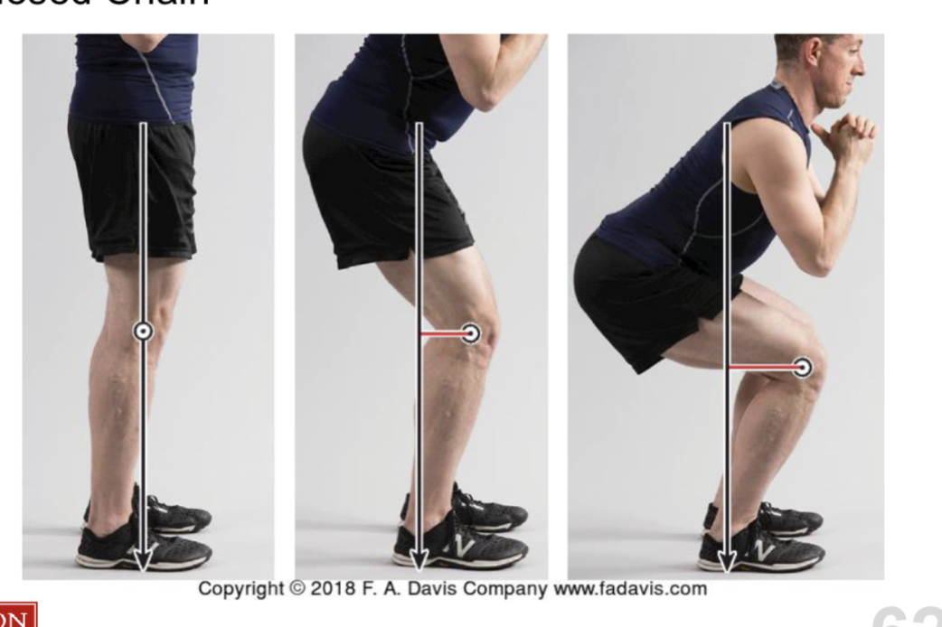
knowt flashcard image
48
New cards

Implications for Exercise

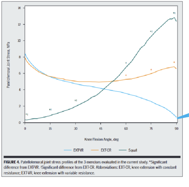
(Where should our patients with PFPS be exercising in closed chain?)

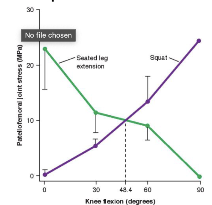
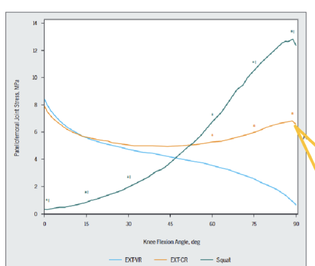
< 45 flexion → less PF stress in squat

> 45 flexion → more PF stress in squat
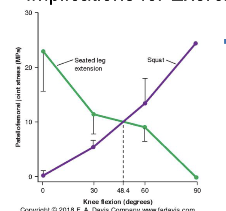
<p>&lt; 45 flexion → less PF stress in squat  </p><p>&gt; 45 flexion → more PF stress in squat</p><p></p>
49
New cards
<p>Why is the slope getting small going from 30 -60</p>

Why is the slope getting small going from 30 -60

b/c MA of Quad is greatest → not needing a lot of force → less of PFJ force
50
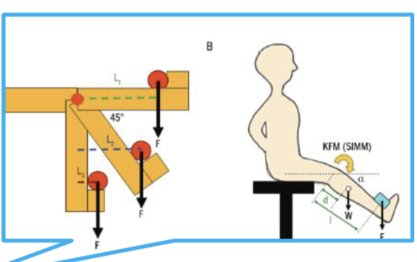
New cards
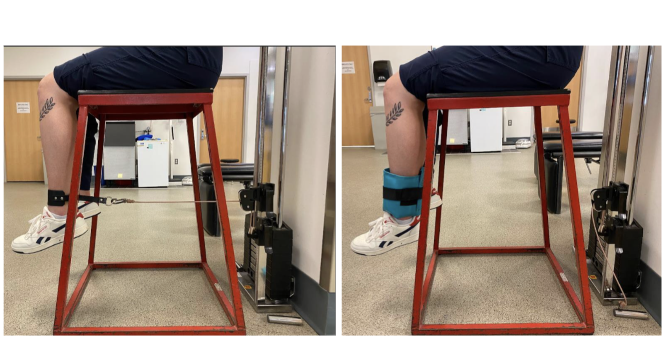
<p>For these open chain, which would require more quad force at this position</p>

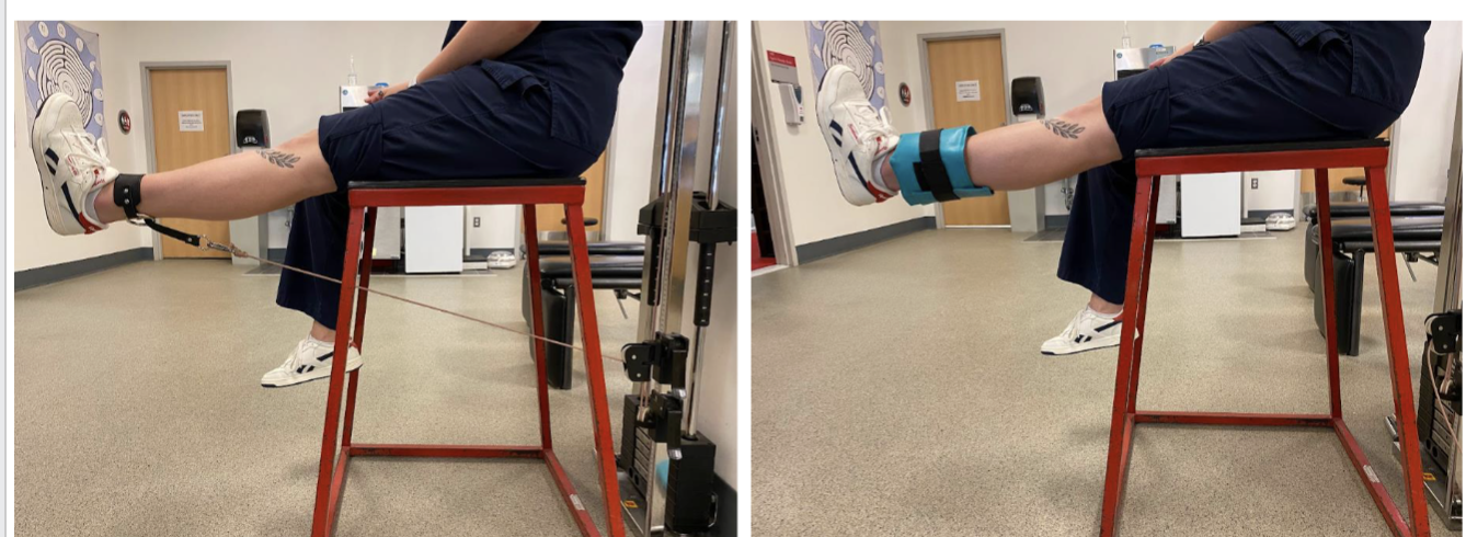
For these open chain, which would require more quad force at this position

  • pulley b/c it’s @ 90 dg flexion meaning more PFJ stress

    • rope makes 90 dg angle to tibia, external moment is most

  • NOT ankle weight

    • NONE of force is perpendicular to tibia → NO external moment

    • its external moment increases when going up
51
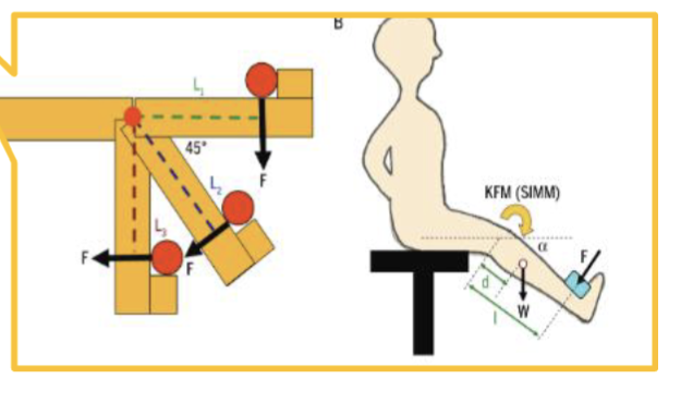
New cards
<p>For these open chain, which would require more quad force at this position</p>

For these open chain, which would require more quad force at this position

  • ankle weight b/c

    • gravity vector is perpendicular to tibia

    • more quad force → more PFJ stress

  • NOT pulley b/c it’s NOT @ 90 dg to tibia

    • less from quad force → less PFJ stress
52
New cards
<p>Cuff weight</p>

Cuff weight

cuff weight always going straight down being @ mercy of gravity

  • NO pulley changing force vector

<p>cuff weight always going straight down being @ mercy of gravity </p><ul><li><p>NO pulley changing force vector </p></li></ul><p></p>
53
New cards
<p>pulley </p>

pulley

  • resistance always making 90 dg to leg (tibia) regardless of ROM

  • good challenge to quad throughout range

  • quad force is pretty much the same

<ul><li><p>resistance always making 90 dg to leg (tibia) regardless of ROM</p></li><li><p>good challenge to quad throughout range</p></li><li><p>quad force is pretty much the same </p></li></ul><p></p>
54
New cards

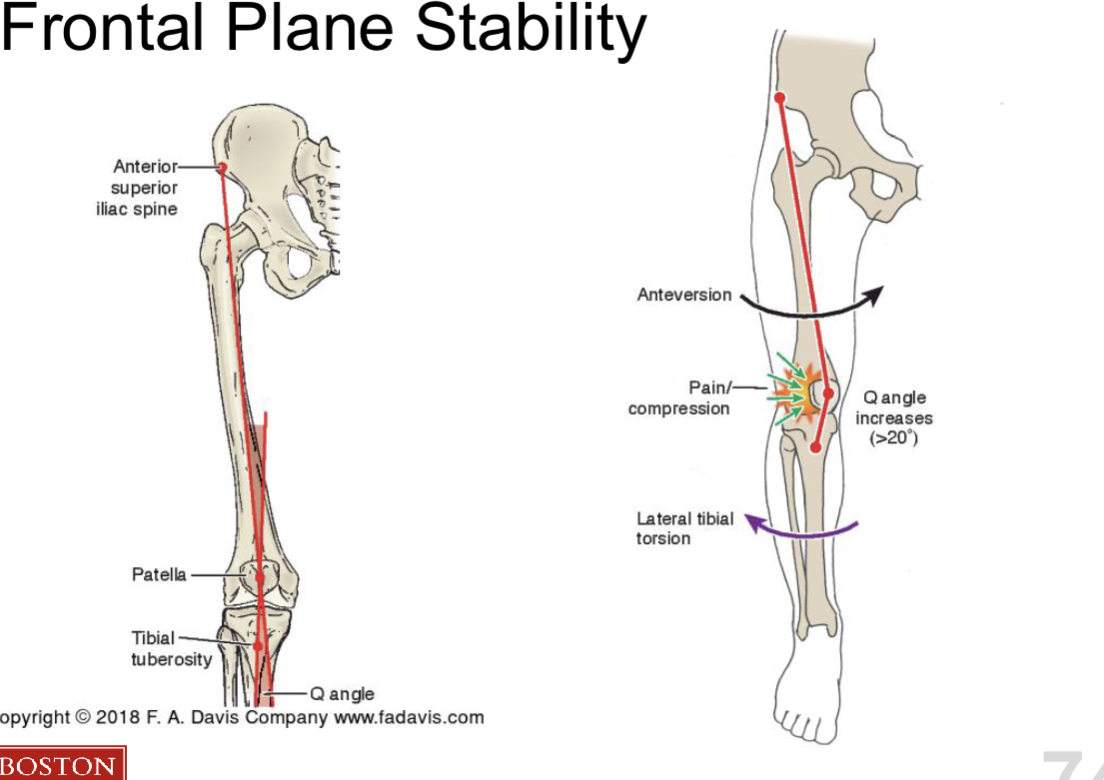
More of Frontal Plane stability (Q angle)

  • line of ASIS → patella

  • line of patella → tibial tuberosity

Anteversion → lateral tibial torsion → increase Q angle

Increase Q angle is NOT indication of PFJ pain
<ul><li><p>line of ASIS → patella </p></li><li><p>line of patella → tibial tuberosity </p></li></ul><p></p><p>Anteversion → lateral tibial torsion → increase Q angle </p><p></p><p>Increase Q angle is NOT indication of PFJ pain</p><p></p>