IMAGE EVAL #2
 Call Kai
 Learn
Learn Practice Test
 Spaced Repetition
 Match
Match Flashcards
 Knowt Play
1/68

Earn XP
Description and Tags
chap 3 and chap 2 images only
Name | Mastery | Learn | Test | Matching | Spaced | Call with Kai | Chat |
|---|
No analytics yet
Send a link to your students to track their progress
69 Terms
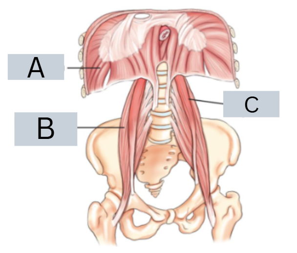
label anatomy
A = diaphragm
B = right psoas major muscle
C = left psoas major muscle

what are the arrows directed towards
psoas muscles

label anatomy
extra: what are the main organs and accessory organs of digestive system?
1 = oral cavity 2 = pharynx
3 = esophagus 4 = stomach
5 = small intestine 6 = large intestine
7 = liver 8 = spleen 9 = pancreas
main = oral cavity, pharynx, esophagus, stomach, small i, large i
accessory = liver, pancreas, gallbladder (NOT spleen. belongs to lymphatic system)

what does the white and grey/black represent
white = barium
grey/black = gas

label anatomy
1 = esophagus
2 = cardia
3 = fundus
4 = body of stomach
5 = greater curvature
6 = pylorus
7 = duodenum
8 = lesser curvature
9 = lower esophageal sphincter

label anatomy
1 = small i
2 = duodenal bulb
3 = duodenum
4 = jejunum (2/5)
5 = ileum (3/5)
6 = stomach

label radiograph
1 = duodenum
2 = connivent valves
3 = jejunum
4 = ileum

label anatomy
1 = liver 8 = appendix
2 = large i 9 = spleen
3 = hepatic flexure (right colic f) 10 = splenic flexure (left colic f)
4 = transverse colon 11 = descending colon
5 = ascending colon 12 = sigmoid colon
6 = ileocecal valve 13 = rectum
7 = cecum 14 = anus
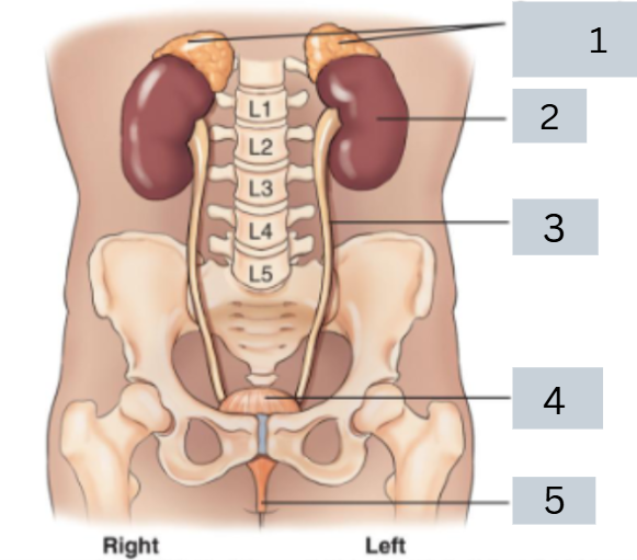
label anatomy
1 = adrenal (suprarenal) glands
2 = kidneys
3 = ureters
4 = (urinary) bladder
5 = urethra

label radiograph
1 = right renal pelvis
2 = right ureter
3 = urinary bladder
4 = left kidney
5 = ureteropelvic junction UPJ
6 = ureterovesical junction UVJ

what kind of radiograph is this called?
IVP
intravenous pyelogram
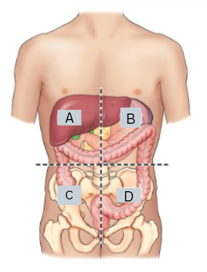
label the quadrants
what are important organs in these quadrants?
A = RUQ right upper quadrant
B = LUQ left upper quadrant
C = RLQ right lower quadrant
D = LLQ left lower quadrant

label the nine abdominal regions
what are the important organs in these regions?
1 = right hypochondriac
2 = epigastric
3 = left hypochondriac
4 = right lateral (lumbar)
5 = umbilical
6 = left lateral (lumbar)
7 = right inguinal (iliac)
8 = pubic (hypogastric)
9 = left inguinal (iliac)
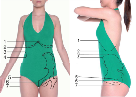
label topographic landmarks
1 = xiphoid process (T9-10)
2 = inferior costal margin (L2-3)
3 = iliac crest (L4-5)
4 = anterior superior iliac spine (ASIS)
5 = greater trochanter
6 = symphysis pubis
7 = ischial tuberosity

label anatomy
1 = greater trochanter
2 = symphysis pubis
3 = ischial tuberosity
4 = ASIS
5 = iliac crest

position + projection
AP projection, supine position
full name = AP supine abdomen (KUB)

label radiograph
what type of graph is this depending on the anatomy shown?
1 = liver
2 = 12 rib
3 = edge of psoas major muscle
4 = sacrum
5 = coccyx
6 = symphysis pubis
7 = transverse process L4
8 = left iliac crest
9 = obturator foramen
10 = left femur
AP supine abdomen (KUB) = can see kidneys, ureter, bladder
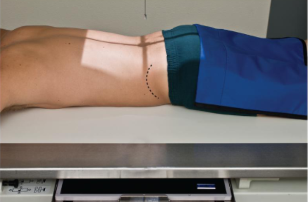
position + projection
PA projection, prone position
full name = PA prone abdomen (KUB)

label radiograph
1 = kidney
2 = 3rd lumbar vertebra L3
3 = pelvis ilium
4 = liver
5 = transverse process
6 = sacrum

position + projection
AP projection, lateral decubitus position
could either be right or left, depends on which side is the patient laying on
full name for this pic = Left AP lateral decubitus

what type of position is in this radiograph?
Left lateral decubitus
can tell by the right marker, indicating the right side is up and the left side is what the patient is laying on
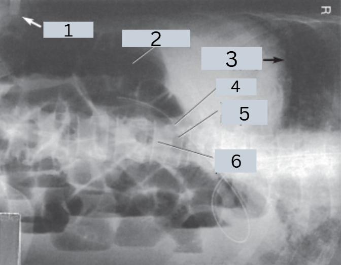
label radiograph
1 = iliac wing
2 = intestinal gas
3 = diaphragm dome
4 = pedicle
5 = vertebral body
6 = spinous process

position + projection
AP erect abdomen
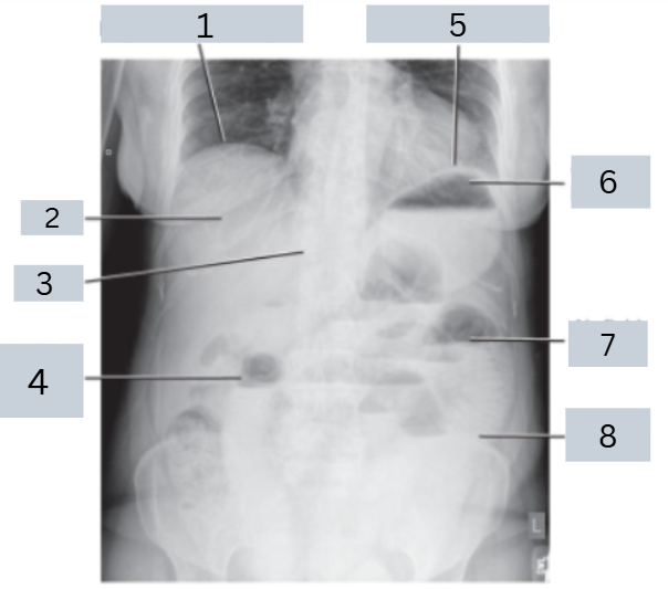
label radiograph
1 = right hemidiaphragm
2 = liver
3 = T12
4 = air-filled intestine (bowel)
5 = left hemidiaphragm
6 = gastric bubble
7 = air fluid level
8 = left iliac crest
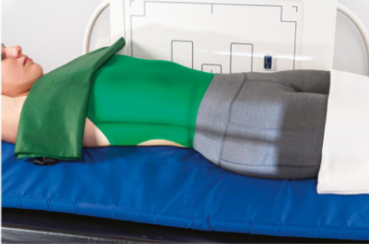
position + projection
dorsal decubitus position R/L lateral abdomen
for this pic = L lateral dorsal decubitus

label radiograph
1 = iliac wings
2 = gas in intestines
3 = prevertebral region

position + projection
R lateral dorsal decubitus

label anatomy
A - clavicle (2)
B - scapula (2)
C - ribs (12 pairs)
D - thoracic vertebrae (12)
E - xiphoid process (inferior)
F - body (center)
G - manubrium (superior)
H - sternum (3 parts, G/F/E, top to bottom)

What are the 3 positioning landmarks for chest x-ray?
vertebra prominens
spinous process of C7, may be easier to find if neck is flexed, good for PA chest positioning
jugular notch
deep notch on superior portion of sternum, good for AP chest positioning
xiphoid process
corresponds to level of T9-T10 + anterior portion of diaphragm, not reliable due to body habitus

label anatomy
A - trachea
B - diaphragm (right/left hemi diaphragms)
C - lungs
D - bronchi
E - pharynx
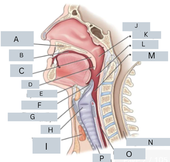
label anatomy
A = hard palate B = oral cavity
C = soft palate D = uvula E = hyoid bone
F = epiglottis G = thyroid cartilage of larynx
H = larynx I = thyroid gland
J = pharynx K = nasopharynx
L = oropharynx M = laryngopharynx
N = region of spinal cord
O = esophagus P = trachea
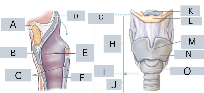
label anatomy
A = hyoid bone B = thyroid cartilage
C = cricoid cartilage D = epiglottis
E = true vocal cords F = trachea
G = level of C3 H = larynx
I = level of C6 J = trachea
K = epiglottis L = hyoid bone
M = laryngeal prominence (level of C5)
N = thyroid cartilage
O = cricoid cartilage

label anatomy
A = larynx B = level of C6
C = trachea D = level of T4/T5
E = right bronchi (3) F = left bronchi (2)
G = region of thymus gland
H = parathyroid glands
I = thyroid gland J = thyroid cartilage

label radiograph
A = trachea + larynx
B = esophagus
C = thyroid gland region
D = thymus gland region
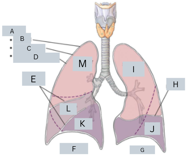
label anatomy
A = pleura B = parietal
C = pleural cavity D = pulmonary visceral
E = fissures F = right lung
G = left lung H = fissure
I = superior lobe J = inferior lobe
K = inferior lobe L = middle lobe
M = superior lobe

label anatomy
A = trachea (windpipe)
B = pleura (lung lining)
C = lung
D = pleural effusion (fluid bt pleural space)
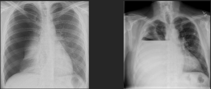
what are the lung pathology for L and R?
L = pneumothorax
R = pleural effusion hemothorax

projection?
label radiograph
PA
A = trachea
B = apex of lung
C = carina
D = base of lung
E = diaphragm
F = costophrenic angle of lung
G = hilum

position?
label radiograph
L lateral
A = apex of lung
B = upper lobe of lung (superior)
C = hilum
D = lower lob of lung (inferior)
E = base
F = diaphragm
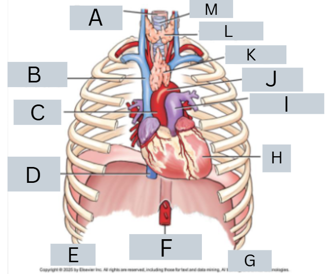
label anatomy
A = esophagus
B = superior vena cava
C = ascending aorta
D = inferior vena cava
E = right F = abdominal aorta
G = left H = heart
I = pulmonary artery J = arch of aorta
K = thymus gland L = thyroid gland
M = trachea

label radiograph
A = superior vena cava
B = right atrium
C = inferior vena cava
D = left ventricle
E = left atrial appendage
F = main pulmonary artery
G = aorta
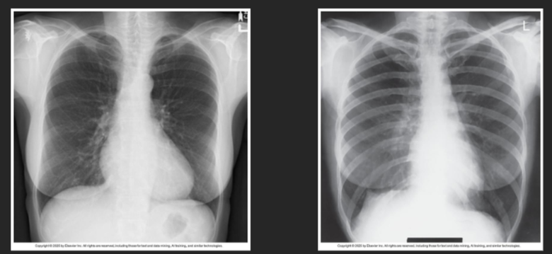
determine body habitus
L = PA chest for a sthenic patient
R = PA chest for hyposthenic patient

which is expiration and which is inspiration?
L = inspiration
R = expiration

projection + position
PA chest

label radiograph
1 = apex
2 = clavicle
3 = hilum
4 = R hemidiaphragm
5 = R costophrenic angle
6 = air filled trachea
7 = 4th rib
8 = aortic arch
9 = heart

position + projection
lateral chest
pic = L lateral chest

label radiograph
1 = apices
2 = thoracic vertebrae
3 = posterior ribs
4 = costophrenic angles
5 = body of sternum
6 = heart
7 = diaphragm

position + projection
AP supine chest
angled caudad 5 degrees

position + projection
AP semierect chest
angled caudad 5 degrees

position + projection
AP (R/L) lateral decubitus position
this pic = AP left lateral decubitus

label radiograph
1 = lung
2 = heart
3 = air fluid level

position + projection
L lateral decubitus AP
right marker indicating right side is up, left side is towards table

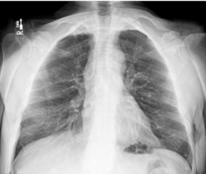
chest critique
possible effusion = fluid in pleural space
a lot of grey area in lower lung area
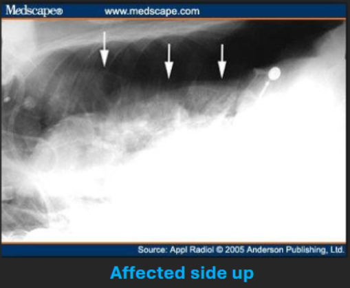
chest critique
possible pneumothorax = air in pleural space
grainy grey area in upper lung area

position + projection
AP lordotic chest

position + projection
AP semiaxial chest
if patient cannot assume erect lordotic position, supine position with semiaxial projection
position + projection
AP lordotic chest
can see that clavicles are not in the way of apices

position + projection
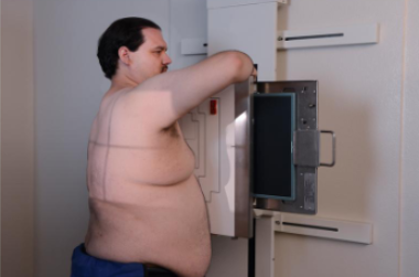
RAO, PA projection
right anterior oblique

position + projection
RAO position, PA
L marker indicating outer part, R side closer to IR
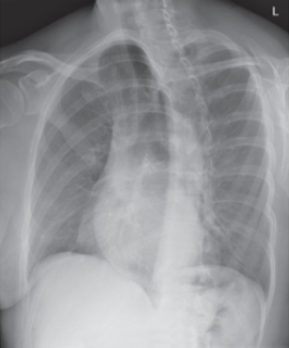
position + projection
LAO position, PA projection
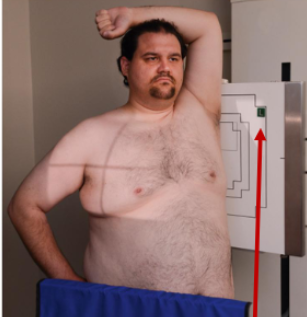
position + projection
LAO, PA
position + projection
LPO, AP
left posterior oblique

label radiograph
1 = trachea
2 = carina
3 = heart
4 = left clavicle
5 = left costophrenic angle
6 = right clavicle
7 = scapula
8 = right lung
9 = left apex
10 = heart
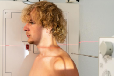
position + projection
R lateral upper airway
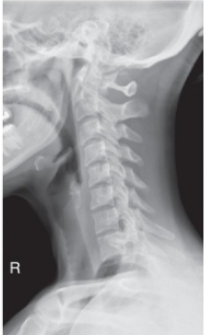
position + projection
R lateral upper airway
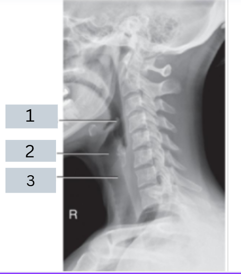
label radiograph
1 = epiglottis
2 = larynx
3 = trachea

position + projection
AP upper airway

position + projection
AP upper airway
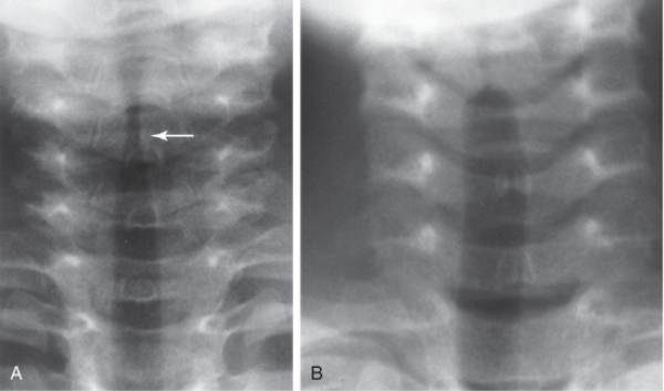
arrow
steeple sign