Postmenopausal Vaginal Bleeding and Endometrial Thickening
 Call Kai
 Learn
Learn Practice Test
 Spaced Repetition
 Match
Match Flashcards
 Knowt Play
1/51

There's no tags or description
Looks like no tags are added yet.
Name | Mastery | Learn | Test | Matching | Spaced | Call with Kai | Chat |
|---|
No analytics yet
Send a link to your students to track their progress
52 Terms
A common indication for postmenopausal sonography is —
postmenopausal vaginal bleeding (PMB)
— are among the differential abnormalities that can be linked to vaginal bleeding in the postmenopausal population
Endometrial atrophy, uncontrolled HRT, endometrial hyperplasia, endometrial polyps, submucosal or intracavitary leiomyoma, endometrial carcinoma, and some ovarian tumors
HRT will influence the sonographic appearance of the —
endometrium
Transvaginal sonography has been reported to be highly effective, with a sensitivity of up to 95% for detecting endometrial carcinoma when an endometrial thickness threshold of — is used for the patient with PMB
4 to 5 mm
If the endometrium measures less than —, the bleeding is typically caused by endometrial atrophy
5 mm
if the endometrium exceeds —, endometrial biopsy is warranted
5 mm
The asymptomatic patient, or those with no vaginal bleeding, can have an endometrial thickness of up to —
8 mm
Negative postmenopausal vaginal bleeding: normal endometrial thickness (mm)
< 8
Positive postmenopausal vaginal bleeding: normal endometrial thickness (mm)
< 5
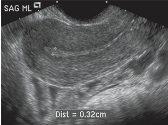
Normal postmenopausal endometrium. This endometrium (between calipers) appears thin and echogenic.
In the postmenopausal patient, the endometrium often bleeds spontaneously secondary to
atrophy
the most common cause of PMB is
endometrial atrophy
with endometrial atrophy the endometrium will appear thin and should not exceed —, although with atrophy the endometrium typically measures 4 mm or thinner
5 mm
A — in the postmenopausal patient usually does not warrant endometrial biopsy
thin endometrial stripe
— is a common cause of abnormal vaginal bleeding, not only in the postmenopausal female but also in the reproductive years.
Endometrial hyperplasia
Endometrial hyperplasia results from the —
unopposed stimulation of estrogen on the endometrium
Endometrial hyperplasia may also be caused by —
polycystic ovary syndrome, obesity, tamoxifen therapy for breast cancer, or estrogen-producing ovarian tumors, such as the ovarian thecoma or granulosa cell tumor
There seems to be an increased risk for one form of endometrial hyperplasia (—) progressing into endometrial carcinoma more often in the postmenopausal woman.
atypical adenomatous hyperplasia
an endometrial biopsy is typically warranted to rule out —
endometrial carcinoma
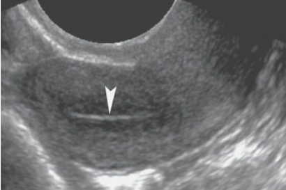
Endometrial atrophy. Longitudinal transvaginal sonogram revealing a thin endometrium (arrowhead) measuring only 2 mm in a postmenopausal woman with vaginal bleeding, which is indicative for endometrial atrophy.
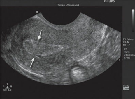
A thickened endometrium (between arrows) is demonstrated in this patient with endometrial hyperplasia.
clinical findings of endometrial hyperplasia
abnormal uterine bleeding (any age)
PCOS
obesity
tamoxifen therapy
sonographic findings of endometrial hyperplasia
Thickened echogenic endometrium
Small cystic spaces within the endometrium
— is the most common female genital tract malignancy, with PMB being the most common clinical presentation.
Endometrial carcinoma
Endometrial carcinoma is most often in the form of —
adenocarcinoma
endometrial carcinoma has been linked with
unopposed estrogen therapy, nulliparity, obesity, chronic anovulation, Stein–Leventhal syndrome, estrogen-producing ovarian tumors, and the use of tamoxifen
peak incidence at the time of diagnosis for endometrial carcinoma is
50 and 65 yeras of age
Tumors with penetration into the surrounding myometrium have a — than those confined to the endometrium
poorer prognosis
clinical findings of endometrial carcinoma
postmenopausal bleeding
intermenstrual bleeding
enlarged uterus
elevation of CA-125
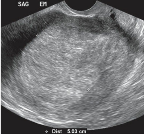
ultrasound of Endometrial carcinoma
sonographic findings of endometrial carcinoma
Thickened endometrium
Heterogeneous uterus
Enlarged uterus with lobular contour
Endometrial fluid
Polypoid mass within the endometrium
Pulsed Doppler of the uterine cavity may indicate — in the presence of endometrial carcinoma
low-impedance flow
— are small nodules of hyperplastic endometrial tissue that may cause abnormal vaginal bleeding in both postmenopausal or perimenopausal woman
Endometrial polyps
Endometrial polyps have been linked with
infertility during the reproductive period
Endometrial polyps can have many different shapes, including a —
broad base, or can be pedunculated and, if large, may prolapse through the cervix.
Polyps are better visualized with the use of —
saline infusion sonohysterography
Treatment for endometrial polyps is typically a —
polypectomy with the use of hysteroscopy
A key clinical feature of patient with an endometrial polyp is —
intermenstrual bleeding.
clinical findings of endometrial polyps
asymptomatic
menometrorrhagia
intermenstrual bleeding
has been linked with infertility in reproductive-aged groups
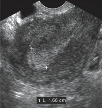
A polyp is suspected in a patient with focal thickening of the endometrium

Saline (S) infusion sonohysterography better depicts the evidence of an endometrial polyp (arrow).
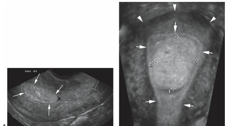
Three-dimensional endometrial polyp. A. Longitudinal image of a rounded mass (between arrows) within the endometrial cavity suggesting a polyp. B. Coronal reconstruction of the uterus (arrowheads) revealing the presence of a polyp (between calipers) within the endometrial cavity (arrows).
estrogen-producing tumors such as the thecoma, which is mostly benign, have been shown to cause —
abnormal thickening of the endometrium with subsequent vaginal bleeding
the malignant serous and mucinous cystadenocarcinomas have been associated with —
vaginal bleeding
— are often elevated in women who have some forms of cancer
Serum levels of CA-125
elevation of CA 125 has been linked with cancers of the —
ovary, endometrium, breast, gastrointestinal tract, and lung
CA125 may also be elevated in some benign conditions such as —
endometriosis, pelvic inflammatory disease, fibroids, and even pregnancy
— is a breast cancer drug that inhibits the effects of estrogen on the breast, thus slowing the growth of malignant breast cells.
Tamoxifen
Tamoxifen can also be used in the treatment of —
female infertility as well
tamoxifen use has been linked with the development of —
endometrial hyperplasia, endometrial polyps, and endometrial carcinoma.
Sonographically, tamoxifen will cause cystic changes to occur within the endometrium, and it produces a more —
heterogeneous and thickened endometrial appearance