topic 2 J) coordination and response
1/28
There's no tags or description
Looks like no tags are added yet.
Name | Mastery | Learn | Test | Matching | Spaced |
|---|
No study sessions yet.
29 Terms
why do organisms need to respond to change
changes in both external and internal environments need responses
respond to coordinate the activities of different organisms
internal conditions have to be kept relatively constant to function properly and efficiently - have different control and communication systems to do this
physiological control systems maintain internal environment within restricted limits through HOMEOSTASIS
homeostasis very important - ensures maintenance of optimal conditions for enzyme action and cell function
examples of physiological factors controlled by homeostasis in mammals:
core body temperature
metabolic waste
blood pH
blood glucose concentration
water levels of blood
concentration of respirator gases in blood (CO2 and O2)
homeostatic mechanisms in mammals require information to be transferred between parts of body
two communication systems in mammals to do this:
nervous system
endocrine system
what is homeostasis
control or regulation of internal conditions of a cell or organism
eg:
water content of cell or body fluid
temperature
pH
blood glucose concentration
blood pressure
important to keep these within set limits to stay healthy and maintain optimum conditions so organism can function to respond to external and internal changes
organism may die if limits are exceeded
homeostasis means optimal conditions for enzyme activity and all cell functions, ensures reactions in body cells can function
e.g. of homeostasis in humans:
control of body temperature - around 37 degrees, regulated by thermoregulatory centre in base of brain, contains receptors that measure temperature of blood that passes through, skin also has temperature receptors and sends nervous impulses to thermoregulatory centre, brain coordinates cooling or heating response.
control of blood water content - water loss via lungs or skin cannot be controlled, but volume of water lost in production of urine can be controlled by kidneys, water can be reabsorbed more or less controlled by ADH
how is a response coordinated
homeostasis is under automatic control, so brain stem - non conscious part of brain, and spinal cord are involved in maintaining homeostasis - not conscious
may involve nervous or chemical responses (hormones)
all control systems that carry out coordinated responses need:
a stimulus (change in environment)
receptor - cells that detect stimuli
coordination centre - brain, spinal cord, pancreas - receives and processes information from receptors
effector (muscle/gland) - brings about response to restore optimum levels
how do plants respond to stimuli
plants need to grow in response to stimuli e.g. light for photosynthesis and gravity so shoots are up, roots are down
directional growth responses in response to light and gravity are tropisms
if growth is towards stimulus, tropism is positive
if growth is away from stimulus, tropism is negative
geotropic and phototropic responses in plants
phototropism - response to light
geotropism - response to gravity
shoot grows upwards, away from gravity, towards light - shoots show positive phototropic response and negative geotropic
roots grow downwards into soil, away from light and towards gravity - roots show negative phototropic responses and positive geotropic response
role of auxin in phototropism
auxin = plant growth regulators, coordinate and control directional growth responses
made mostly in tips of growing shoots, diffuses down to region where cell division occurs - just below tip
only region behind tip of shoot is able to contribute to growth by cell division and elongation
auxin stimulates cells in this region to elongate - more auxin, faster the growth
if light shines around tip evenly, auxin distributed evenly throughout and shoot grows at same rate
when light shines on shoot predominantly from one side, auxin produced in tip concentrates on the shaded side, making cells on that side elongate and grow faster than cells on sunny side
unequal growth on either side causes shoot to bend and grow in direction of light
control systems in humans
nervous system
hormonal/endocrine system
changes in external or internal environment are stimuli
nervous and hormonal systems coordinate suitable response to stimuli - allow us to make sense of our surroundings, to respond to changes and coordinate and regulate body functions
what is a nervous system
information sent as electrical impulses - electrical signals that pass along nerve cells called neurones
impulses travel along neurones at very high speeds, allowing rapid responses
nervous system coordinates activities of sensory receptors, decision-making centres in the CNS and effectors e.g. muscles and glands
nervous system used to control functions that need instant or very rapid responses
structure of endocrine system
information is send through endocrine system as chemical substances called hormones
hormones carried by blood and can circulate around whole body
hormones transmit information from one part of organism to another, bring about a change (provide signal that triggers a response)
alter the activity of one or more specific target organs
hormones are used to control functions that do not need instant responses, produced by endocrine glands
endocrine glands are collectively known as endocrine system
gland is a group of cells that produces and releases one or more substances (secretion)
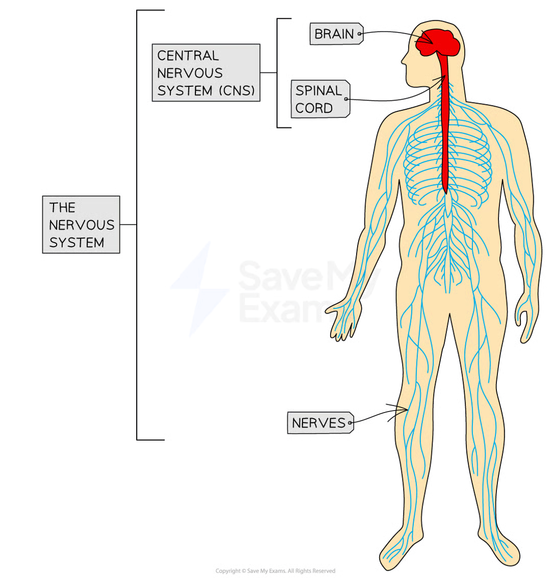
structure and function of human nervous system
consists of:
central nervous system - brain and spinal cord
peripheral nervous system - all the nerves in the body
bundle of neurones = nerve
nerves spread out from the CNS to all other regions of the body and to all the sense organs
CNS acts as a central coordinating centre for the impulses that come in from any part of the body
pathway through nervous system:
stimulus
sensory neurone
relay neurone
motor neurone
effector
response
stimulus is received by sensory (receptor) neurone, producing electrical impulses
impulses travel along sensory neurone to CNS
in CNS impulses are passed on to relay neurone
relay neurone links to motor neurone, impulses travel until they reach the effector
effector carries out response (may be muscle or gland)
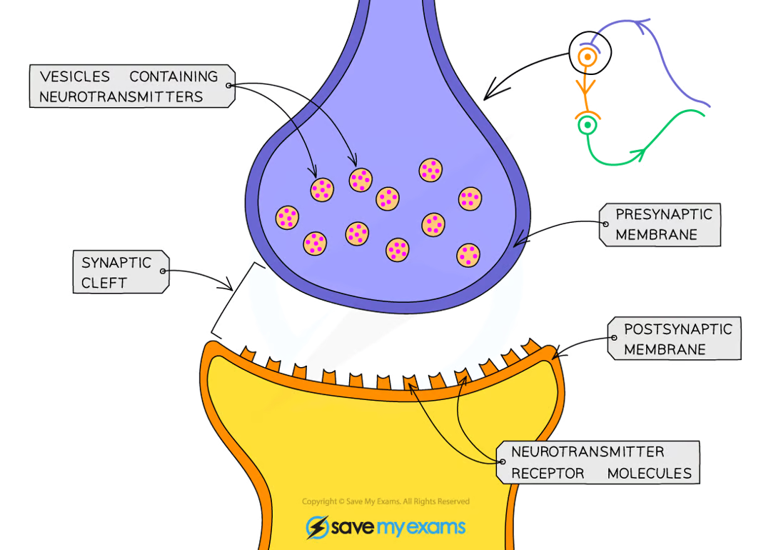
role of neurotransmitters
neurons do not come into direct contact with each other
when dendrites of two neurones meet to make a connection between neurones, a synapse is formed
at the synapse, there is a very small gap between neurones called synaptic cleft/gap
electrical impulses cannot travel directly from one neurone to the next due to the gap
so electrical signal is briefly converted to a chemical signal that can cross cleft - chemical signalling molecules used to transfer signal between neurones at synapse are known as neurotransmitters
once neurotransmitters cross cleft and meet other neurone, signal is converted back into electrical impulse
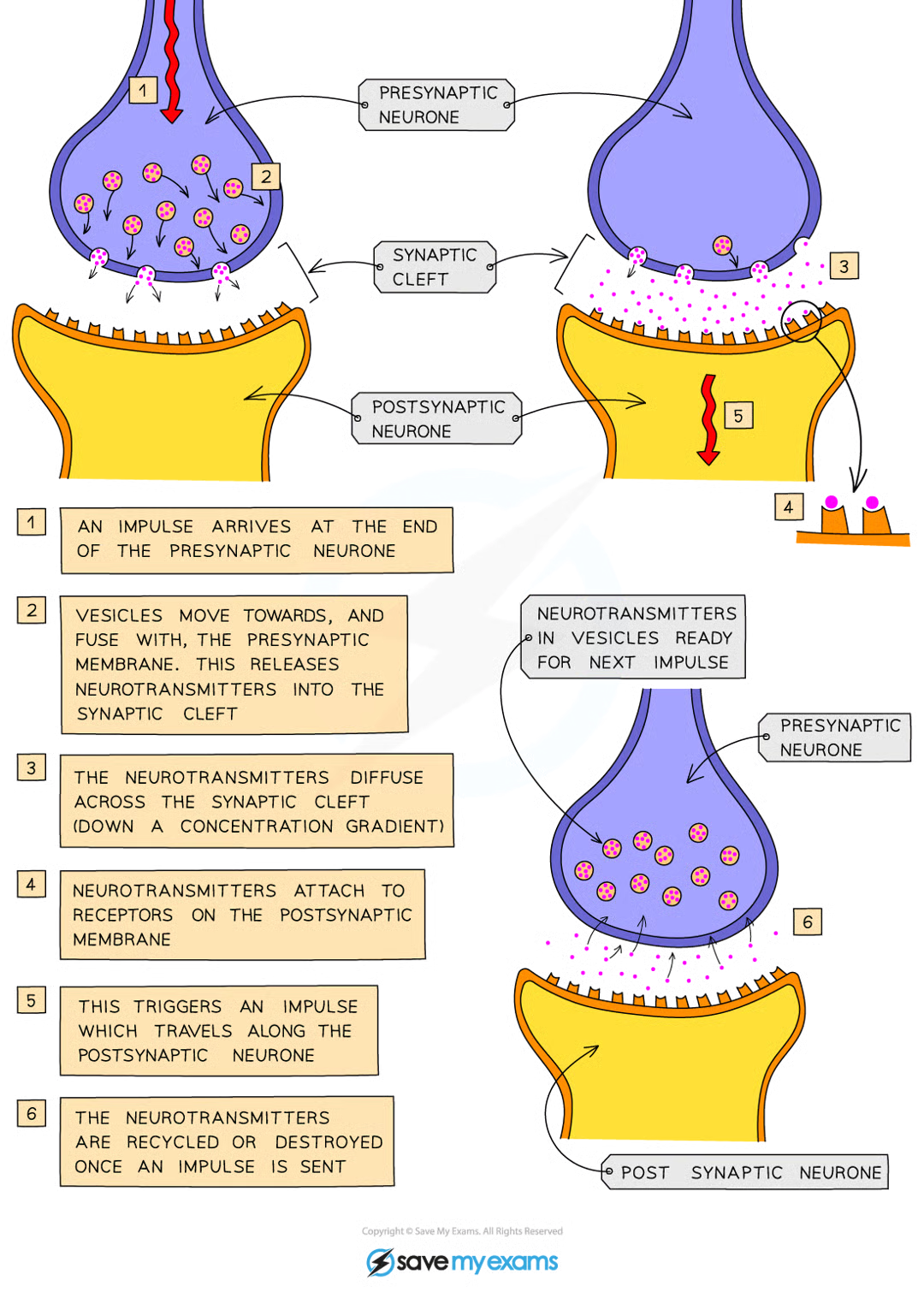
how does an impulse pass across a synapse
electrical impulse travels along the axon of the first neurone (presynaptic neurone)
triggers the end of the presynaptic neurone to release neurotransmitters from vesicles
vesicles fuse with the presynaptic membrane, releasing their contents into the synaptic cleft
neurotransmitters diffuse across the synaptic cleft and bind with receptor molecules on the membrane of the second neurone (postsynaptic membrane)
This stimulates the second neurone to generate an electrical impulse (then travels down the second axon)
The neurotransmitters are then destroyed to prevent continued stimulation of the second neurone (otherwise the neurotransmitters would cause repeated impulses to be sent)
Synapses ensure that impulses only travel in one direction, avoiding the confusion that would be caused within the nervous system if impulses were able to travel in both directions
simple reflex arc
reflex response/involuntary response does not involve conscious part of the brain as the coordinator
awareness of response happens after response has been carried out
responses are automatic and rapid - helps minimise damage to body, aids survival
pain-withdrawal, blinking and coughing help avoid serious injury e.g. damage to eye/choking
reflex arc
pathway of a reflex response e.g. pain-withdrawal reflex when smn steps on pin
The pin (stimulus) is detected by a (pain/pressure/touch) receptor in the skin on the person's foot
A sensory neurone sends electrical impulses to the spinal cord (the coordinator)
An electrical impulse is passed to a relay neurone in the spinal cord (part of the CNS)
A relay neurone synapses with a motor neurone
A motor neurone carries an impulse to a muscle in the leg (the effector)
When stimulated by the motor neurone, the muscle will contract and pull the foot up and away from the sharp object (the response)
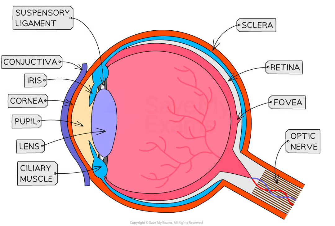
structure of the eye
highly specialised sense organ with receptor cells that detect stimulus of light
retina has two types of receptors::
rods - sensitive to light
cones - sensitive to colour
Cornea - transparent lens that refracts light as it enters the eye
Iris - controls how much light enters the pupil
Lens - transparent disc that can change shape to focus light onto the retina
Retina - contains light receptor cells – rods (detect light intensity) and cones (detect colour)
Optic nerve - sensory neuron that carries impulses between the eye and the brain
Pupil - hole that allows light to enter the eye
Conjunctiva - a clear membrane that covers the white of the eye and the inside of the eyelids; it lubricates the eye and provides protection from external irritants
Ciliary muscle - a ring of muscle that contracts and relaxes to change the shape of the lens
Suspensory ligaments - ligaments that connect the ciliary muscle to the lens
Sclera - the strong outer wall of the eyeball that helps to keep the eye in shape and provides a place of attachment for the muscles that move the eye
Fovea - a region of the retina with the highest density of cones (colour detecting cells) where the eye sees particularly good detail
Blind spot - the point at which the optic nerve leaves the eye, where there are no receptor cells
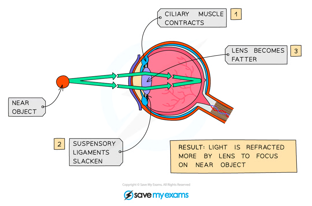
function of eye focusing on near and distant objects
lens brings about fine focusing called accommodation
lens is elastic and shape can be changed by suspensory ligaments to be tight/loose
changes are brought about by contracting or relaxing ciliary muscles
when object is near:
ciliary muscles contract
suspensory ligaments loosen and stop pulling on lens
lens becomes fatter
light refracted more
when object is far:
ciliary muscles relax
suspensory ligaments tighten and pull on lens
lens becomes thinner
light refracted less
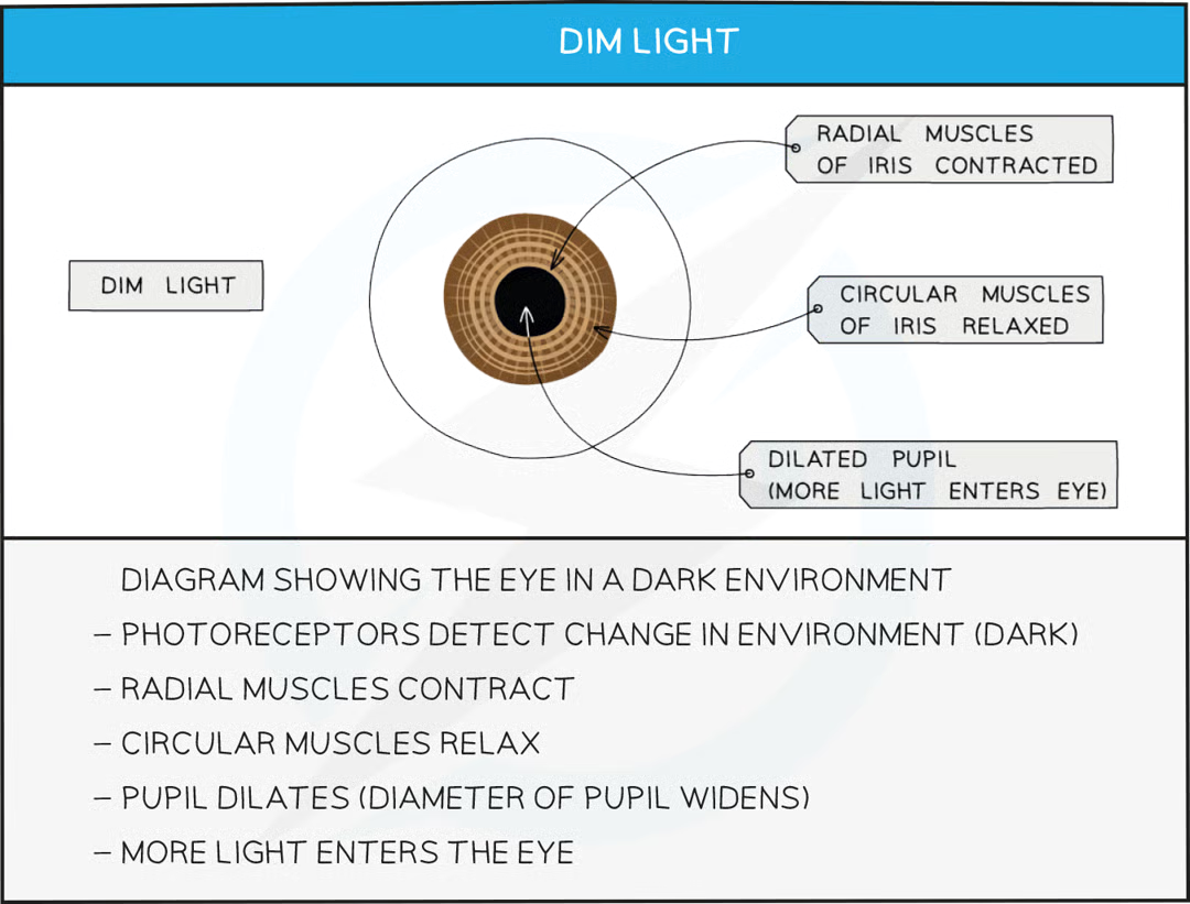
function of eye responding to bright/dim light
pupil reflex is carried out to protect retina from damage
dim light - pupil dilates (widens) to allow as much light into eye as possible to improve vision
bright light - pupil constricts (narrows) to prevent too much light from entering and damaging retina
radial and circular muscles relax/contract to allow pupil size to change
Stimulus
Radial muscles
Circular muscles
Pupil size
Light entering eye
Bright light
Relaxed
Contracted
Narrow
Less
Dim light
Contracted
Relaxed
Wide
More
role of skin in temperature regulation
skin is largest sense organ, contains many different receptors that enable detection of external stimuli
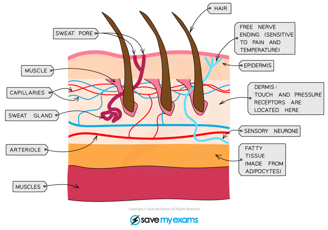
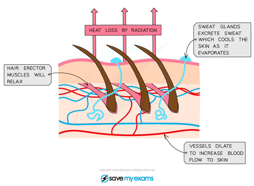
cooling mechanism in humans
vasodilation of skin capillaries
Heat exchange (both during warming and cooling) occurs at the body's surface as this is where the blood comes into closest proximity to the environment
One way to increase heat loss is to supply the capillaries in the skin with a greater volume of blood, which then loses heat to the environment via radiation
Arterioles (small vessels that connect arteries to capillaries) have muscles in their walls that can relax or contract to allow more or less blood to flow through them
During vasodilation, these muscles relax, causing the arterioles near the skin to dilate (get wider) and allowing more blood to flow through capillaries
Sweating
Sweat is secreted by sweat glands
This cools the skin by evaporation which uses heat energy from the body to convert liquid water into water vapour
Flattening of hairs
The hair erector muscles in the skin relax, causing hairs to lie flat
This stops them from forming an insulating layer by trapping air and allows air to circulate over the skin and allows heat to leave by radiation
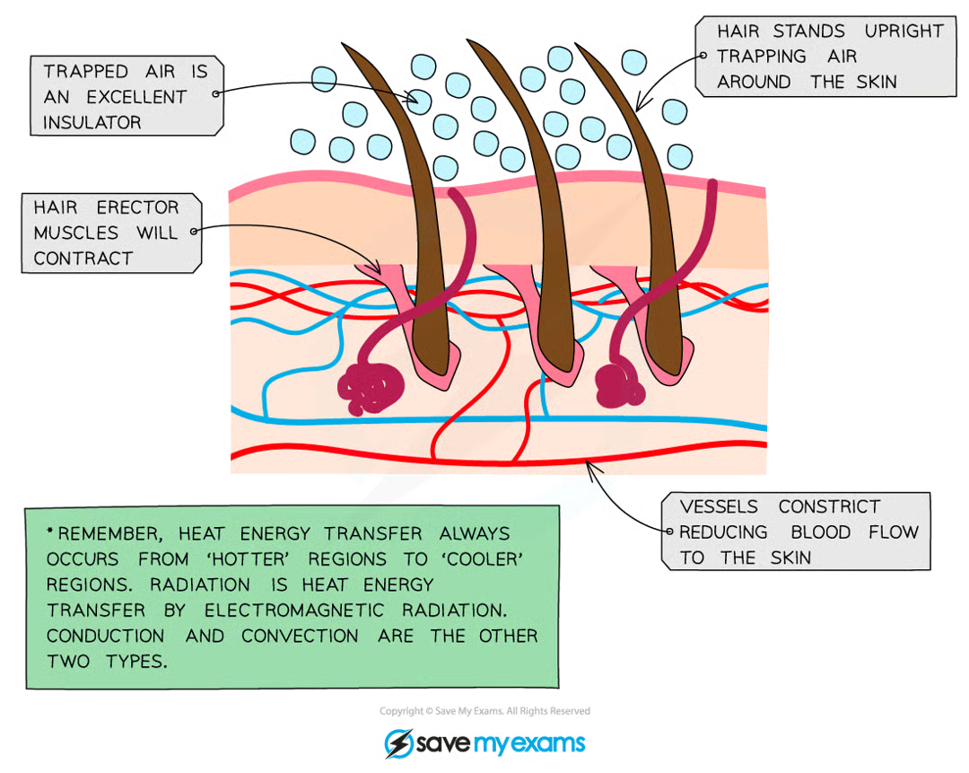
warming mechanisms in humans
Vasoconstriction of skin capillaries
One way to decrease heat loss is to supply the capillaries in the skin with a smaller volume of blood, minimising the loss of heat to the environment via radiation
During vasoconstriction, the muscles in the arteriole walls contract, causing the arterioles near the skin to constrict (get smaller) and allowing less blood to flow through capillaries
Vasoconstriction is not a 'warming' mechanism as it does not raise the temperature of the blood but instead reduces heat loss from the blood as it flows through the skin
Shivering
reflex action in response to a decrease in core body temperature
Muscles contract in a rapid and regular manner
The exothermic metabolic reactions required to power this shivering generate sufficient heat to warm the blood and raise the core body temperature
Erection of hairs
hair erector muscles in the skin contract, causing hairs to stand on end
forms an insulating layer over the skin's surface by trapping air between the hairs and stops heat from being lost by radiation
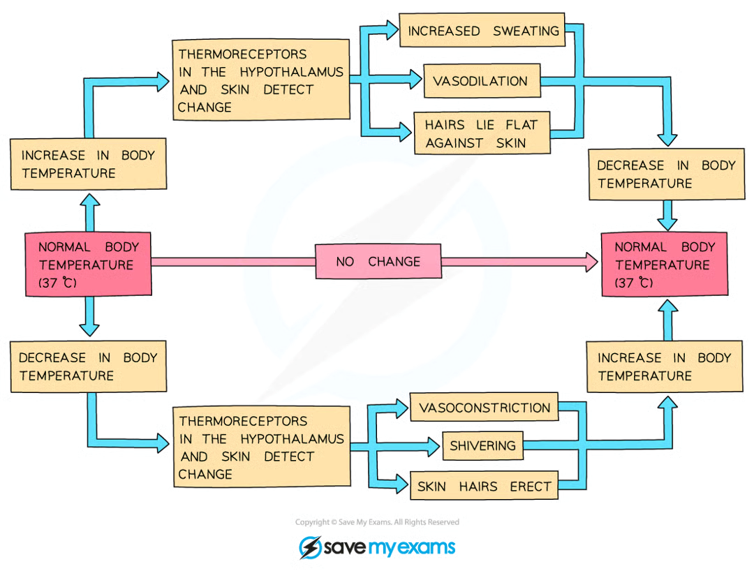
response to change in core body temperature
core body temp - 37
what is a hormone
a chemical substance produced by a gland and carried out by blood, alters activity of one or more specific target organs
chemicals that transmit information from one part of the organism to another and bring about a change
source, role and effect of adrenaline
fight or flight hormone, produced in situations where body might be in danger
things it causes prepare body for movement
increased heart rate - ensures glucose and oxygen can be delivered to muscle cells at a faster rate
divert blood flow to muscles to ensure increased supply of respiration reactants
dilation of blood vessels inside muscles - more blood circulating through to supply O2 and glucose
break down stored glycogen to glucose in liver and muscles - higher blood glucose concentration for increased respiration in muscle cells, more energy for movement
source, role and effect of insulin
controls blood glucose concentration - otherwise cells in body lose or gain too much water by osmosis/brain receives too little glucose for respiration
controlled by pancreas and liver
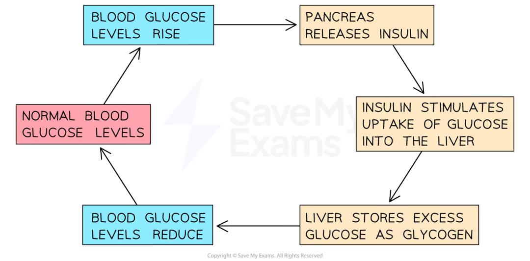
if blood glucose too high:
Cells in the pancreas detect the increased blood glucose levels
The pancreas produces the hormone insulin, secreting it into the blood
Insulin stimulates muscles and the liver to take up glucose from the bloodstream and store it as glycogen (a polymer of glucose)
This reduces the concentration of glucose in the blood back to normal levels, at which point the pancreas stops secreting insulin
source, role and effect of testosterone
produced in male testes
responsible for development of secondary sexual characteristics in males
source, role and effect of progesterone
produced in female ovaries
maintains uterine lining during pregnancy
source, role and effect of oestrogen
produced in female ovaries
responsible for development of secondary sexual characteristics in females and regulating menstrual cycle
source, role and effect of ADH
If the water content of the blood falls below a certain level:
The blood is too concentrated
Receptors detect this and stimulate the pituitary gland to release more ADH
This causes the collecting ducts of the nephrons to become more permeable to water
This leads to more water being reabsorbed from the collecting ducts
The kidneys produce a smaller volume of urine that is more concentrated (contains less water)
If the water content of the blood rises above a certain level:
The blood is too dilute
Receptors detect this and stimulate the pituitary gland to release less ADH
This causes the collecting ducts of the nephrons to become less permeable to water
This leads to less water being reabsorbed from the collecting ducts
The kidneys produce a larger volume of urine that is less concentrated (contains more water)
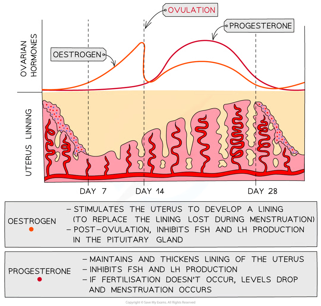
hormonal control of menstrual cycle
FSH (follicle-stimulating hormone)
LH (luteinising hormone)
O and P involved in maintaining uterus lining
O stimulates uterus to develop lining, P maintains and thickens lining
FSH is released by the pituitary gland and causes an egg to start maturing in the ovary
It also stimulates the ovaries to start releasing oestrogen
The pituitary gland is stimulated to release LH when oestrogen levels have reached their peak
LH causes ovulation to occur and also stimulates the ovary to produce progesterone