019 Posterior Segment in Pediatrics
 Call Kai
 Learn
Learn Practice Test
 Spaced Repetition
 Match
Match Flashcards
 Knowt Play
1/95

There's no tags or description
Looks like no tags are added yet.
Name | Mastery | Learn | Test | Matching | Spaced | Call with Kai | Chat |
|---|
No analytics yet
Send a link to your students to track their progress
96 Terms
What are the five primary ocular warning signs that guide the differential diagnosis of pediatric eye disease?
Leukocoria (white pupillary reflex)
Macular problems
Optic nerve problems
Tumors
Retinal hemorrhages
Why is it important to recognize key pediatric ocular signs rather than memorizing individual diseases?
Recognizing major signs helps generate a focused differential diagnosis and allows more efficient clinical problem-solving when a child presents with visual abnormalities.
What is leukocoria, and why is it clinically important?
Leukocoria is a white pupillary reflex. It is a major pediatric ocular red flag because it may indicate vision-threatening or life-threatening disease, including retinoblastoma.
What are the major causes of leukocoria, grouped by pathophysiology?
Media opacity: Cataract, Persistent Hyperplastic Primary Vitreous (PHPV)
Inflammation/Exudation: Coats disease, Toxocariasis, Posterior uveitis
Retinal detachment: Advanced ROP, advanced Coats disease, advanced toxocariasis
Tumor: Retinoblastoma
What tumor must always be considered in a child presenting with leukocoria?
Retinoblastoma until proven otherwise.
What is the underlying defect in Persistent Hyperplastic Primary Vitreous (PHPV)?
Failure of normal regression/development of the fetal vitreous system, resulting in persistent embryologic vitreous tissue.
How does PHPV typically present clinically?
Usually unilateral
Presents with leukocoria and/or strabismus
What are the characteristic anterior and posterior findings of PHPV?
Anterior: Microphthalmia, microcornea, whitish vascularized membrane behind the lens
Posterior: Vitreous membranes and retinal folds
What is the typical management of PHPV?
Referral for surgical management, typically lensectomy and vitrectomy.
What is Coats disease, and who is most commonly affected?
Idiopathic, non-hereditary retinal vascular disorder
About 75% occur in males
Usually unilateral
What is the underlying pathophysiology of Coats disease and its major complication?
Retinal telangiectasias (abnormal retinal vessels) cause intraretinal and subretinal exudation
Progressive exudation can lead to exudative retinal detachment (RD)
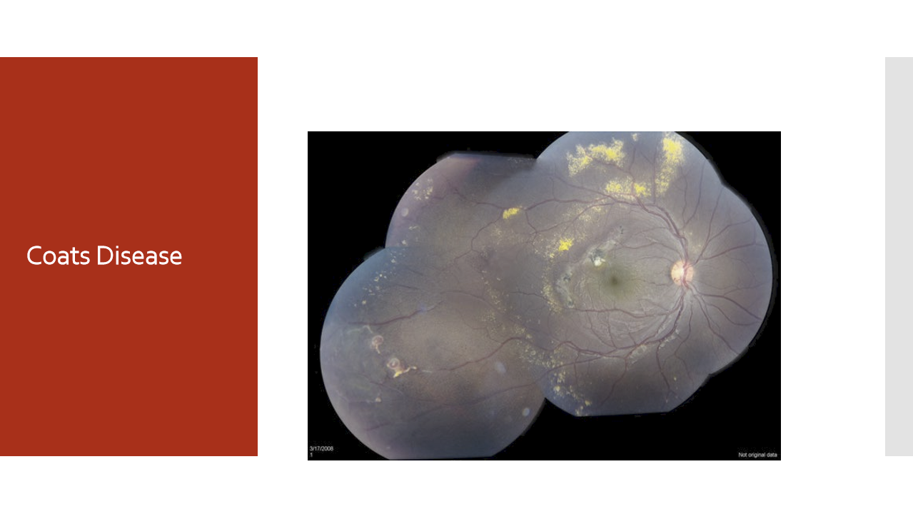
How can Coats disease present as leukocoria?
Accumulation of retinal exudates and/or progression to exudative retinal detachment can produce a white pupillary reflex (leukocoria), making it an important differential diagnosis of retinoblastoma.
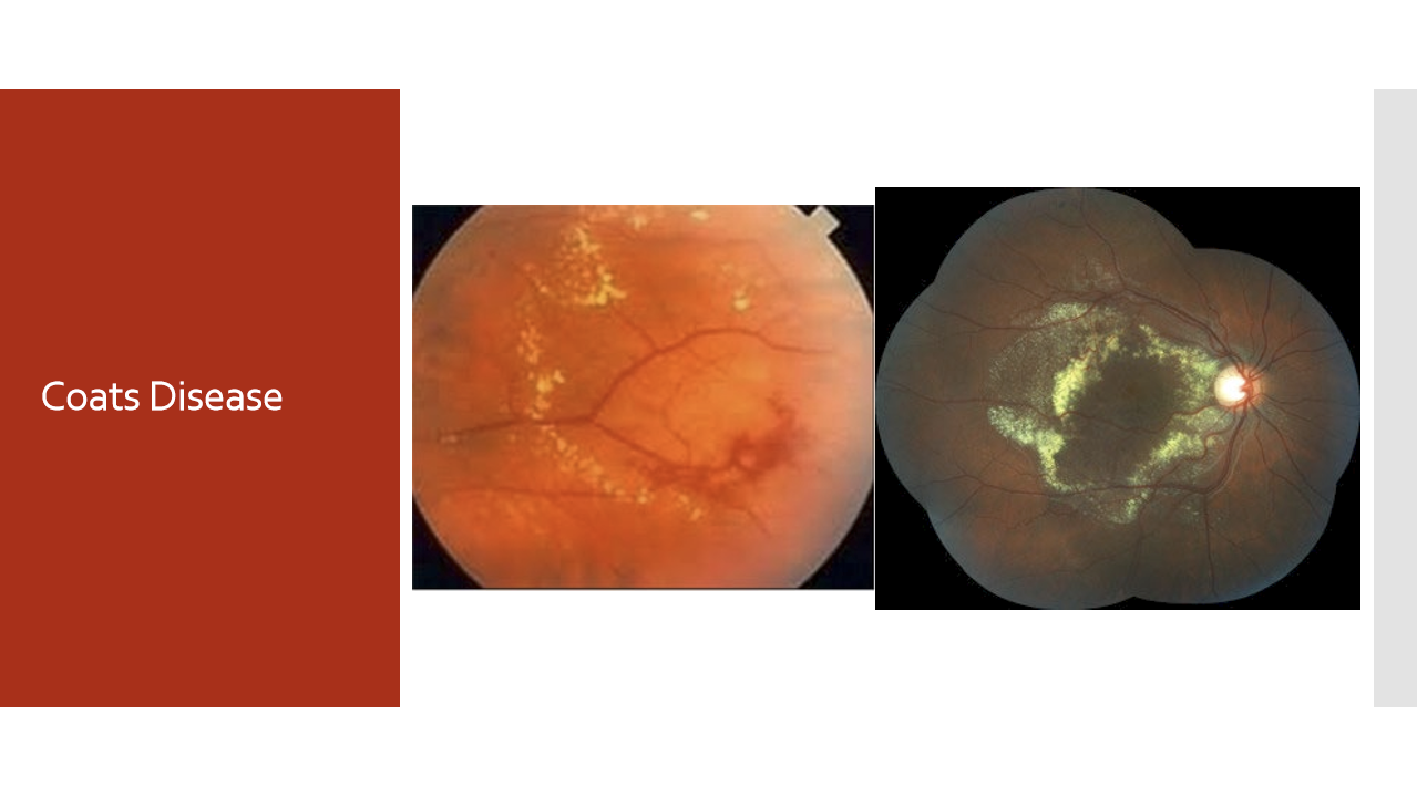
How is Coats disease treated, and why is early diagnosis important?
Goal: treat before retinal detachment develops
Laser photocoagulation → treats retinal telangiectasias
Cryotherapy → used when retinal detachment is present or disease is more advanced
Delayed treatment increases risk of neovascular glaucoma
What is the inheritance pattern of Stickler syndrome, and what are the major systemic manifestations?
Autosomal dominant disorder
Facial findings: flat nasal bridge, maxillary hypoplasia
Skeletal findings: arachnodactyly, arthropathy, joint hyperextensibility
May have Robin sequence (micrognathia, small tongue, cleft soft palate, high-arched palate)
Associated with deafness and mitral valve prolapse
What are the characteristic ocular findings of Stickler syndrome?
Abnormal vitreous
High myopia
Predisposition to retinal detachment (RD)
Why is Stickler syndrome important in pediatric ophthalmology?
It is the most common inherited cause of retinal detachment.
What is the risk and timing of retinal detachment in Stickler syndrome?
Retinal detachments occur in approximately 65% of patients
Can occur as early as the first 5 years of life and continue through young adulthood
What are the major risk factors for retinopathy of prematurity (ROP)?
Prematurity: < 36 weeks gestation
Low birth weight: < 1500 g (especially < 1250 g)
Supplemental oxygen therapy
How does supplemental oxygen contribute to the development of ROP?
High oxygen levels suppress normal peripheral retinal vessel development → avascular retina
When oxygen is reduced, retinal ischemia stimulates pathologic neovascularization
What are the characteristic examination findings in ROP?
Avascular peripheral retina
Dilated, tortuous retinal vessels (Plus disease)
Poor pupillary dilation
Engorged iris vessels
Usually bilateral
What complications can develop in advanced ROP?
Neovascularization
Retinal hemorrhage
Retinal detachment (RD)
Leukocoria
How is ROP classified anatomically by retinal zones?
Zone I: Circle centered on the optic nerve with radius equal to 2× the disc-to-macula distance
Zone II: Extends from Zone I to the nasal ora serrata
Zone III: Remaining temporal crescent anterior to Zone II
Besides retinal zone, what other parameter is used to describe the severity of ROP?
Extent, measured in clock hours of retinal involvement.
What are the stages of Retinopathy of Prematurity (ROP)?
Stage 1: Flat demarcation line between vascular and avascular retina
Stage 2: Elevated ridge
Stage 3: Ridge with fibrovascular proliferation
Stage 4A: Partial retinal detachment, macula on
Stage 4B: Partial retinal detachment, macula off
Stage 5: Total retinal detachment
What is Plus Disease in ROP, and why is it important?
Dilated, tortuous retinal vessels
May have retinal hemorrhages
Indicates more severe/active disease
Can occur at any stage of ROP
Which ROP stages involve retinal detachment?
Stage 4A: RD with macula attached
Stage 4B: RD with macula detached
Stage 5: Total RD
When are infants at risk for ROP typically screened?
Before discharge from the NICU or children's hospital.
What follow-up schedule is recommended for very low birth weight infants (<2 lb 12 oz / <1250 g)?
Every 2 weeks until 14 weeks
Every 1 month until 6 months
Every 6 months thereafter
How is ROP managed based on stage?
Stages 1-2: Observation; 80–90% regress spontaneously
Stage 3: Laser photocoagulation and/or anti-VEGF therapy
Stages 4-5: Surgical repair (scleral buckle, vitrectomy, or both)
Which ROP stages usually do not require treatment?
Stages 1 and 2, because most cases (80-90%) regress without intervention.
When are laser photocoagulation and anti-VEGF therapy indicated in ROP?
Stage 3 ROP, when fibrovascular proliferation develops and risk of progression increases.
What surgical options are used for advanced ROP?
For Stages 4-5 with retinal detachment:
Scleral buckle
Vitrectomy
Combination of both
What are the major long-term visual sequelae seen after treatment for Retinopathy of Prematurity (ROP)?
High myopia
Reduced peripheral vision
Reduced central visual acuity (from macular dragging)
Strabismus and amblyopia
Cataracts
Why can patients treated for ROP have decreased vision even if the retina is preserved?
Visual impairment may result from:
Macular dragging → decreased central visual acuity
Loss of peripheral retina → decreased peripheral vision
Associated refractive errors, especially high myopia
Development of amblyopia or strabismus
What is the newest major medical treatment for ROP?
Intravitreal anti-VEGF therapy (bevacizumab/Avastin)
Used particularly for severe neovascular ROP
Suppresses VEGF-driven pathologic retinal neovascularization
What is the mechanism of action of bevacizumab (Avastin) in ROP?
Bevacizumab is an anti-VEGF monoclonal antibody that inhibits VEGF-mediated abnormal retinal vessel growth, reducing neovascularization and disease progression.
What are the major concerns with anti-VEGF treatment in premature infants?
Because VEGF is important for normal development throughout the body:
Systemic absorption may occur
Potential impact on neurodevelopment and organ development
Reported concerns include:
Increased risk of motor impairment in some studies
Possible association with pulmonary hypertension
Why is there interest in using very low doses of bevacizumab for ROP?
To maintain retinal efficacy while minimizing systemic VEGF suppression and potential developmental side effects.
What are the three major inherited macular disorders commonly considered in pediatric patients?
X-linked (Juvenile) Retinoschisis
Best Vitelliform Macular Dystrophy
Stargardt Disease
What is the inheritance pattern and typical age of presentation of X-linked juvenile retinoschisis?
X-linked inheritance
Prevalence: ~1:5,000 to 1:25,000
Usually presents between 5 to 10 years of age
What is the characteristic macular finding in X-linked juvenile retinoschisis?
Macular "spoke-wheel" pattern caused by splitting (schisis) of the retinal layers.
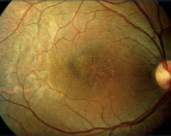
What are the major retinal complications of X-linked juvenile retinoschisis?
Approximately 50% develop peripheral retinoschisis
May develop:
Retinal tears (RT)
Retinal detachment (RD)
What is the classic ERG finding in X-linked juvenile retinoschisis?
Reduced b-wave with relatively normal a-wave ("electronegative ERG").
What is the typical visual acuity range in X-linked juvenile retinoschisis?
Variable vision loss, typically around 20/60 to 20/200.
What are the key features of Best (Vitelliform) macular dystrophy?
Autosomal dominant
Onset before driving age (childhood)
Vision often remains relatively preserved early (~20/40–20/200)
Tritan (blue-yellow) color vision defect
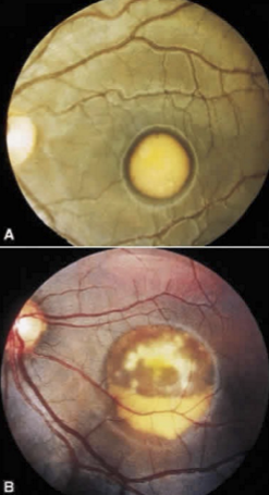
What are the characteristic electrophysiologic findings in Best disease?
Normal ERG
Reduced EOG
What is the classic fundus appearance of Best disease?
Vitelliform ("egg-yolk") lesion in the macula.
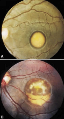
What are the stages of Best vitelliform dystrophy?
0. Abnormal EOG, no retinal findings
Pigment mottling
Vitelliform ("egg-yolk") lesion
Lesion begins to reabsorb
"Scrambled egg" stage with vision decline
Atrophy
How is Best disease managed?
Genetic counseling
Vision rehabilitation/supportive care
Long-term monitoring
When does Stargardt disease typically present, and how does vision change over time?
Onset in the 1st or 2nd decade
Progressive central vision loss
Vision may decline to ~20/200
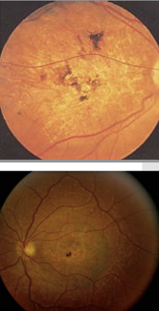
What are the characteristic fundus findings in Stargardt disease?
Early foveal mottling
"Beaten bronze" appearance of the macula
Yellow-white pisciform flecks surrounding the macula
Eventual geographic atrophy
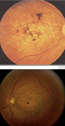
What angiographic finding is classically associated with Stargardt disease?
Dark choroid on fluorescein angiography.
What are the inheritance patterns and typical presentation of cone dystrophy?
May be autosomal dominant, autosomal recessive, or X-linked
Presents from teenage years through adulthood
Progressive decrease in visual acuity (often to ~20/200)
What are the hallmark symptoms of cone dystrophy?
Decreased central vision
Abnormal dark adaptation
Hemeralopia (poor vision in bright light)
What are the characteristic electrophysiologic findings in cone dystrophy?
Abnormal photopic (cone) ERG
Scotopic (rod) responses relatively preserved until late disease
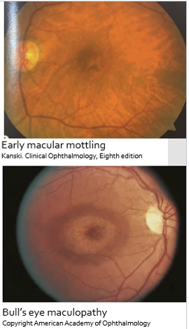
What retinal findings are associated with cone dystrophy?
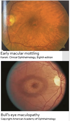
Early macular mottling
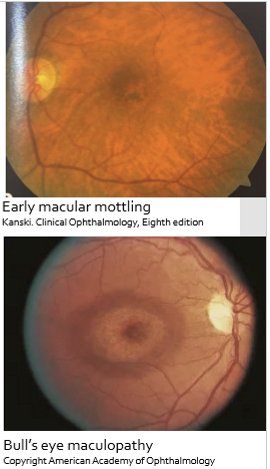
Bull's-eye maculopathy
Fluorescein angiography may show a hyperfluorescent window defect surrounding a hypofluorescent center
How do X-linked retinoschisis, Best disease, Stargardt disease, and cone dystrophy differ?
X-linked retinoschisis: Spoke-wheel macula, ↓ b-wave with normal a-wave.
Best disease: Egg-yolk lesion, normal ERG + abnormal EOG, AD.
Stargardt disease: Beaten-bronze macula, yellow flecks, dark choroid.
Cone dystrophy: Hemeralopia, abnormal photopic ERG, bull's-eye maculopathy.
What are the key inheritance patterns and epidemiology of retinitis pigmentosa (RP)?
Most common hereditary fundus disorder
Can be autosomal dominant, autosomal recessive, or X-linked
Symptoms typically begin in the 1st to 3rd decades of life
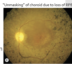
What is the classic fundus triad of retinitis pigmentosa?
Bone-spicule pigmentation
Arteriolar attenuation
Waxy optic disc pallor
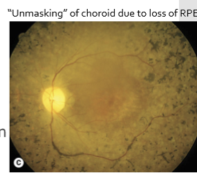
What are the characteristic electrophysiologic findings in retinitis pigmentosa?
Early: abnormal scotopic (rod) ERG
Photopic ERG relatively preserved initially
Reduced EOG
What future treatment strategy is being explored for retinitis pigmentosa?
Gene therapy, with multiple ongoing efforts targeting inherited retinal disorders.
What are the major congenital and acquired optic nerve disorders covered in pediatric ophthalmology?
Optic nerve pit
Optic nerve hypoplasia
Optic atrophy
Optic nerve coloboma
What is an optic nerve pit and where is it typically located?
Congenital excavation of the optic disc
Appears as a round or oval pit
Usually located in the inferotemporal (lower temporal) quadrant of the optic disc
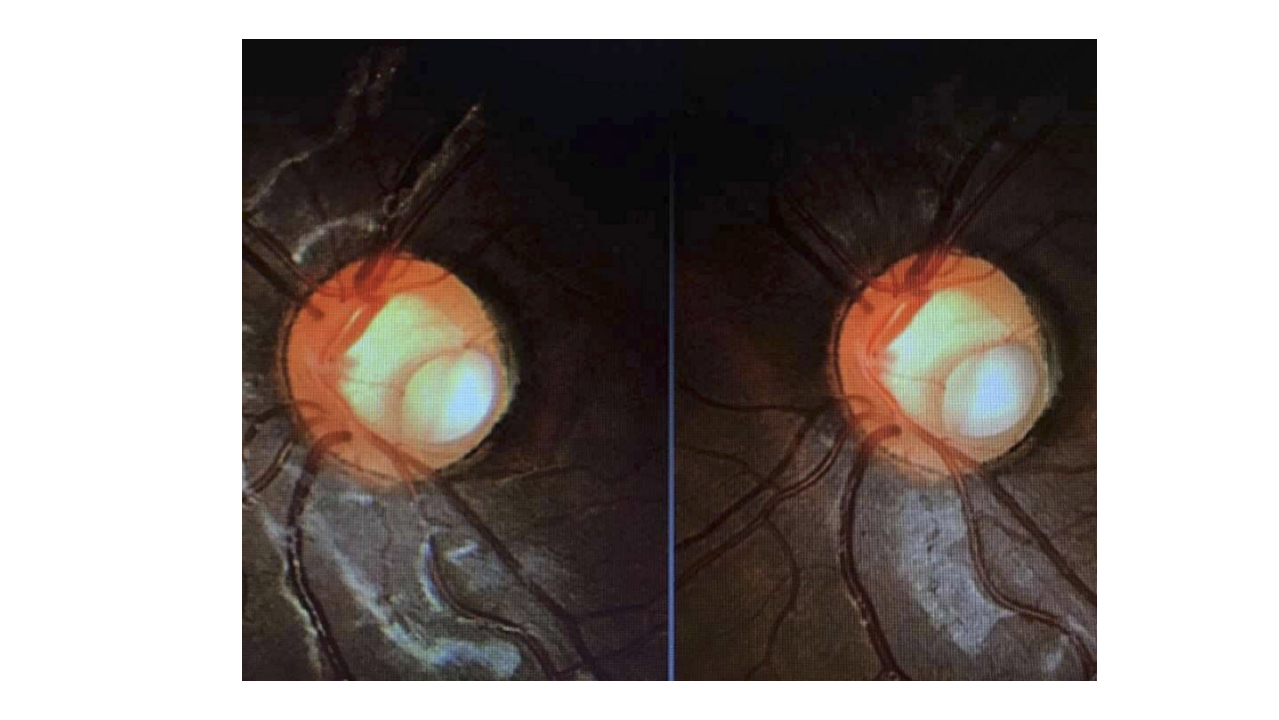
What visual findings can occur with an optic nerve pit?
Visual field defects
May mimic glaucoma on visual field testing
Optic disc is often larger than normal
What is the most important complication of an optic nerve pit?
Serous macular retinal detachment
Occurs in approximately 45% of patients
Median age of occurrence ≈ 30 years
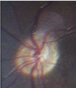
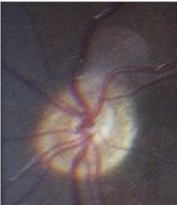
What is optic nerve hypoplasia, and how common is it?
Congenital underdevelopment of the optic nerve
Incidence ≈ 2 per 100,000 births
May be unilateral or bilateral
Why is bilateral optic nerve hypoplasia particularly important to recognize?
Bilateral disease may be associated with central nervous system abnormalities and warrants neuroimaging.
Classic association:
Septo-optic dysplasia (de Morsier syndrome)
Absence of the septum pellucidum
Agenesis of the corpus callosum
What maternal exposures have been associated with optic nerve hypoplasia?
Possible association with medications or alcohol exposure during gestation.
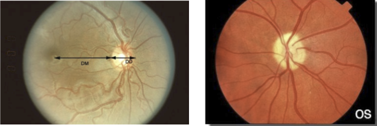
How does visual function vary in optic nerve hypoplasia?
Visual acuity ranges widely:
Near normal vision
To NLP (no light perception)
Visual field defects may also occur
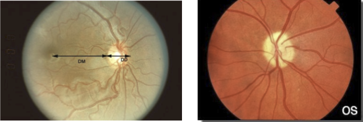
What is the classic fundus appearance of optic nerve hypoplasia?
Small optic nerve with a "double-ring sign"
Inner ring = chorioretinal atrophy
Outer ring = expected normal disc margin
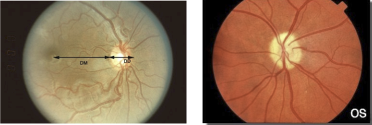
How is optic nerve hypoplasia assessed using the disc-macula/disc-diameter ratio?
Measure:
DM:DD = Disc-Macula distance : Disc Diameter
Ratio > 3:1 suggests optic nerve hypoplasia
What are the two major pediatric intraocular tumors emphasized in this section?
Retinoblastoma (malignant)
Choroidal hemangioma (benign vascular tumor)
What are the key epidemiologic and clinical features of retinoblastoma?
Occurs in approximately 1 in 20,000 births
Most common intraocular malignancy of childhood
Mean age at diagnosis: 18 months
Almost all cases diagnosed before 4 years of age
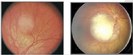
How does retinoblastoma typically present?
Leukocoria (white pupillary reflex) – most common presentation
Strabismus
Occasionally decreased vision or other signs of intraocular mass
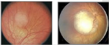
What imaging characteristic helps identify retinoblastoma?
Calcification within the tumor
High calcium content
Highly reflective on B-scan ultrasonography
What percentage of retinoblastoma cases are bilateral, and what is the prognosis?
Approximately 30% are bilateral
Survival rate exceeds 90% with modern treatment
What are the two major growth patterns of retinoblastoma?
Endophytic: Growth toward the vitreous with vitreous seeding
Exophytic: Growth into the subretinal space
What are the major vision- and life-threatening complications of retinoblastoma?
Optic nerve invasion
Metastatic spread
Why is RB1 genetic testing important in retinoblastoma?
Identifies hereditary disease
Helps guide family counseling
Patients with germline RB1 mutations have increased risk of additional non-ocular malignancies
What treatment modalities are used for retinoblastoma?
Chemotherapy
Radiation therapy
Cryotherapy
Laser photocoagulation
Enucleation
Treatment depends on tumor size, laterality, vision potential, and extent of spread.
What are the key features of a choroidal hemangioma?
Benign vascular tumor of the choroid
Appears as a reddish-orange lesion
Usually observed unless vision is threatened
When should a choroidal hemangioma be treated and how?
Treat only if it:
Affects vision
Causes retinal detachment
What are the major causes of retinal hemorrhages in children?
Abuse (nonaccidental trauma)
Anemia
Diabetic retinopathy
Leukemia
What systemic diseases should be considered when retinal hemorrhages are identified?
Hematologic disease (anemia, leukemia)
Vascular disease (diabetic retinopathy)
Trauma, especially child abuse
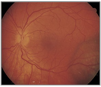
Why are retinal hemorrhages particularly concerning in children younger than 2 years?
In children < 2 years old, retinal hemorrhages should raise strong concern for nonaccidental trauma (abuse).
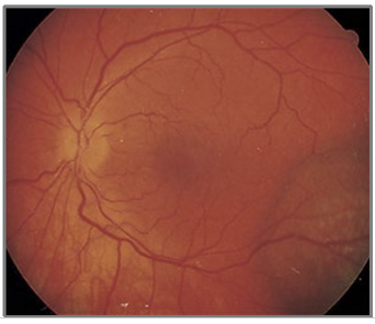
What retinal hemorrhage findings increase suspicion for child abuse?
Retinal hemorrhages in a young child
Bilateral retinal hemorrhages
Hemorrhages that are difficult to explain by accidental trauma
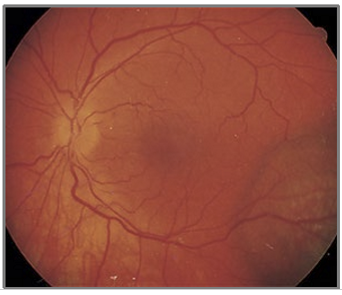
What alternative causes of retinal hemorrhages should be considered before concluding abuse?
Birth-related retinal hemorrhages (typically resolve within 1 month)
Falls
Accidental trauma
Medical causes (anemia, leukemia, diabetic retinopathy)
How can anemia present in a pediatric patient with retinal hemorrhages?
Low red blood cell count
May cause retinal hemorrhages
Systemic symptoms include:
Lethargy
Pallor
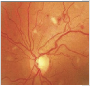
When retinal hemorrhages are seen with pallor and lethargy, what hematologic condition should be considered?
Anemia
Key clues:
Low RBC count
Pallor
Fatigue/lethargy
Retinal hemorrhages
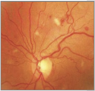
How common is diabetic retinopathy in children, and when should screening begin?
Rare before 10 years of age
Children should receive annual eye examinations after age 10
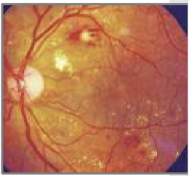
How does diabetic retinopathy behave in pediatric patients?
If present, diabetic retinopathy in children tends to be:
More severe
More rapidly progressive than in many adult patients
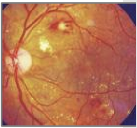
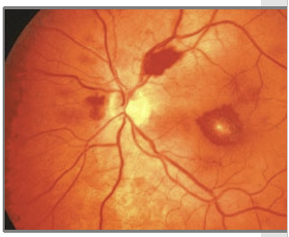
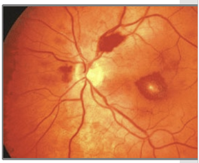
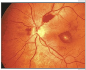
What retinal findings are commonly seen in leukemia?
Intraretinal hemorrhages
Roth spots
Cotton-wool spots
Dilated and tortuous retinal vessels
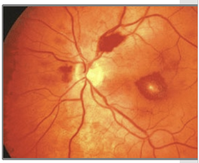
What is the most common childhood leukemia, and how frequently does ocular involvement occur?
Acute Lymphocytic/Acute Lymphoblastic Leukemia (ALL)
Accounts for approximately 90% of childhood leukemias
Ocular involvement occurs in about 75–80% of cases
What are the major systemic signs and symptoms of childhood leukemia?
Pallor
Fever
Easy bruising
Recurrent infections
Joint pain
Petechial hemorrhages
Why is early recognition of childhood leukemia important?
Can be rapidly fatal if untreated
Often responds well to:
Chemotherapy
Bone marrow transplantation
What are the major causes of retinal hemorrhages in children, and what clues help distinguish them?
Abuse: Infant <2 years, often bilateral hemorrhages
Anemia: Pallor, lethargy, low RBC count
Diabetic retinopathy: Usually after age 10; can progress rapidly
Leukemia: Roth spots, pallor, fever, bruising, infections