301: Anterior Abdominal Wall
 Call Kai
 Learn
Learn Practice Test
 Spaced Repetition
 Match
Match Flashcards
 Knowt Play
1/162

There's no tags or description
Looks like no tags are added yet.
Name | Mastery | Learn | Test | Matching | Spaced | Call with Kai | Chat |
|---|
No analytics yet
Send a link to your students to track their progress
163 Terms
External oblique
Internal oblique
Transversus abdominis
What are the 3 flat muscles of the anterolateral wall?
Rectus abdominis
Pyramidalis
What are the 2 vertical muscles of the anterolateral wall?
diaphragm
what is the roof of the abdomen?
femoral branch of genitofemoral n.
ilioinguinal n.
afferent innervation for cremaster m.:
genital branch of genitofemoral n.
efferent innervation for cremaster m.:
no floor
what is the floor of the abdomen?
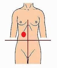
right upper quadrant
identify the region of the abdomen:
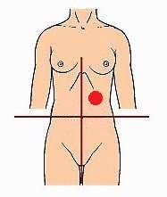
left upper quadrant
identify the region of the abdomen:
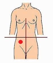
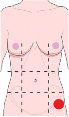
right lower quadrant
identify the region of the abdomen:
left lower quadrant
identify the region of the abdomen:
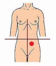
right hypochondrium region
identify the region of the abdomen:
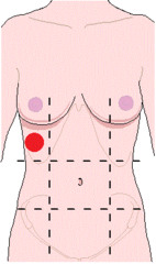
left hypochondrium region
identify the region of the abdomen:
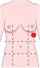
epigastric region
identify the region of the abdomen:
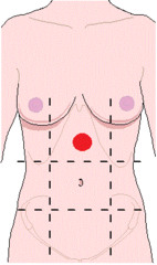
right lumbar (flank) region
identify the region of the abdomen:
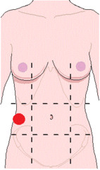
left lumbar (flank) region
identify the region of the abdomen:
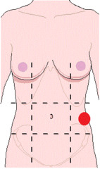
umbilical region
identify the region of the abdomen:
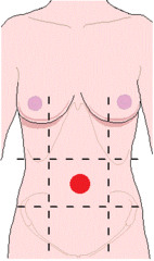
right inguinal (groin) region
identify the region of the abdomen:
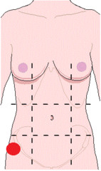
left inguinal (groin) region
identify the region of the abdomen:
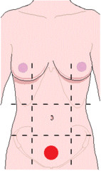
hypogastric region
identify the region of the abdomen:
median plane (midsagittal)
the line that separates the right and left quadrants is the:
transumbilical plane
the line that separates the upper and lower quadrants is the:
subcostal plane
the line that separates the hypochondriac and lumbar regions is the:
intertuberculer plane
the line that separates the inguinal and lumbar regions is the:
midclavicular planes
the two lines that carve out the epigastric, umbilical and hypogastric regions are called:
superficial Camper's fascia
what layer of the abdominal wall lies directly under the skin?
Scarpa's fascia
what layer of the abdominal wall lies directly under the Campers fascia?
Skin
Superficial fascia - Camper's with adipose tissue, paraumbilical veins & Scarpa's /membranous
- Deep (investing) fascia - CT sheathing muscles & Transversalis fascia
- Extraperitoneal (endoabdominal) fat
- Parietal peritoneum
From superficial to deep, what are the layers of the anteriolateral abdominal wall, excluding the muscles?
External oblique
Internal oblique
Transverse abdominis
From superficial to deep, what are the muscle layers of the anteriolateral abdominal wall?
external oblique m.
if you pierce the right side of the abdomen with a needle to the right of the midclavicular line, what muscle will you run into 1st?
internal oblique m.
if you pierce the right side of the abdomen with a needle to the right of the midclavicular line, what muscle will you run into 2nd?
transversus abdominis m.
if you pierce the right side of the abdomen with a needle to the right of the midclavicular line, what muscle will you run into 3rd?
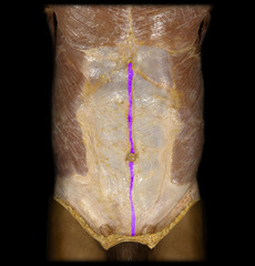
linea alba
Identify the structure
external oblique m.
Identify the structure
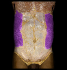
ribs 5-12
what ribs does the external oblique m. attach to?
thoracoabdominal (intercostal) n. - T7-T11
Subcostal n. - T12
innervation for external oblique m.:
midclavicular lines
the external oblique m. will continue anteriorly as the external oblique aponeurosis at the:
linea alba, pubic tubercle, anterior half of iliac crest
Where are the insertions for the external oblique m?
Muscular part of external oblique m.
Aponeurotic part of external oblique m.
What are the two parts of the external oblique m?
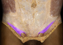
Inguinal ligament
Identify the structure
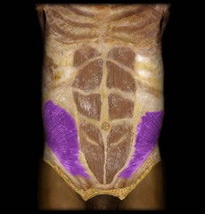
internal oblique m.
Identify the structure
thoracoabdominal (intercostal) nn. - T7-T12
iliohypogastric nn. & ilioinguinal nn. - L1
innervation for the internal oblique m.:
Thoracolumbar fascia, iliac crest, anterior superior iliac spine, iliopsoas fascia
Origin for the internal oblique m.:
10th to 12th ribs (lower borders), linea alba
Insertion for the internal oblique m.:
External oblique
Identify the structure
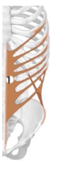
Internal oblique
Identify the structure
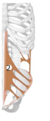
Internal oblique and internal oblique aponeurosis
What are the two parts of the internal oblique?
transversus abdominis m.
Identify the structure
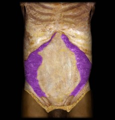
thoracoabdominal (intercostal) nn. - T7-T12
iliohypogastric nn. & ilioinguinal nn. - L1
innervation for the transversus abdominis m.:
7th-12th costal cartilages, thoracolumbar fascia, iliac crest, anterior superior iliac spine, iliopsoas fascia
What are the origins of the transversus abdominis muscle?
linea alba, pubic crest
What are the insertions of the transversus abdominis muscle?
Transversus abdominis m.
Identify the structure
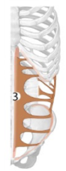
rectus abdominis m.
Identify the structure
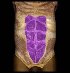
pyramidalis m.
Identify the structure
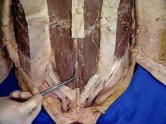
thoracoabdominal (intercostal) n. - T5-T12
innervation for the rectus abdominis m.:
Pubis
Origin for rectus abdominis m.:
5th to 7th intercostal cartilages, xiphoid process of sternum
Insertion for rectus abdominis m.:
O: Pubis
I: Linea alba
Origin and insertion of pyramidalis:
subcostal, T12
Innervation of pyramidalis:
Rectus abdominis m.
Identify the structure
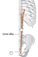
subcostal n.
innervation for the transversus abdominis m.:
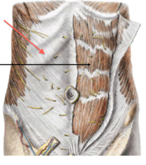
Rectus sheath
Identify the structure
arcuate line
what separates the difference in aponeurosis around the rectus abdominis m.?
above
in a dissection, you notice 1½ anterior and 1½ posterior layers of aponeurosis around the rectus abdominis. You must be _______ the arcuate line.
below
in a dissection, you notice 3 anterior and 0 posterior layers of aponeurosis around the rectus abdominis. You must be _______ the arcuate line.
T7-T11
the thoracoabdominal n. originates form what spinal nerve roots?
T12
the subcostal n. originates form what spinal nerve roots?
L1
the iliohypogastric n. originates form what spinal nerve roots?
L1
the ilioinguinal n. originates form what spinal nerve roots?
Between the second and third muscle layers (internal oblique and transverse adominis mm.)
Where do the neurovascular bundles of the anterolateral abdominal wall primary run between?
T10
What vertebra is found at the belly button level?
musculophrenic a.
superior epigastric a.
what are the two branches of the internal thoracic artery that supply the abdomen?
Rectus muscle
What muscle do the superior and inferior epigastric arteries anastomose with?
superior epigastric a.
Identify the structure
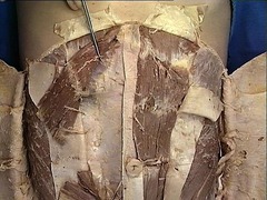
musculophrenic a.
Identify the structure
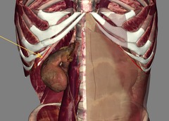
inferior epigastric a.
Identify the structure
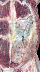
superficial epigastric a.
Identify the structure
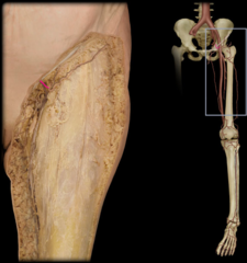
inferior and superior epigastric aa.
what two arteries lie superficial to the rectus abdominis and anastomose?
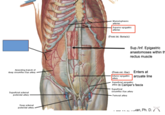
Inferior epigastric artery
What artery enters at the arcuate line?
Anastomoese with lower intercostal, subcostal, and lumbar arteries
What is shown at the blue box?
Superior epigastric veins
Thoracoepigastric vein
Paraumbilical veins in round ligament of liver
Inferior epigastric veins
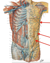
What are four of the important veins to focus on of the anterolateral abdominal wall?
Superior epigastric veins
Identify the structure
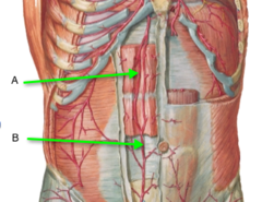
Thoracoepigastric vein
Identify the structure
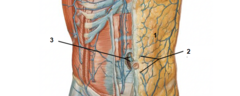
Paraumbilical veins in round ligament of liver
Identify the structure #3
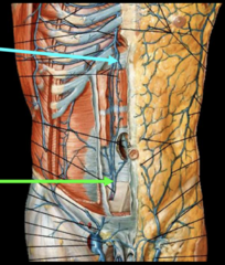
Inferior epigastric veins
Identify the structure at the green arrow
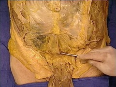
median umbilical fold/ligament
Identify the structure
medial umbilical folds/ligaments
#2
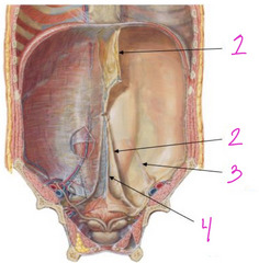
lateral umbilical folds/ligaments
#3
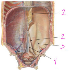
lateral umbilical folds
which umbilical folds house the inferior epigastric vessels?
medial umbilical folds
which umbilical folds house the fetal (obliterated) umbilical arteries?
median umbilical fold
which umbilical folds house the remnant of the fetal urachus?
inferior epigastric vessels
inguinal ligament
lateral boarder of rectus abdominis m.
what are the boarders on the Hesselbach triangle?
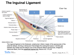
Inguinal ligament
Poupart's ligament, a fibrous band formed by the inferior border of the aponeurosis of the Ex.Ob. That extends from the ASIS to the pubic tubercle
Inguinal ligament
the thickened, underturned, inferior margin of the aponeurosis of the external oblique, forming a retinaculum that bridges the subinguinal space. A slit-like gap between the medial and the lateral crura of the external oblique aponeurosis, bridged by intercrural fibers, form the superficial inguinal ring.
superficial inguinal ring
A slit-like gap between the medial and the lateral crura of the external oblique aponeurosis, bridged by intercrural fibers, form the:
Inguinal canal
• The obliquely directed passage through the layers of the lower abdominal wall that transmits the spermatic cord in the male and the round ligament in the female.
• Deep (internal) inguinal ring
• Superficial (external) inguinal ring
Spermatic cord (male)
What structure is in the inguinal canal in males?
Round ligament of the uterus (female)
What structure is in the inguinal canal in females?
Deep inguinal ring
What structure is at #1?
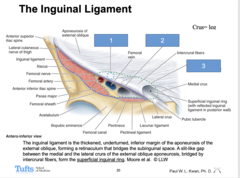
Sublinguinal space
What structure is at #2?
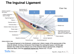
Myopectinaeal orifice
What structure is at #3?