Equine pelvic limb arthrology and synovial structures
1/17
There's no tags or description
Looks like no tags are added yet.
Name | Mastery | Learn | Test | Matching | Spaced |
|---|
No study sessions yet.
18 Terms
what are the joints of the stifle
femeropatellar
medial femorotibial
lateral femorotibial
what two joints of the stifle have natural communication
femeropatellar
medial femorotibial joint
does each joint of the stifle have its own synovial compartment
yes
if cut what can cause a complication during stifle surgery
infrapatellar fat pad
What are the components of the patellar locking mechanisms
patella
Intermediate patellar ligament
medial patellar ligament
medial trochlear ridge with tubercle
what is upward fixation of the leg
it is where the medial patellar ligament is locked in place and the patella cannot be moved
what is gonitis
inflammation of the stifle joint
what are the synovial compartments of the tarsus
tarsocrural
proximal intertarsal
distal intertarsal
tarsometatarsal
which tarsal joint has the greatest movemnt
tarsocrural
which tarsal joints have 100% communication between them
tarsocrural
proximal intertarsal
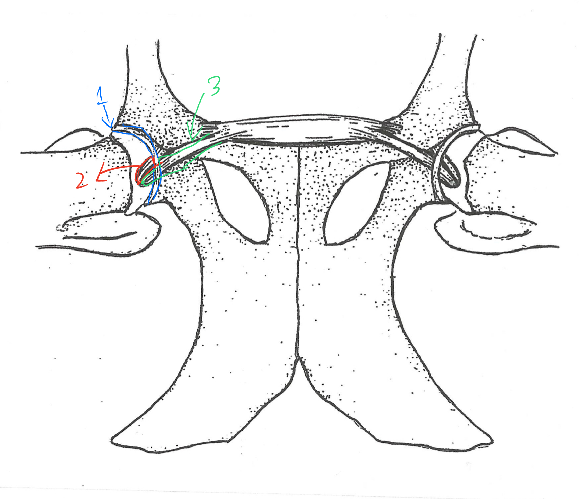
label the ligaments of the hip
transverse acetabular
round lig of head of the femur
accessory lig of hip abdominus
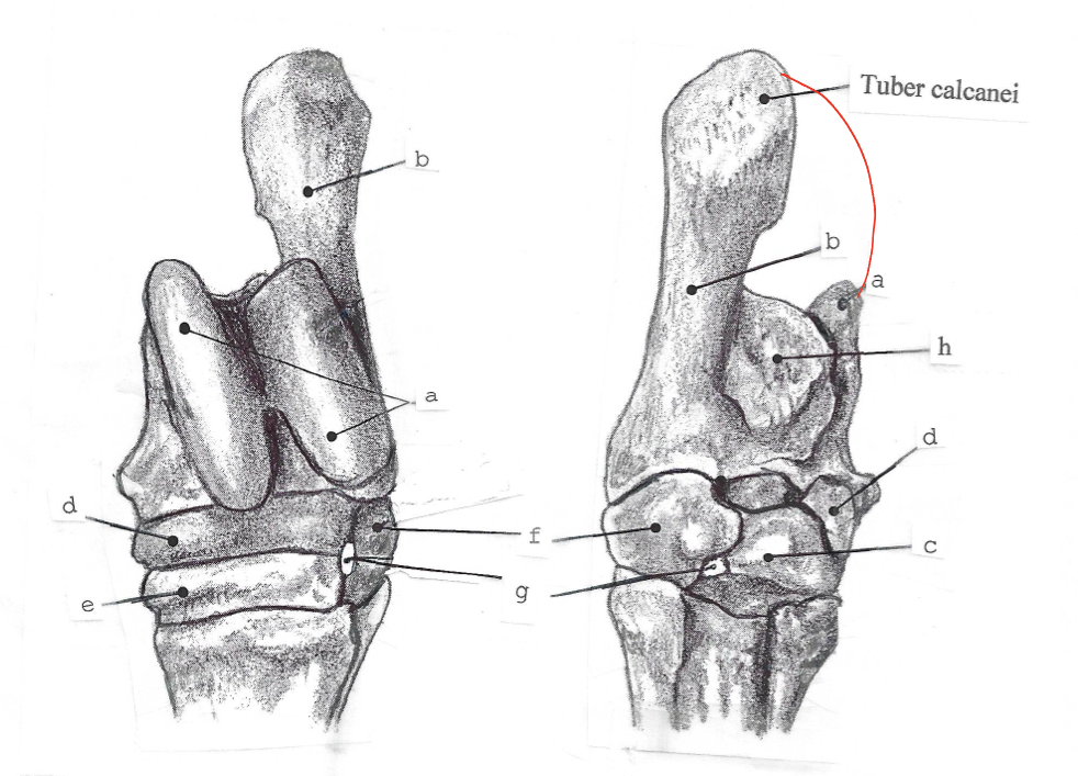
label the bones of the equine tarsus
a. talus
b. calcaneus
c. T1 and T2
d. central tarsal bone
e. t3
f. t4
g tarsal canal
h. tarsal groove
the tarsal groove and flexor retinaculum makes the ______
flexor canal
what artery travels through the tarsal canal
the perforating tarsal artery
name the synovial joint pouches of the hock
dorsal
medioplantar
lateroplantar
the subtendinous bursa is a cushion for the tendon of what muscle
superficial digital flexor
which bursa is inconsistent and generally arises as a result of strain to the part of the hock
subcutaneous bursa
what tendon and bursa combo are present of the median portion of an equine hock
cunean