Microscopic Urinalysis Vocabulary
1/61
Earn XP
Description and Tags
Vocabulary flashcards covering key cells, casts, crystals, and concepts encountered during microscopic urinalysis of urine sediment.
Name | Mastery | Learn | Test | Matching | Spaced |
|---|
No study sessions yet.
62 Terms
MICROSCOPIC
EXAMINATIOn
• Many abnormalities in urine that can be found on microscopic
examination can not be detected by strips or physical examination.
– Bacteria, crystals, and other nasties!
• Important final step in a complete urinalysis.
• Can help recognize diseases of the urinary tract, and verify dipstick
findings for occult blood.
Urine Sediment
– The “pellet” of solids formed after centrifugation
– We look at centrifuged sediment to perform the microscopic analysis
• Most species = little to no sediment
• Evaluate the pellet before you re-suspend your urine!
Pellet
Should be evaluated for:
-Size, Small? Medium? Large?XL?-more subjective
-Color, White? Red? Brown? Spotted?-A little less subjective
• Technically speaking, every urine sample has a pellet. With normal
urine, it should be very small, or not visible at all upon gross
inspection.
Microscopic Sample Preparation
• Place 5 to 10 ml of a well-mixed sample in a centrifuge tube
• Spin for 3 to 5 minutes at approximately 1000 to 2000 rpm
After centrifugation:
– record the size/color of pellet
– gently pour off the supernatant (liquid portion above the pellet)
• leave approximately 0.5 ml of urine in the bottom of the tube
Microscopic Slide Preparation
• Re-suspend the sediment by gently flicking the bottom of the
centrifuge tube or mixing gently with a pipette
Sample Preparation-Staining
• Sediment may be examined stained or unstained
• Staining sediment can often introduce artifacts like precipitates and
bacteria.
• Staining also dilutes the sample, therefore recording results from
stained slides is not as accurate.
• Stain works best for verifying presence of epithelial cells and other
abnormalities after looking at the unstained urine first
Unstained Preparation
• Place a small drop of the re-suspended sediment on a clean glass slide,
• Cover with a cover slip
• Examine immediately!
Stained preparation
• Mix 1 drop of stain with the suspended sediment
• Place a drop of sediment/stain mix on a microscope slide
• Cover with coverslip.
Reading and Reporting
• Scan for large artifacts and debris on 4x and 10x – note any large
cells/rafts- large sheets of epithelial cells
• Read on 40x (DIM LIGHT!)
• Reporting is done like platelets, average over 10-15 fields:
– Cells (WBC/RBC/Epi) = #seen per hpf
– Casts = #seen per hpf
– Crystals = #seen per hpf
– Bacteria = +, ++, +++ (subjective)
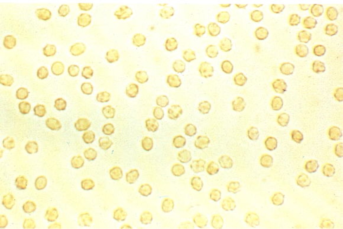
RBCS
• Normal Value = 0-5 per hpf
• High amounts indicate bleeding along the urinary tract
• Iatrogenic causes must be ruled out!
Red blood cells
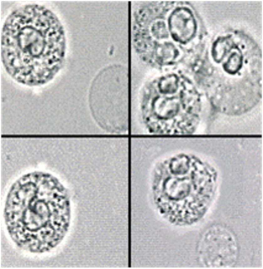
White Blood Cells
• Normal Value = 0-3 per hpf
• High amounts indicate infection
• May be seen near or actually phagocytizing bacteria with bad
infections
• “Glitter” appearance
White Blood Cells
Epithelial Cells
• 3 Kinds
– Renal
– Transitional (round and caudate forms)
– Squamous
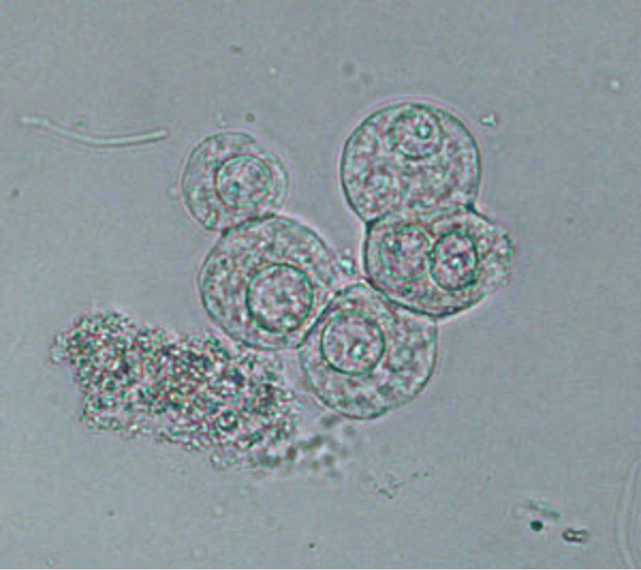
Renal Epithelial Cells
• Very rare, usually do not see any
• Slightly bigger than WBCs
• Presence indicates damage to the renal tubules
Renal Epithelial Cells
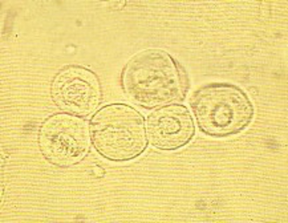
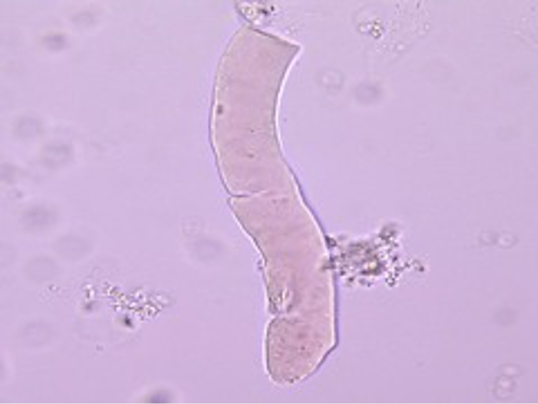
Transitional Epithelial Cells
• Can be perfectly round or have a tail (caudate form)
– Slightly larger than renal epithelial cells
• May see 0-1 per hpf normally, but usually only occasional
• More than normal can indicate damage to bladder wall, cancerous
conditions
• Damage from poor catheterization technique can result in greater
exfoliation of these cells from the bladder
Transitional Epithelial Cell(round)
Transitional Epithelial cell (caudate)
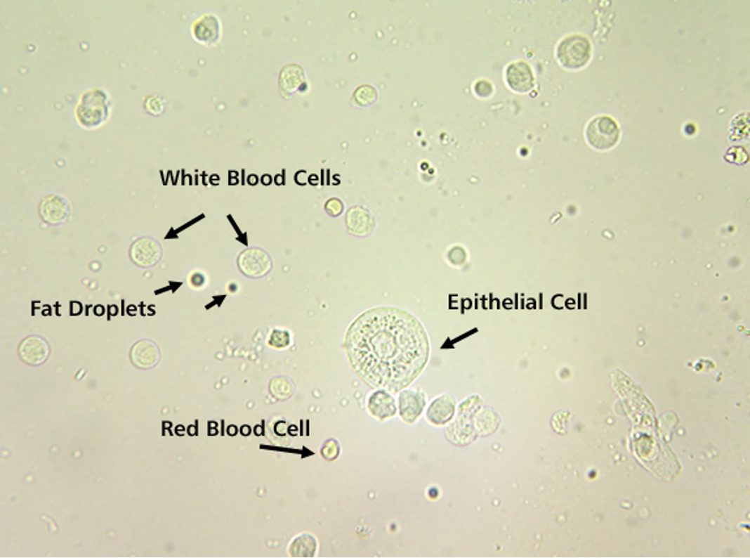
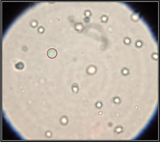
Various Cells
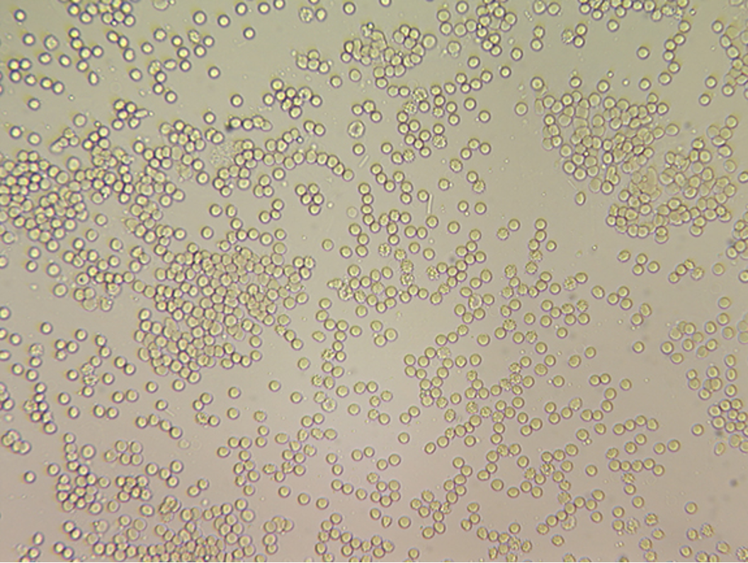
RBCs-TNTC
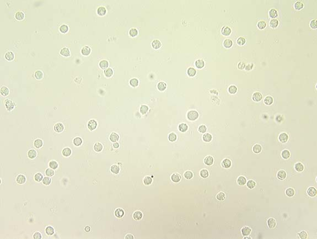
Lots of WBCS
Crenated RBCS and WBCS
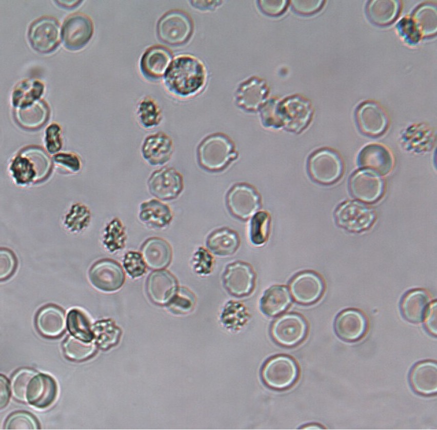
RBCS/WBCS
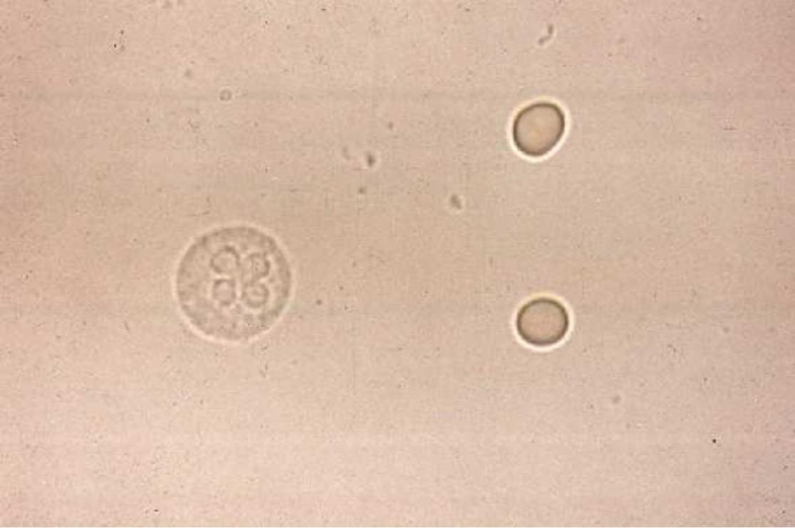
Air Bubbles(vary in size)
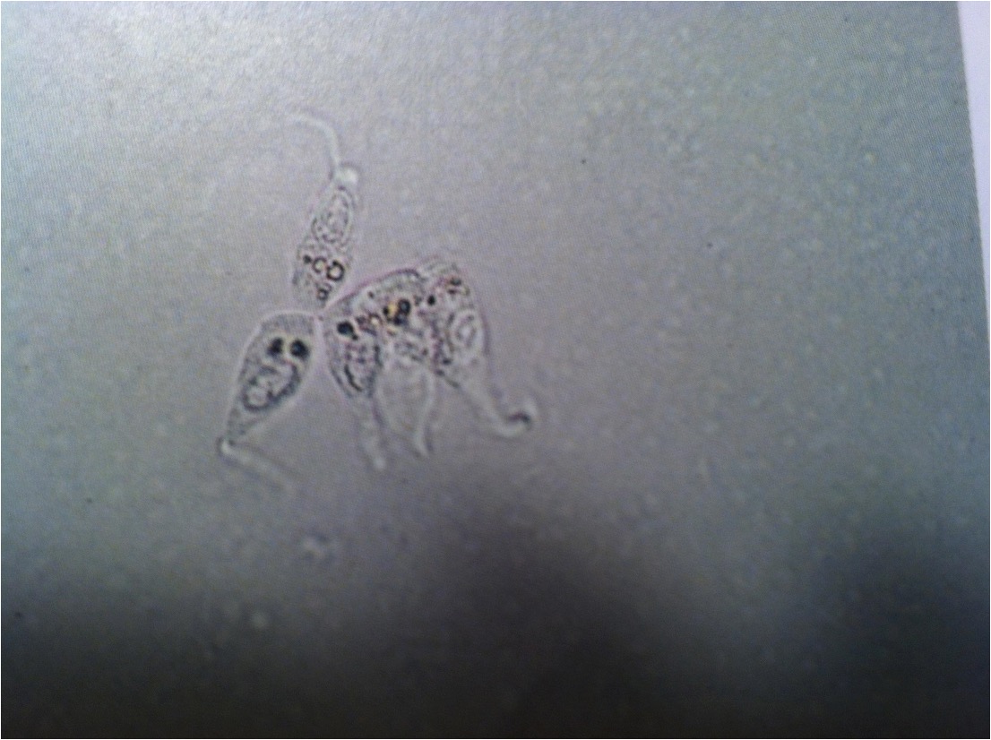
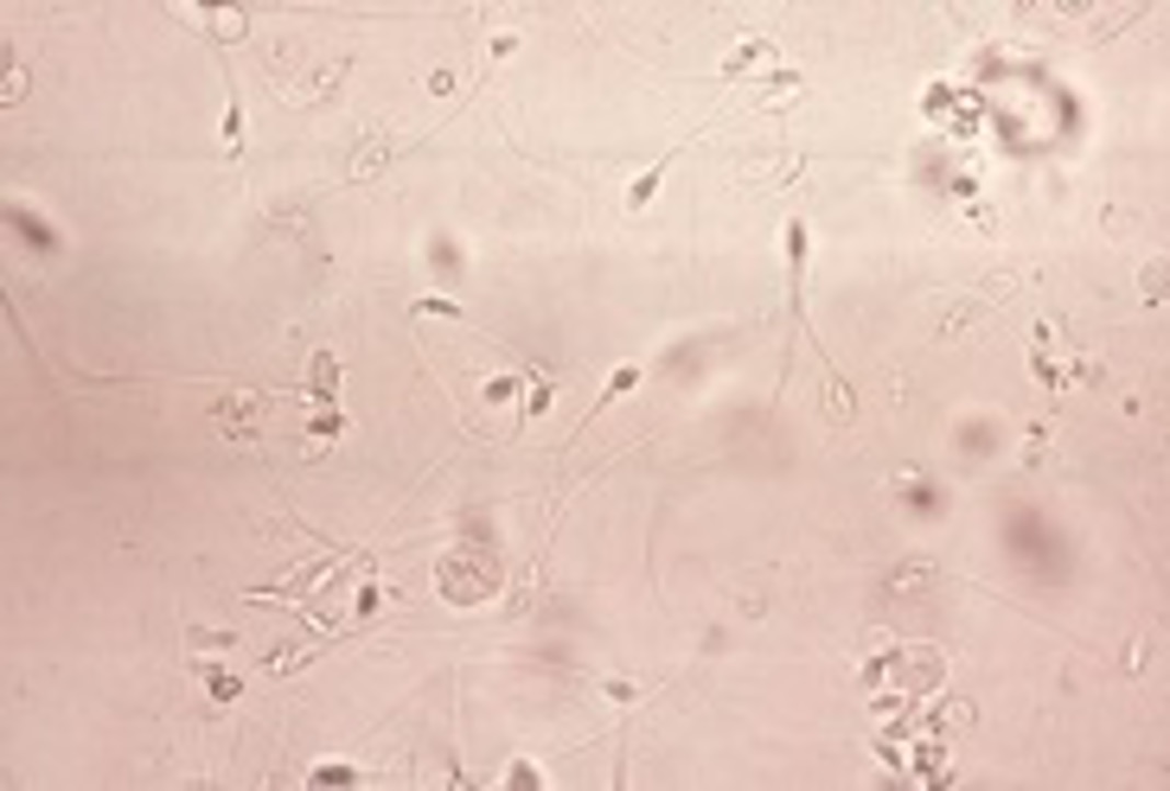
Sperm
Fat Droplets(vary in size)
Bacteria
Squamous Epithelial Cells
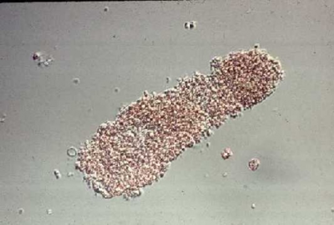
Granular Cast
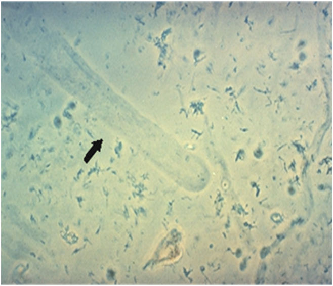
Waxy Cast
Hyaline Cast
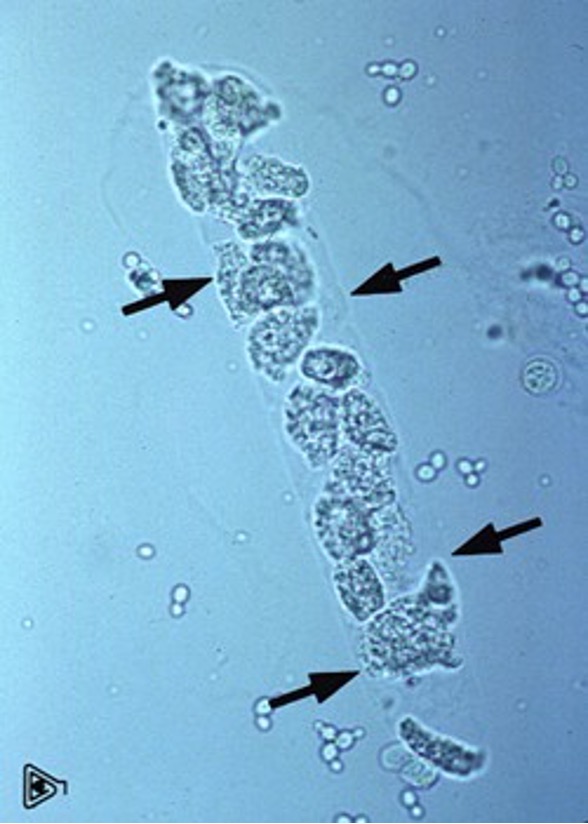
Cellular Cast
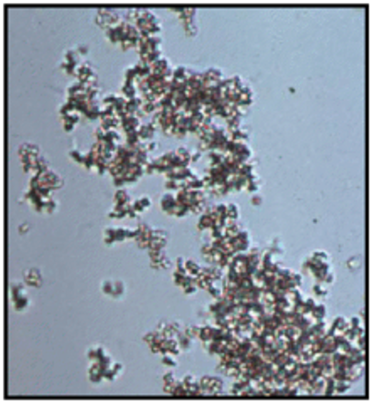
Amorphous Crystals
Triple Phosphate Crystals(struvite)
Calcium oxalate Dehydrate (Calcox)
Calcium Oxalate Monohydrate
Bilirubin Crystals
Calcium Carbonate Crystals
Calcium Carbonate crystals
Biurate crystals (“Thorn Apple”)
Biurate crystals (“Thorn Apple”)
Sulfonamide Crystals
Uric Acid Crystals
Leucine Crystals
Tyrosine Crystals
Cystine Crystals
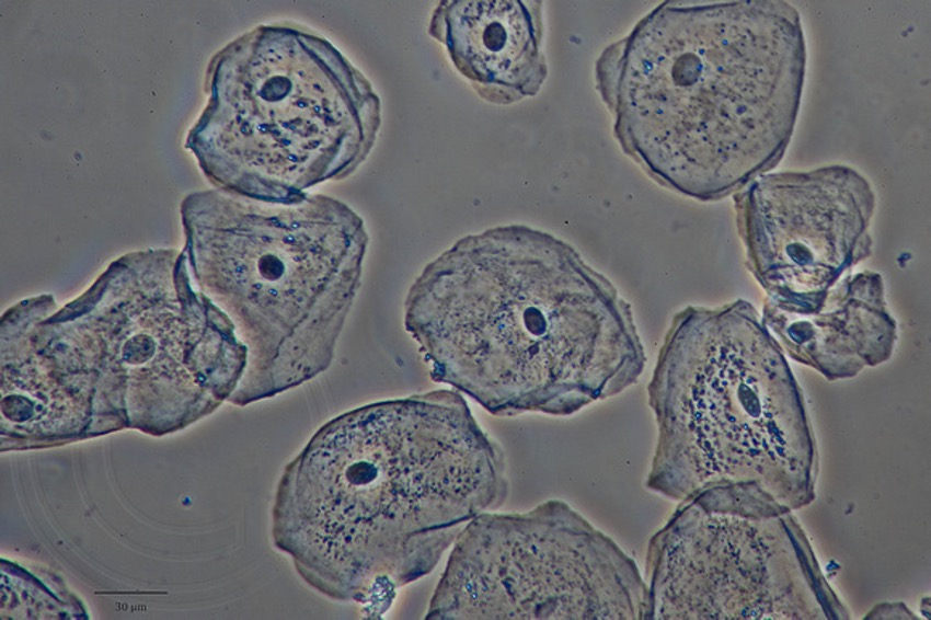
SQUAMOUS EPITHELIAL CELLS
• Most common, largest
• Look like cornflakes!
• 0-3 per hpf can be seen
– Higher amounts on catheter samples
• Higher numbers can indicate damage to lining of urinary tract,
cancerous conditions
Casts
• Formed by sediment resting in the renal tubules
• Made of protein matrix
• Can indicate damage or infection of the kidneys
• Come in many forms: granular, waxy, cellular (many types), and hyaline
• Normal to see presence in very low numbers (may see 1-2 in 10 hpf)
With renal tubular injury, epithelial cells slough into the lumen of the
renal tubules and are caught up within a mucoprotein matrix made
from Tamm-Horsfall protein (hyaline cast). This forms a cellular cast.
With time, the epithelial cells degenerate and can no longer be
recognized as cells within the hyaline matrix, thus forming coarsely
granular, then finely granular, casts. Waxy casts are the final step in the
formation of casts and indicate tubular injury (acute or chronic).
Crystals in urine
• AKA Crystalluria
• Can be caused by many factors: pH, medications, disease process, diet,
etc.
• May indicate presence of stones
• Can form in healthy urine with no disease process, especially if sample
ages
Amorphous Crystals
Found in all pH of urine
– Amorphous phosphate in alkaline
– Amorphous urate in acidic
• Most common crystals seen, usually look like “trash” or “junk”
• Mostly harmless
• May form large amounts in older samples
Triple Phosphate Crystals (struvite)
• Most commonly form in alkaline urine
– Can form in neutral and acidic as well
• “Coffin lid” appearance
• May occasionally look leaf-like in groups
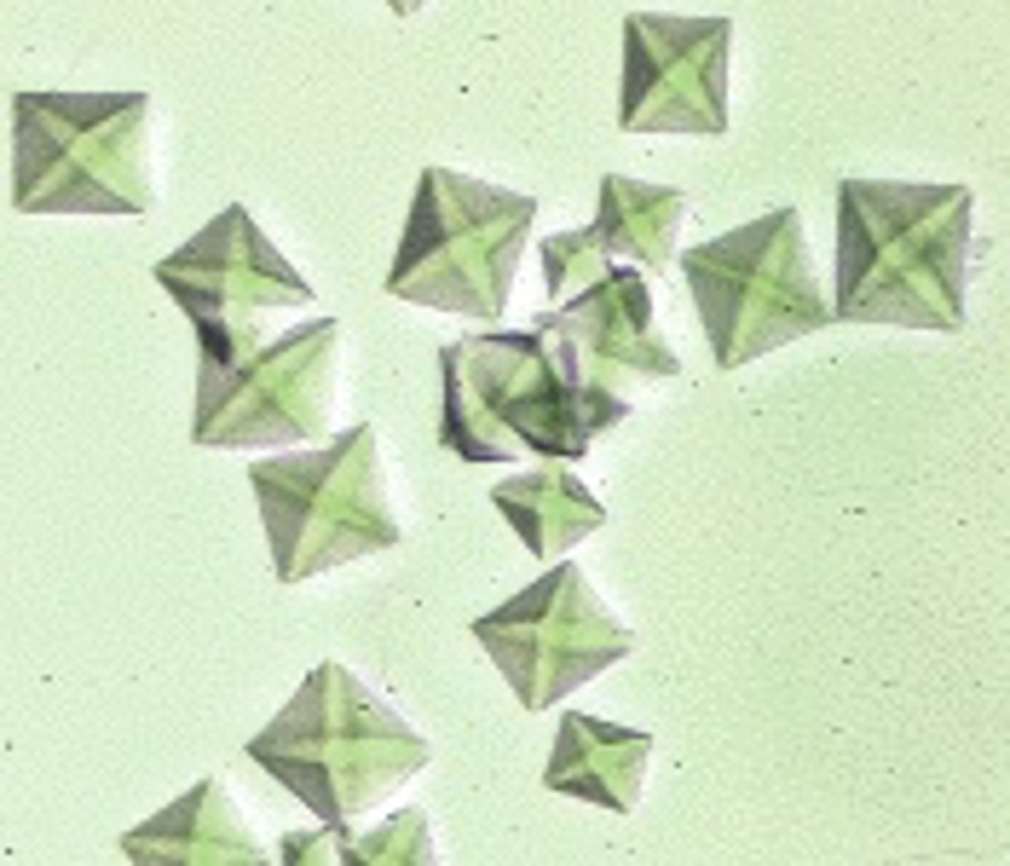
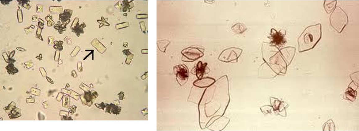
Calcium Oxalate Crystals
• Most commonly form in acidic urine
– Can form in neutral and alkaline as well
• Dihydrate Form = “diamonds”
– Usually form due to pH
• Monohydrate Form = “picket fence”
– Antifreeze toxicity!
Bilirubin Crystals
• Acidic urine
• May appear as needles or plates
• Indicate bilirubin dumping into urine
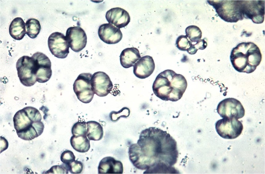
Calcium Carbonate Crystals
• Form in alkaline urine
– Normal to see in horses and rabbits!
• Dumbbell or oval shaped
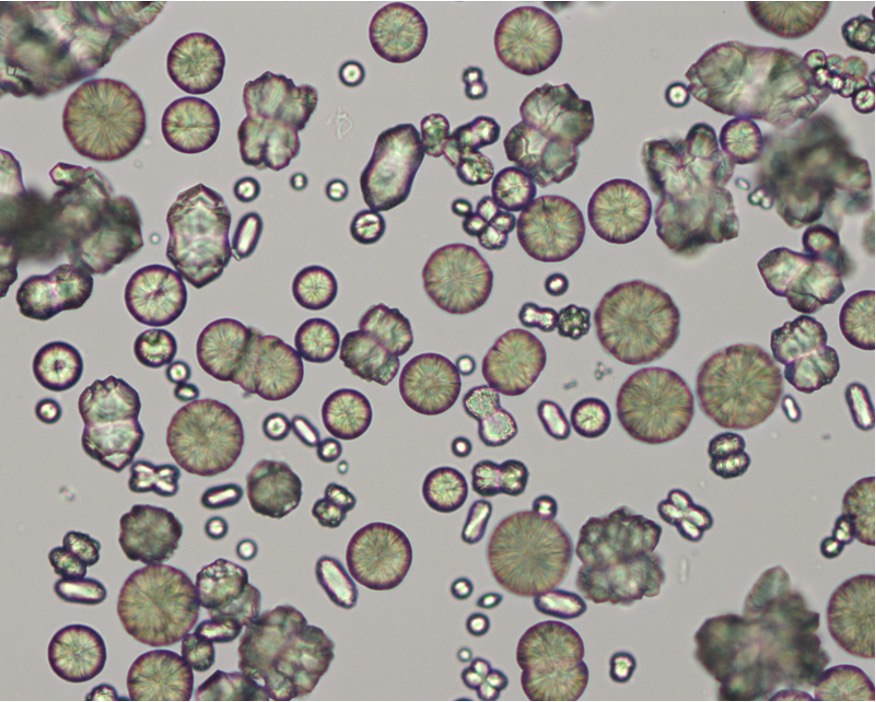
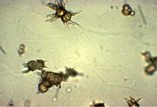
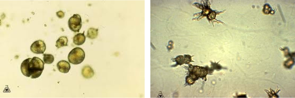
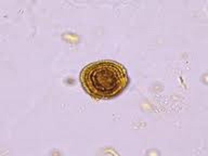
Biurate Crystals(“thorn apple”)
• Are brown and round, with long irregular spicules (thorn apple shape)
• Often the spicules fracture; remaining crystal is brown, with fine
radiating lines
• Seen in slightly acidic, neutral, or alkaline urine
Sulfonamide Crystals
-Are round, usually dark, with individual crystals radiating from the
center
• Seen in Acidic-neutral urine
Uric acid Crystals
• Uric acid crystals take on a variety of shapes but are usually diamond
or rhomboid
• Not commonly found in dogs and cats (Except Dalmations)
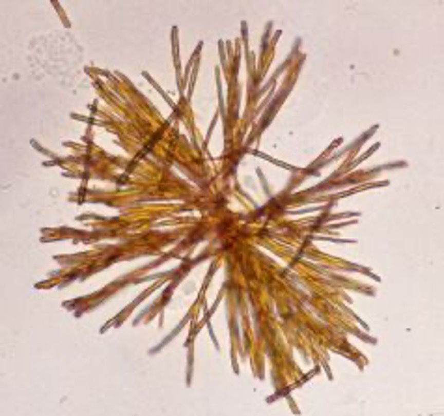
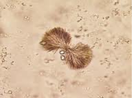
Leucine crystals
-Leucine crystals are wheel or “pin cushion” in shape and are yellow or
brown
• Seen with acidic urine
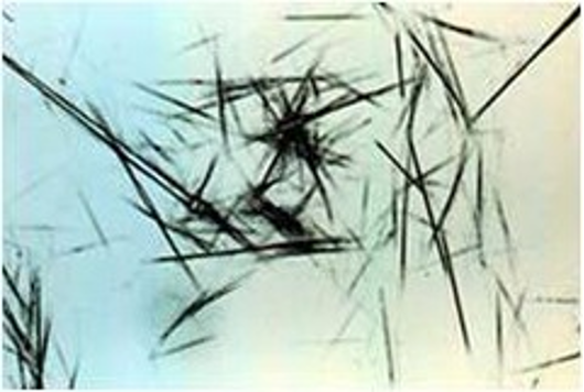
Tyrosine Crystals
• Tyrosine crystals are dark, have needlelike projections, and are highly
refractile
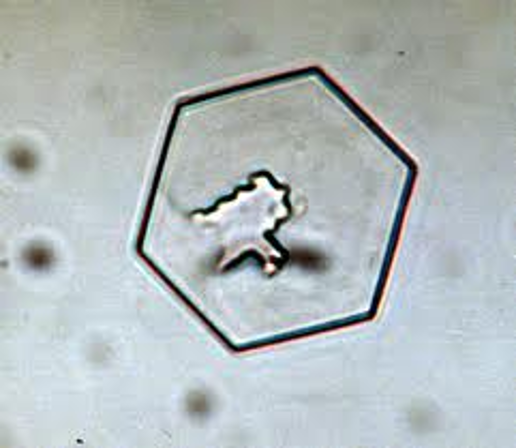
Cystine crystals
• Cystine crystals appear flat, hexagonal, and are colorless and thin
• Acidic/ Neutral Urine.
• Rare inherited error of amino acid metabolism affecting many breeds
of dogs. (Pitbulls)
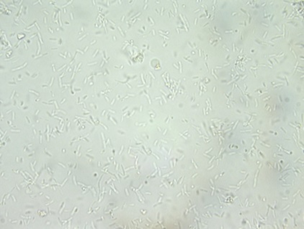
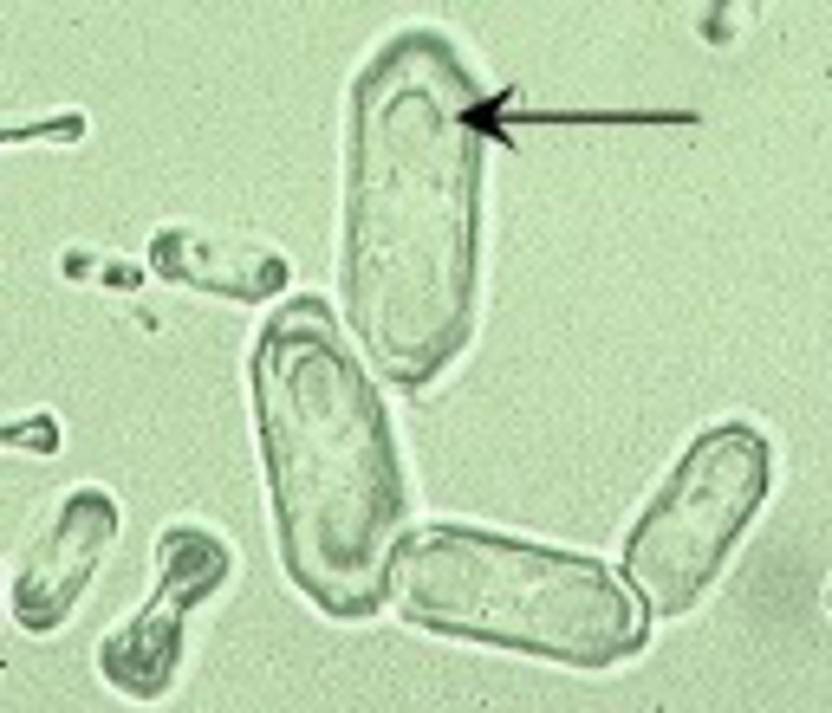
Bacteria
• Small and consistent in shape
• May appear as spheres (cocci) or rods (bacilli)
• Urine normally sterile -- Large numbers indicate UTI
• Can be introduced to a sterile sample through poor technique or
catch method
– Free-Catch is least sterile!