MSK & Breast
1/34
There's no tags or description
Looks like no tags are added yet.
Name | Mastery | Learn | Test | Matching | Spaced | Call with Kai |
|---|
No analytics yet
Send a link to your students to track their progress
35 Terms
When do we perform infant hip evaluations?
4 - 6 weeks of age
What is hip dysplasia?
Inability of hip socket to properly support femoral head
What are the symptoms of hip dysplasia?
Hip click on physical exam
Limited abduction
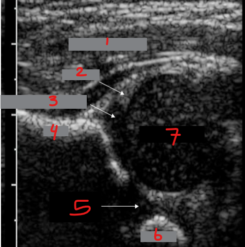
Identify this image.
Gluteal muscles
Labrum
Acetabulum
Ilium
Triradiate cartilage
Ischium
Femoral head
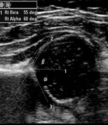
Which angle is used to evaluate hip dysplasia?
Alpha
What are the classifications for hip dysplasia?
Type I or normal: Alpha angle > 60
Type II or normal from newborn to three months: Alpha angle 44 - 60
Type III or dislocation: Alpha angle < 43
Type IV or gross dislocation: Alpha angle not measurable
What is transient arthritis?
Inflammation of hip joint associated with recent respiratory infections
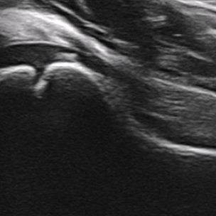
Identify this image.
Normal pediatric hip
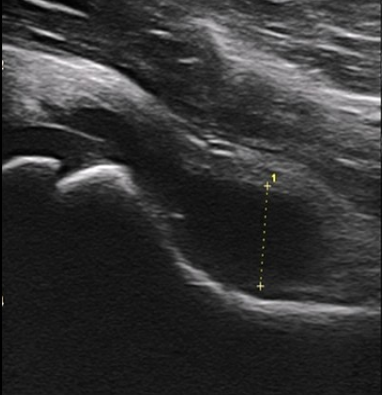
Identify this image.
Transient arthritis seen as effusion in joint
What is septic arthritis?
Inflammation of hip joint due to bacterial infection that is considered a MEDICAL EMERGENCY
What is the most common tendon to rupture?
Achilles tendon
What is the sonographic appearance of a tear in tendon?
Focal thinning
Discontinuity of internal fibers
Hematoma
Effusion
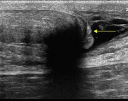
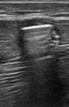
Identify this image.
Achilles tendon tear
What is tendinosis?
Degenerative changes in tendon with no signs or symptoms of damage
What is tendinitis?
Acute inflammation of tendon with symptoms
What is bursitis?
Inflammation and fluid accumulation of a joint bursa
What is a Baker’s cyst?
Cystic structure located in medial popliteal fossa
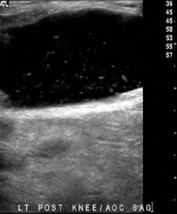
Identify this image.
Baker’s cyst
What is the most common hand tumor?
Ganglion cyst
What is a ganglion cyst?
Cystic formation adjacent to joint
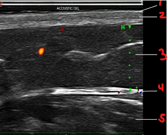
Identify this image.
Layers of skin
Epidermis
Dermis
Hypodermis or subcutaneous adipose tissue
Fascia
Muscle
Identify this image.
Layers of skin
Epidermis
Dermis
Hypodermis or subcutaneous adipose tissue
Fascia
Muscle
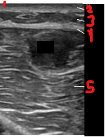
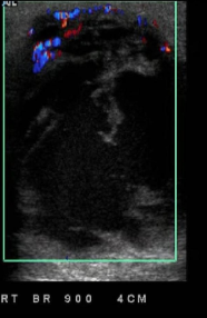
Identify this image.
Foreign body located in muscle
What is reactive hyperemia?
Hypervascularity around foreign body with inflammation
What is a sebaceous cyst?
Epidermal inclusion cyst located in inframammary fold, axillary area, and near areola
Identify this image.
Sebaceous cyst
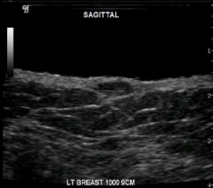
What is acute mastitis?
Inflammation of breast that most commonly occurs during lactation (puerperal mastitis)
Identify this image.
Acute mastitis
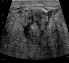
What is a breast abscess?
Complex cystic structure that causes fever and swelling
Identify this image.
Breast abscess
What is the most common benign breast mass?
Fibroadenoma
What is the most common malignant breast mass?
Invasive ductal carcinoma
What sonographic characteristics increase suspicion of malignancy in a breast mass?
Irregular margins and indistinct borders
Hypoechoic
Heterogenous
Posterior shadowing
Spiculations
Fine, linear branching calcifications
Vertical orientation
Fixed position
What is a galactocele?
Localized accumulation of milk caused by obstruction of lactiferous ducts
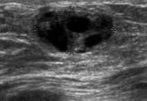
Identify this image.
Galactocele