35. MAXILLARY FRACTURES. TREATMENT.
1/17
There's no tags or description
Looks like no tags are added yet.
Name | Mastery | Learn | Test | Matching | Spaced | Call with Kai |
|---|
No analytics yet
Send a link to your students to track their progress
18 Terms
what are the aims of treatment in maxillary fractures?
restore anatomical form, function, aesthetics and symmetry
prevent any complications such as brain injuries, upper airway occlusion and massive blood loss
what is the aims of treating a maxillary fracture?
1. Reposition the fragments
Put the maxilla back where it belongs.
2. Restore occlusion
The teeth must meet correctly.
3. Immobilize the fracture
Prevent movement while healing occurs.
4. Maintain airway and control associated injuries
Especially:
Traumatic brain injury
Airway obstruction
Blood loss
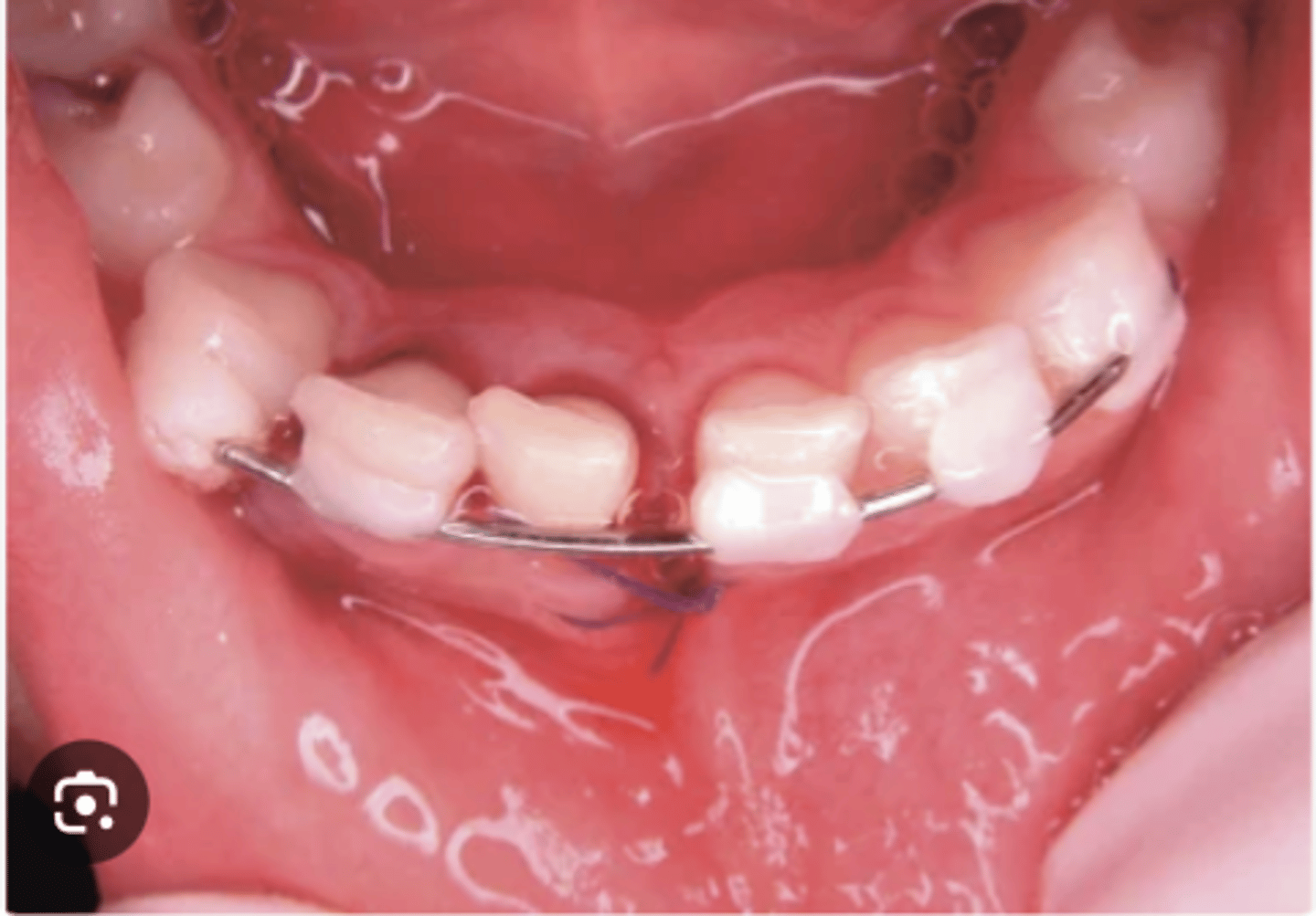
how do you treat an alveolar ridge fracture?
Step 1: Reposition
reduce the fragment back into its normal anatomical position.
Step 2: Immobilize
Use:
Arch bars
Splints
Wiring
to stabilize the teeth and attached alveolar segment.
splinting
wiring
arch bars
how do you treat a sagittal (ombredan) fracture
Closed reduction
Manually reduce the fragment.
Immobilization
Can be performed with:
Occlusal plate (acrylic splint over teeth)
Submental bandage (a sling for the lower jaw)
Antibiotics:
Frequently required because of communication with:
Nasal cavity
Oral cavity
Maxillary sinus
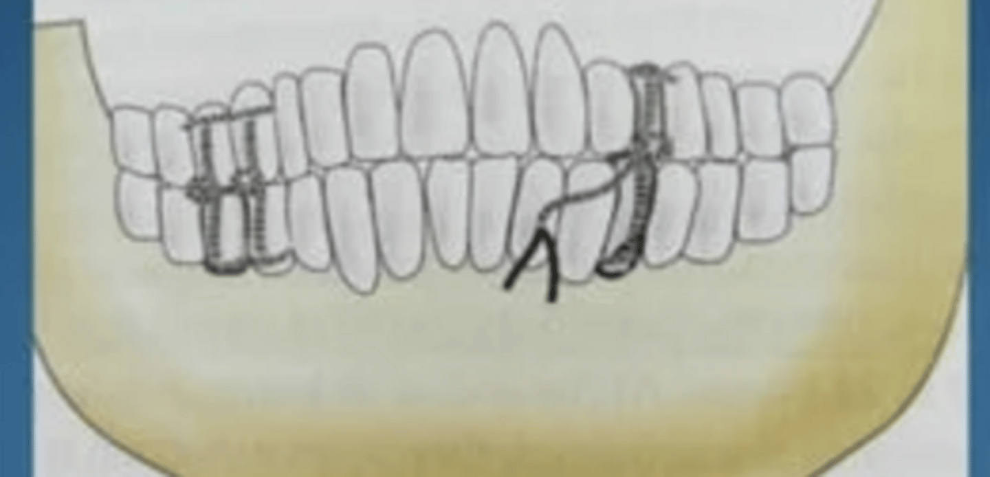
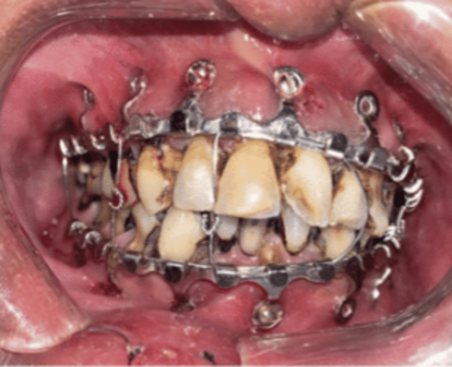
what are the methods of treatment for total maxillary fractures?
1. orthopaedic methods
2. orthopaedic surgical methods
3. surgical methods
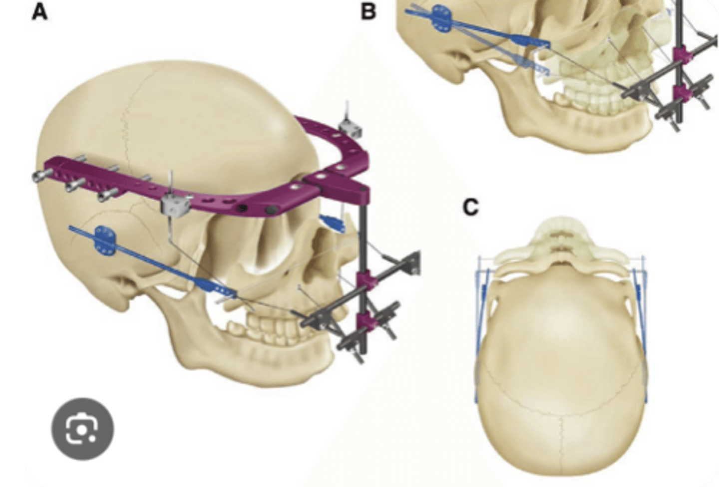
what are the orthopaedic methods for treatment?
not commonly used use of special external fixation devices to:
- reduce fracture fragments into correct anatomical position + restore occlusion
- fixate fragments by anchoring device to stable cranial structures
what are the components of the external fixation device?
1. Reducing Part
Repositions fracture fragments.
Restores normal occlusion.
Often uses a mandibular arch bar.
2. Fixation Part
Anchors the reduced maxilla to the cranium.
Consists of:
Cranial fixation elements → attached to skull.
Maxillary fixation elements → attached to maxilla.
Connecting rods and wires → connect cranial and maxillary components.
what are the orthopaedic surgical methods?
1. Adam's approach
2. Klisarov's approach
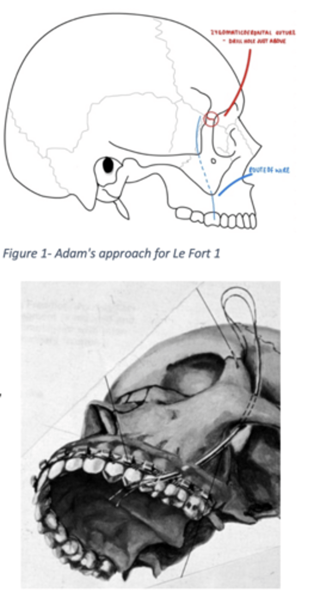
what is Adam's approach?
general anaesthesia
skin incision
- from the distal part of the eyebrow for access to the supraorbital rim
- drill a hole
- above the zygomaticofrontal suture
place 0.5mm ligature wire through the hole,
- guide it under the zygomatic arch until it reaches the buccal sulcus of the first molar
- both wires on each side tied to same maxillary splint (a modified version as author recognised potential damage to first molar)

what are the fixation points for the le fort fractures?
Le Fort 1:
Zygomaticoalveolar crest
(because this area remains stable.)
Le Fort II:
Infraorbital rim
because it is above the fracture.
Le Fort III:
Supraorbital rim
because almost the entire midface is detached and only structures above the orbit remain stable.
what is the Klisarov approach?
modified approach of Adam
guide the wire around the zygomatic arch and underneath the skin of the forehead
do this on the other side
fasten both wires to the splnt + IMF
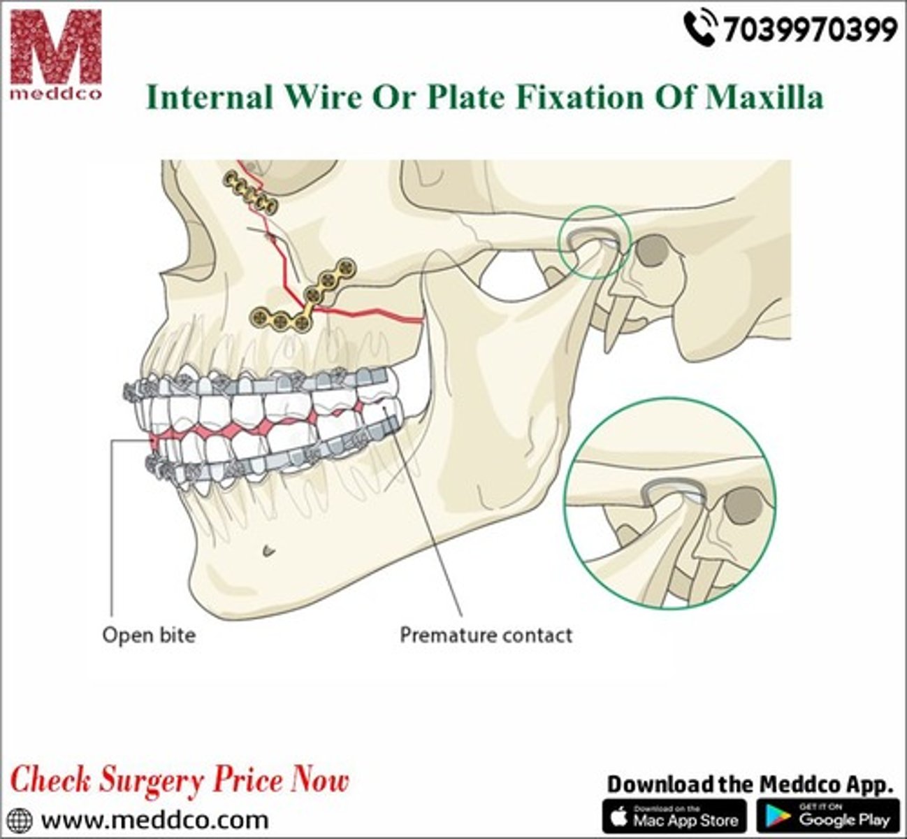
what are the main principles of surgical methods?
Open Reduction:
Open the fracture site
↓
Directly visualize fragments
↓
Reposition anatomically
Osteosynthesis:
Fixing bone fragments together.
what are the 3 types of osteosynthesis that can be performed?
A- wire osteosynthesis
= drill holes ~1cm from fracture
= steel wire passed through and tied in x/z/n shape
B- Plate osteosynthesis
= Ti plates + screws
C- Miniplate Osteosynthesis
= miniplate screws to fixate
what are the indications and advantages of Ti plates + screws?
indications:
Fragments are significantly displaced
Fracture passes through pathological bone
Strong stabilization is needed
advantages:
Rigid fixation
Better healing
Better stability
Still learning (17)
You've begun learning these terms. Keep up the good work!