Organisms responds to changes in environments
1/142
There's no tags or description
Looks like no tags are added yet.
Name | Mastery | Learn | Test | Matching | Spaced | Call with Kai |
|---|
No analytics yet
Send a link to your students to track their progress
143 Terms
Define stimulus
A detectable change in the an organism’s internal or external environment
Why is it important that organisms can respond to stimuli?
To increase their chance of survival
What are stimuli detected by?
Receptors
What is a taxis?
A simple response in which an organism will move its entire body towards a favourable stimulus (positive taxis) or away from an unfavourable stimulus (negative taxis)
What is a kinesis?
A non-directional response to presence and intensity of a stimulus in which an organism changes the speed of movement and the rate it changes direction so it can quickly return to a favourable environment
What is a tropism? Explain the 2 types
The growth of a plant in response to a directional stimulus
Positive tropism: grows toward stimulus
Negative tropism: grows away from stimulus
What are plant growth factors? Where are they produced? Give an example
Chemicals that regulate plant growth response to directional stimuli (tropisms)
They are produced in plant growing regions and can diffuse to other cells
Eg IAA (indoleacetic acid)
Summarise the role of growth factors in flowering plants
Specific growth factors move via phloem or diffusion from growing regions where they’re produced to other tissues where they regulate tropisms
Explain phototropism in shoots of flowering plants
Cells in tip of shoot produce IAA
IAA diffuses down shoot
Light causes IAA to move to the shaded side of the shoot
A higher concentration of IAA build up on the shaded side
In shoots, IAA causes cell elongation
Cells on shaded side elongate more, and faster
This causes the shoot tip to bend towards the light
This is positive phototropism
Explain phototropism in roots of flowering plants
Cells in tip of root produce IAA
IAA diffuses down root
Light causes IAA to move to shaded side of root
In roots, a high concentration of IAA inhibits cells elongation
Root cells elongate more on the lighter side
Root bends away from light
This is negative phototropism
Explain gravitropism in shoots of flowering plants
Cells in tip of shoot produce IAA
IAA diffuses down shoot
Gravity causes IAA move to the lower side of shoot - increasing concentration at lower side
This stimulates cell elongation at lower side
Shoots bend upwards - away from gravity
This is negative gravitropism
Explain gravitropism in roots of flowering plants
Cells in tip of root produce IAA
IAA diffuses down root
Gravity causes IAA to move to lower side of root - concentration increases at lower side
This inhibits cell elongation at lower side
Due to greater elongation of cells on the upper side, roots bend downwards towards gravity
This is positive gravitropism
According to the acid growth hypothesis, explain the role of IAA in elongation growth
IAA causes active transport of H+ ions into cell wall
Disruption to H bonds between cellulose molecules and action of expansins make cell more permeable to water
These cells elongate faster due to a higher turgor pressure and increased flexibility
What are the 2 major divisions of the nervous system?
The central nervous system (CNS) - made up of brain and spinal cord
The peripheral nervous system (PNS) - made up of pairs of nerves that originate from either the brain or the spinal cord
What is a reflex?
An involuntary response to a sensory stimulus
Outline the pathway of nerve impulses involved in a reflex arc
Receptor detects stimulus
Sensory neurone
Relay neurone in CNS coordinates response
Motor neurone
Response by effector
Explain 3 advantages of a reflex arc
Involuntary - doesnt have to be learnt or considered by brain
Fast - short neurone pathway (only 3 neurones and few synapses (which are typically slow in transmitting nerve impulses))
Protects from harmful stimuli
What 2 features are common to all sensory receptors?
Specific to a single type of stimulus
Act as energy transducers - convert the energy of the stimulus into a nervous impulse known as a generator potential
What is a generator potential?
Depolarisation of the membrane of a receptor cell as a result of a stimulus
What type of stimuli do Pacinian corpuscles respond to?
Mechanical stimuli eg pressure
Where are pacinian corpuscle receptors found?
Deep in the skin, mainly in fingers, soles of feet and external genitalia
Describe the structure of a Pacinian corpuscle
Single sensory neurone surrounded by layers of tissue, each separated by gel
Sensory neurone has stretch-mediated sodium channels in its plasma membrane
Contained in a capsule
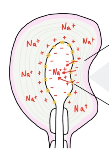
Describe how a generator potential is established in a Pacinian corpuscle
Mechanical stimulus eg pressure deforms the Pacinian corpuscle
This stretches and widens the Na+ channels
Na+ ions diffuse into the sensory neurone
This causes depolarisation, leading to a generator potential
If generator potential reaches threshold potential, it triggers an action potential
Name the 2 types of photoreceptor cell located in the retina
Rod cells
Cone cells
Where are rod and cone cells located in the retina?
Rod cells: mostly around the periphery of the retina. NOT in central fovea
Cone cells: mainly at central fovea, fewer at periphery of retina
State why rod cells and cone cells act as transducers
They covert light energy into the electrical energy of a nerve impulse
Explain the differences in sensitivity to light for rods and cones in the retina
Rods are more sensitive to light - several rods are connected to a single neurone in the optic nerve which means that there is a greater chance that the threshold value is exceeded to create a generator potential and then an an action potential due to spatial summation
Also, to create a generator potential, the pigment in rod cells (rhodopsin) must be broken down - there is enough energy from low-intensity light to cause this breakdown
Cone cells are less sensitive to light - each one is connected to a single neurone so no spatial summation. The stimulation of a number of cone cells cannot be combined to help excess the threshold value and create a generator potential
Also, the pigment in cone cells (iodopsin) requires a higher light intensity for its breakdown and creation of a generator potential
Explain the differences in visual acuity for rods and cones in the retina
Rods give lower visual acuity - several rods are connected to a single neurone so several rods send a single impulse to the brain so the brain cannot distinguish between the separate sources of light that stimulated each rod
Cones give higher visual acuity - each cone is connected to a single neurone so send separate impulses to brain which are distinguished as separate sources of light
Explain the differences in sensitivity to colour for rods and cones in the retina
Rods cannot distinguish between different wavelengths of light so only see images in black and white, due to there only being 1 type of rod with 1 type of pigment
Cones allow colour vision as there are 3 types of cone cells each containing a specific type of iodopsin which absorb different wavelengths of light
What is the autonomic nervous system? Name and explain the 2 divisions of the autonomic nervous system
The systems that controls the involuntary activities of muscles and glands
Sympathetic nervous system - stimulates effectors to speed up activity (involved in ‘fight or flight’ response)
Parasympathetic nervous system - inhibits effectors to slow down activity (involved in normal resting conditions)
Cardiac muscle is myogenic. What does this mean?
It contracts on its own accord rather than by nerve impulses
State the name and location of the 2 nodes involved in heart contraction
Sinoatrial node (SAN) - located within the wall of the right atrium. Known as the pacemaker
Atrioventricular node (AVN) - located in lower end of right atrium, in the wall that separates the 2 atria
Where is the Bundle of His located?
Runs through the septum
Where are the Purkyne fibres located?
In the walls of the ventricles
Describe how heartbeats are initiated and coordinated
The SAN releases a wave of depolarisation across the atria, causing atrial systole
The AVN releases another wave of depolarisation when the first reaches it.
There is a non-conducive layer between the atria and ventricles which prevents the wave of depolarisation travelling down to the ventricles, allowing them to fill
Impulse travels down Bundle of His, which conducts and passes the wave of depolarisation down the septum and branches into the Purkyne fibres in the walls of the ventricles
This causes the ventricles to contract from the apex upwards - there’s a shot delay before this happens, whilst the AVN transmits the second wave of depolarisation
Which part of the brain controls the heart rate, via the autonomic nervous system?
The medulla oblongata
Explain the difference the 2 centres of the medulla oblongata that are concerned with heart rate
Centre which increases heart rate - linked to the sinoatrial node by the sympathetic nervous system
Centre which decreases heart rate - linked to the sinoatrial node by the parasympathetic nervous system
Name the 2 receptors involved in changing heart rate and state what they detect
Where are they found?
Chemoreceptors (detect changes in blood pH)
Baroreceptors (detect changed in blood pressure)
Found in aorta and carotid arteries
Why is it important for chemoreceptors to respond to changes in blood pressure?
If blood pressure is too high, this can damage walls of arteries
If blood pressure is too low, there may be insufficient supply of oxygenated blood to respiring cells and removal of waste
Why is it important for Baroreceptors to respond to changes in blood pH?
If blood pH is too high, enzymes may denature
Describe how receptors in the heart responds to a decrease in blood pH
When blood has a higher than normal concentration of carbon dioxide, its pH is lowered
Chemoreceptors in walls of carotid arteries and aorta detect this and increase the frequency of nervous impulses to the centre in the medulla oblongata that increases heart rate
This centre increases frequency of impulses via the sympathetic nervous system to the SAN
This increases rate of production of electrical waves by SAN, cardiac muscle contracts more and therefore increases the heart rate
This increases blood flow leads to more CO2 being removed by lungs
CO2 concentration returns back to normal, as does pH of blood
Describe how receptors in the heart responds to an increase in blood pH
Chemoreceptors detect fall in blood CO2/rise in blood pH
They send impulses to medulla oblongata
Which sends more frequent impulses to SAN along parasympathetic neurones
So less frequent impulses/electrical waves produced by SAN
Cardiac muscle contracts less
Heart rate decreases
Describe how receptors in the heart respond to an increase in blood pressure
Baroreceptors detect rise in blood pressure
These send more impulses to the centre in the medulla oblongata that decreases heart rate
This centre sends more frequent impulses to SAN along parasympathetic nervous system
Cardiac muscle contracts less
Heart rate decreases
Describe how receptors in the heart respond to a decrease in blood pressure
Baroreceptors detect fall in blood pressure
These send more impulses to the centre in the medulla oblongata that increases heart rate
This centre sends more frequent impulses to SAN along sympathetic nervous system
Cardiac muscle contracts more
Heart rate increases
Give 8 differences between the hormonal system and the nervous system
Hormonal: communication is by chemicals called hormones. Nervous: communication is by nerve impulses
Hormonal: transmission by blood system. Nervous: transmission by neurones
Hormonal: transmission relatively slow. Nervous: transmission very rapid
Hormonal: hormones travel to all parts of the body, but only target cells respond. Nervous: nerve impulses travel to specific parts of the body
Hormonal: response is widespread. Nervous: response is localised
Hormonal: response is slow. Nervous: response is rapid
Hormonal: response is often long-lasting. Nervous: response is short-loves
Hormonal: effect may be permanent and irreversible. Nervous: effect is usually temporary and reversible
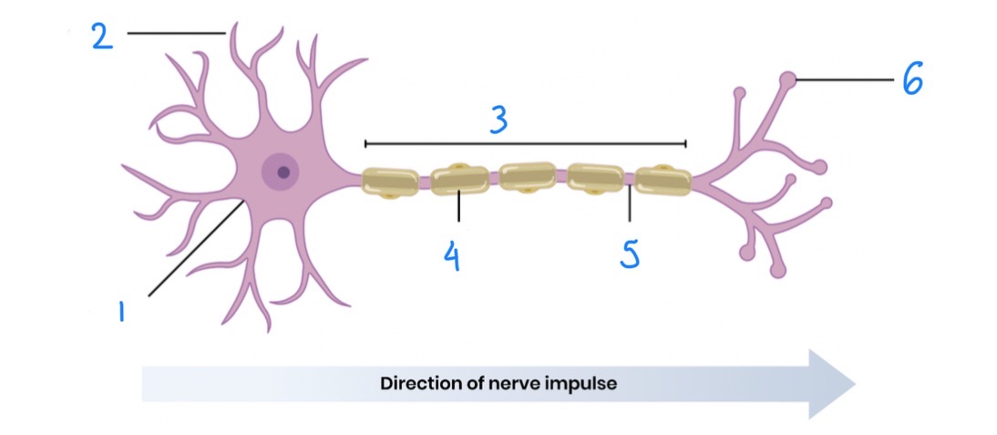
Name and describe the structures of a motor neurone
Cell body - contains all the usual cell organelles. Site of production of proteins and neurotransmitters
Dendrite - (dendrons branch into dendrites) carry nerve impulses to cell body
Axon - single long fibre that carries nerve impulses away from cell body, along the neurone
Myelin sheath - forms a covering to the axon. Made up of Schwann cells
Nodes of Ranvier - gaps between Schwann cells where there is no myelin sheath
Axon terminal
Name 3 processes Schwann cells are involved in
Electrical insulation
Phagocytosis
Nerve regeneration
Describe resting potential
The inside of an axon has a negative charge relative to outside (approx -70mV)
Explain how a resting potential (-70mV) is established across the axon membrane in a neurone
Na+ ions are actively transported out of axon by sodium-potassium pumps
K+ ions are actively transported into the axon by the sodium-potassium pumps
The active transport of sodium ions is greater than that of potassium ions (3 Na+ for every 2 K+)
So there are more Na+ ions in tissue fluid surrounding axon than in cytoplasm and more K+ ions in cytoplasm than in tissue fluid - this creates an electrochemical gradient
Na+ ions hardly diffuse back into axon while K+ ions diffuse back out - most of the gates in sodium ions channels are closed while many gates in potassium ion channels are open. There is a difference in membrane permeability for the 2 ions
This creates a negative charge inside axon relative to outside
When does an action potential occur?
When the neurone’s voltage reaches the threshold, generating a nerve impulse
Name the stages in an action potential
Depolarisation
Repolarisation
Hyperpolarisation
Return to resting potential
What happens during depolarisation?
A stimulus opens some Na+ channels, causing Na+ to enter cell by facilitated diffusion down their electrochemical gradient
Membrane potential becomes less negative
Once threshold (-55mV) is reached, voltage-gated Na+ channels open
This causes a significant influx of Na+ ions, and a reversal of charge across the membrane - membrane potential rises to about +40mV and outside becomes relatively negative
What happens during repolarisation?
Once membrane potential reaches around +40mV, voltage-gated sodium channels close - neurone is at peak depolarisation
Voltage-gated potassium channels open due to electrical gradient (inside more positive than outside)
K+ ions diffuse out of axon
What happens during hyperpolarisation and how does this lead to restoring resting potential?
There’s an overshoot when K+ diffuse out - p.d becomes more negative than resting potential
The gates on the potassium ion channels now close
Sodium-potassium pump starts to act again to restore resting potential
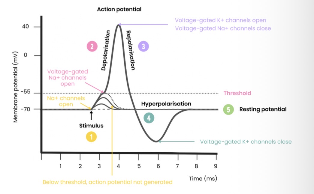
Draw and label a graph showing an action potential
Describe the all-or-nothing principle
For an action potential to be produced, depolarisation must exceed threshold potential
Action potentials produced are always the same size/peak at same potential
Bigger stimuli increase frequency of action potentials (not size)
Explain 2 ways in which organisms can perceive the size of a stimulus
Larger stimuli generate more impulses in a given time so raises membrane to threshold potential more quickly
Different neurones have different threshold values - the brain interprets the number and type of neurone that pass impulses and thereby determines its size
Describe the passage of an action potential along an unmyelinated axon
The action potential passes as a wave of depolarisation
Stimulus leads to influx of Na+ ions in one region of axon - this region depolarises
The localised electrical current established by this influx causes the opening of sodium voltage-gated channels further along the axon
This region depolarises whilst the section behind begins to repolarise
Describe the passage of an action potential along a myelinated axon
Myelin sheath provides electrical insulation
Action potentials only occur at nodes of Ranvier
Action potentials jump from node to node in a process known as saltatory conduction
So no need for depolarisation along whole length of axon
Do action potentials pass along unmyelinated axons or myelinated axons faster? Why?
Myelinated axons
Action potentials don’t happen across the whole length of the axon due to saltatory conduction
Name the 3 factors that affect the speed at which an action potential travels
Myelination
Axon diameter
Temperature
Explain how myelination affects speed of conductance
The myelin sheath provides electric insulation
It prevents an action potential forming in the part of the axon covered in myelin
Depolarisation happens at Nodes of Ranvier only (saltatory conduction)
This increases the speed of conductance
Explain how axon diameter affects the speed at which an action potential travels
The greater the diameter, the faster the speed of conductance
This is due to less leakage of ions and less resistance to flow of ions
Explain how temperature affects the speed at which an action potential travels
Higher temp = higher nerve impulse
Increases rate of diffusion of Na+ and K+ during depolarisation and repolarisation as more kinetic energy
Increases rate of respiration so more ATP for active transport to re-establish resting potential
However, proteins/enzymes denature at a certain temperature
What is the refractory period?
The time taken to restore axon to resting potential when no further action potential can be generated
As sodium voltage-gated channels are closed
Explain 3 reasons why the refractory period is important
Ensures discrete impulses are produced - action potentials don’t overlap
Ensures that action potentials are propagated in one direction only
Limits number of action potentials that pass along an axon in a given time - prevents over reaction to stimulus
What is a synapse?
The point where one neurone communicates with another neurone or with an effector
By what means do synapses transmit action potentials?
As neurotransmitters which diffuse across the synapse
Describe the structure of a synapse
Presynaptic neurone - releases neurotransmitter. It’s axon ends in synaptic knob which contains lots of mitochondria, endoplasmic reticulum (for manufacture of neurotransmitter) and synaptic vesicles (stores neurotransmitter)
Synaptic cleft - gap between neurons
Postsynaptic neurone - has specific receptor proteins to neurotransmitter to receive it
Explain why synaptic transmission is unidirectional
Only presynaptic neurone contains vesicles of neurotransmitter at its axon terminal
Only postsynaptic membrane has complementary receptors at its dendrites
Define summation
The rapid build-up of neurotransmitter in the synapse, allowing threshold to be reached and an action potential to be generated
Name the 2 types of summation
Spatial summation
Temporal summation
Explain the importance of summation
Low frequency action potentials release insufficient neurotransmitter to exceed threshold
Describe spatial summation
Many presynaptic neurones collectively release sufficient neurotransmitter to one postsynaptic neurone to reach threshold and trigger an action potential
Describe temporal summation
A single presynaptic neurone releases neurotransmitter many times over a short period so there is sufficient neurotransmitter to reach threshold and trigger an action potential
What is a cholinergic synapse?
A synapse that uses the neurotransmitter acetylcholine (ACh)
Describe transmission across a cholinergic synapse
Depolarisation of presynaptic membrane causes opening of voltage-gated Ca2+ channels
Ca2+ diffuses into presynaptic neurone
Influx of Ca2+ ions causes synaptic vesicles to fuse with presynaptic membrane and release acetylcholine into synaptic cleft by exocytosis
ACh diffuses across synaptic left and binds to receptors on post-synaptic membrane
This causes Na+ channels to open
Na+ ions diffuse into postsynaptic neurone, causing depolarisation
If threshold is met, action potential generated
Explain what happens to acetylcholine after synaptic transmission and why this is useful
It is hydrolysed by acetlycholinesterase
Hydrolysed into acetyl (ethanoic acid) and choline
These are reabsorbed by the presynaptic neurone - diffuse into it
Reformed when needed using ATP released by mitochondria which is stored in synaptic vesicles
Useful as it stops overstimulation - if not removed, it would keep binding to receptors and continuously generate a new action potential
What are inhibitory synapses?
Synapses that make it less likely that a new action potential will be created on the postsynaptic neurone
Describe inhibition by inhibitory synapses
Presynaptic neurone releases a type of neurotransmitter that binds to chloride ion channels on postsynaptic neurone
Causes chloride ion protein channels to open
Cl- ions move into postsynaptic neurone by facilitated diffusion
Binding of neurotransmitter also causes the opening of K+ protein channels
K+ ions move out of postsynaptic neurone into synapse
Inside of axon has a more negative charge relative to outside - below resting potential (hyperpolarisation)
More Na+ required to enter for depolarisation
Reduces likelihood of threshold being met/action potential formation
Describe the structure of a neuromuscular junction
A synapse that occurs between a motor neurone and a muscle
Receptors are on muscle fibre sarcolemma instead of postsynaptic membrane
Give 4 similarities between neuromuscular junctions and cholinergic synapses
They both have neurotransmitters that are transported by diffusion
They both have receptors, that on binding with the neurotransmitter, causes an influx of Na+ ions
They both use a sodium-potassium pump to repolarise the axon
They both use enzymes to breakdown the neurotransmitter
Give 4 differences between neuromuscular junctions and cholinergic synpases
Neuromuscular junction is always excitatory, cholinergic synapses may be excitatory or inhibitory
Neuromuscular junction only links neurones to muscle, cholinergic synapse links neurones to other neurones or to effectors
In neuromuscular junctions, the action potential ends here (they are the end of a neural pathway), in cholinergic synapses, a new action potential ma be produced along another neurones
In neuromuscular junctions, acetylcholine binds to receptors on membrane of muscle fibre, in cholinergic synapses, acetylcholine binds to receptors on membrane of postsynaptic neurone
How might drugs increase synaptic transmission?
May be similar shape to neurotransmitter
May stimulate release of more neurotransmitter
May inhibit enzyme that breaks down neurotransmitter (eg AChe)
How might drugs decrease synaptic transmission?
May inhibit release of neurotransmitter
May block receptors by mimicking shape of neurotransmitter
May decrease permeability of postsynaptic membrane to ions
May hyperpolarise postsynaptic membrane
Name the 3 types of muscle in the body and where they are located
Cardiac: exclusively found in heart, striated, involuntary
Smooth: walls of blood vessels and intestines, non-striated, involuntary
Skeletal: attached to incompressible skeleton by tendons, striated, voluntary
What does the phrase ‘antagonistic pair of muscles’ mean?
They work in opposition to eachother - when one contracts, the other relaxes
They can only pull so they work in pairs to move bones around joints
Describe the gross structure of skeletal muscle
Muscle fibres are made up of millions of myofibrils
Myofibrils are bundles of fused cells that share nuclei and cytoplasm (sacroplasm) and there is a high number of mitochondria and sarcoplasmic reticulum
Sarcolemma (cell membrane) folds inwards to form traverse (T) tubules
Each muscle fibre is surrounded by endomycium - connective tissue that provides structural support and has many capillaries
Describe the ultrastructure of a myofibril
Made up of 2 types of long protein filaments, arranged in parallel - myosin (thick filament) and actin (thin filament)
Arranged in functional units called sarcomeres (one sarcomere is the distance between adjacent Z-lines)
What 2 types of protein filament are myofibrils made up of?
Actin - thinner and consists of 2 strands twisted around one another
Myosin - thicker and consists of long rod-shapes tails with bulbous heads
Explain the banding pattern to be seen in myofibrils
I-bands - light bands containing only thin actin filaments
A-bands - dark bands containing an overlap of actin and myosin filaments
H-zone - found at centre of A-band (contains only myosin)
Z-line - found at centre of I-band (boundary between sarcomeres)
Describe the changes that occur to a sarcomere when a muscle contracts
I-bands get shorter/narrower
Z-lines move closer together (sarcomere shortens)
H-zone gets shorter/narrower
A-band stays the same
Describe the proteins involved in the sliding-filament mechanism
Myosin is made up of 2 types of protein:
A fibrous protein arranged into a filament made up of several hundred molecules (makes up tail)
A globular protein formed into 2 bulbous structures at each end (makes up heads)
Actin is a globular protein whose molecules are arranged into long chains that are twisted around one another to form a helical strand
Tropomyosin forms long thin threads that are wound around actin filaments
Describe muscle stimulation according to the sliding filament theory
An action potential reaches neuromuscular junctions
This causes voltage-gated calcium ion protein channels to open and Ca2+ ions to diffuse into synaptic knob
These Ca2+ ions cause synaptic vesicles to fuse with presynaptic membrane and release acetylcholine into synaptic cleft
Acetylcholine diffuses across synaptic cleft and binds with receptors on muscle sell surface membrane, causing it to depolarise
Describe muscle contraction according to the sliding filament theory
The action potential travels down sarcolemma via T tubules, causing Ca2+ ions to release from sarcoplasmic reticulum
Ca2+ ions diffuse into myofibrils
Ca2+ ions bind to tropomyosin molecules, that were blocking myosin binding sites on actin filament, causing them to move and expose myosin binding sites
Allows myosin head, with ADP attached, to bind to binding site on actin - forming actinomyosin cross bridges
Once attached, myosin heads change their angle, pulling actin filament along as they do so and releasing a molecule of ADP
A new ATP molecule attaches to myosin head, to detach it from actin filament
Ca2+ ions then activate ATPase which hydrolyses ATP to ADP, providing energy for myosin head to return to its original position
Myosin head, with ADP molecule attached, reattaches itself further along actin file amen an cycle repeats as long as concentration of Ca2+ ions in myofibril remains high
How does sliding filament action cause a myofibril to shorten?
Myosin molecules are joined tail to tail in oppositely facing sets
Each set of myosin heads move in opposite directions
Actin filaments which they are attached to therefore also move in opposite directions - towards eachother
This shortens the distance between adjacent Z-lines
Describe muscle relaxation according to the sliding filament theory
Ca2+ ions are actively transported back into endoplasmic reticulum using energy from the hydrolysis of ATP
This allows tropomysoin to block the binding site on the actin filament again
Myosin heads unable to bind - no actinomyosin cross bridges can form
Explain the role of phosphocreatine in muscle contraction
A source of inorganic phosphate
Rapidly phosphorylates ADP to regenerate ATP
When oxygen for aerobic respiration is limited
Runs out after a few seconds so used for short bursts of vigorous exercise where generating ATP anaerobically is also required
What are the 2 types of muscle fibre?
Slow-twitch fibres
Fast-twitch fibres
Compare the location of slow and fast twitch skeletal muscle fibres
Slow twitch fibres are found mostly in areas of constant, sustained contractions eg calf muscle
Fast twitch fibres are found mostly in areas of short-term, rapid, powerful contraction eg biceps