Topic 18: Vertebrate Systems Development-5 Reproductive Systems
1/59
There's no tags or description
Looks like no tags are added yet.
Name | Mastery | Learn | Test | Matching | Spaced | Call with Kai |
|---|
No analytics yet
Send a link to your students to track their progress
60 Terms
Both the excretory and reproductive systems are derived from?
intermediate mesoderm
Because the excretory and reproductive systems are derived from intermediate mesoderm, both systems are ( )
primitively segmental
Most preconceived notions about kidneys, their shape, and origin are colored by the ( ) condition
mammalian
Recall that those vertebrates that retain the entire kidney derived from every body segment are said to have a ( ) or ( )
holonephros
“holonephric” kidney
Primitively/initially, there is an intermediate mesoderm segment, and thus ( ). Few vertebrates still retain such a long kidney
kidney or gonad segment for every body segment
Only the most primitive remaining vertebrates have a ( )
holonephros
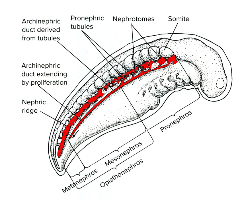
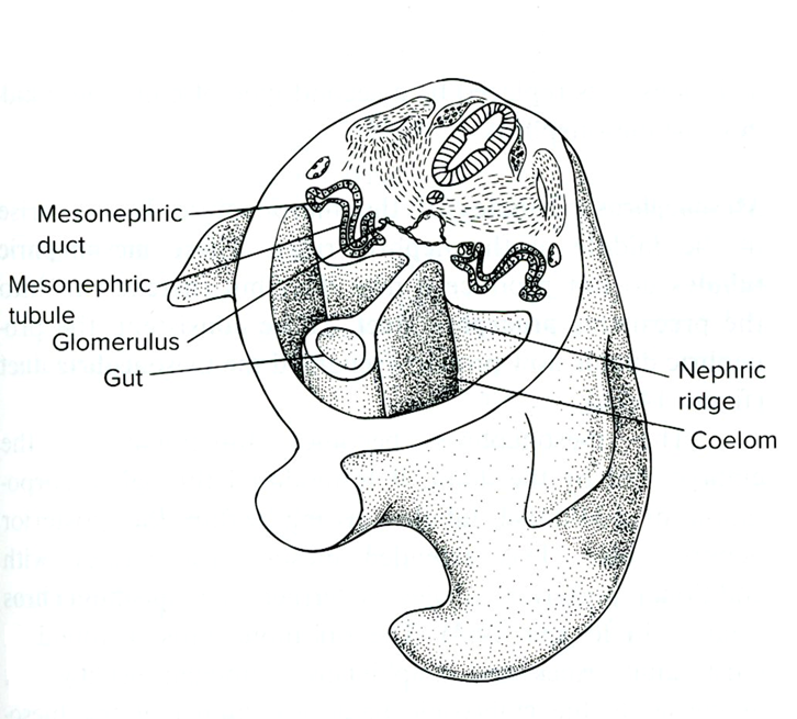
the cranial-most part of the kidney, the ( ) develops first, but is subsequently lost. However, it is critically important, because it induces the development of the more caudal segments of the kidney
pronephros
The remaining portion of the kidney is the, excluding the pronephros, is the ( ). This is found in many fishes, and some amphibians
opistonephros
cool
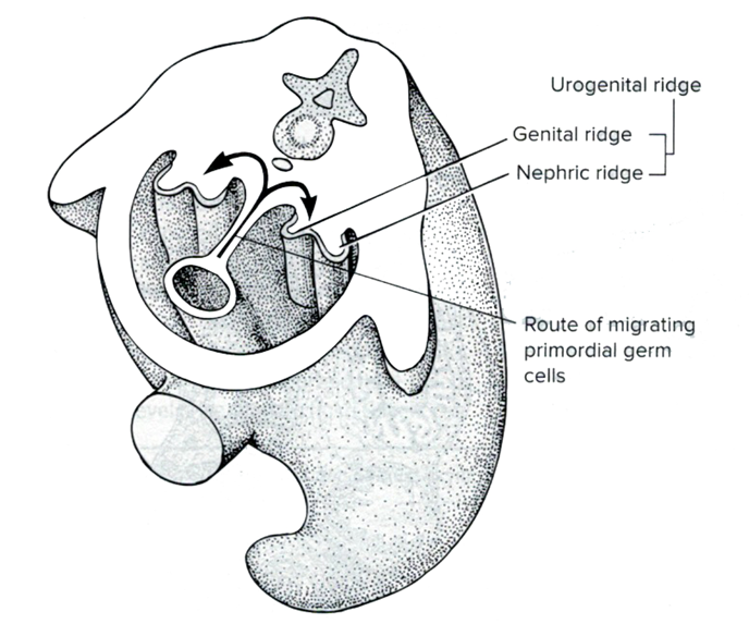
Gonadal materials develop in a ridge that is also derived from ( ), and runs roughly parallel to the ( )
intermediate mesoderm
nephric ridge
The reason we didn’t see gonadal materials prior was because the ( ) expresses only in the ( )
gonadal ridge
more caudal/posterior region of the body
cranial to middle part of the body
more caudal region of the body
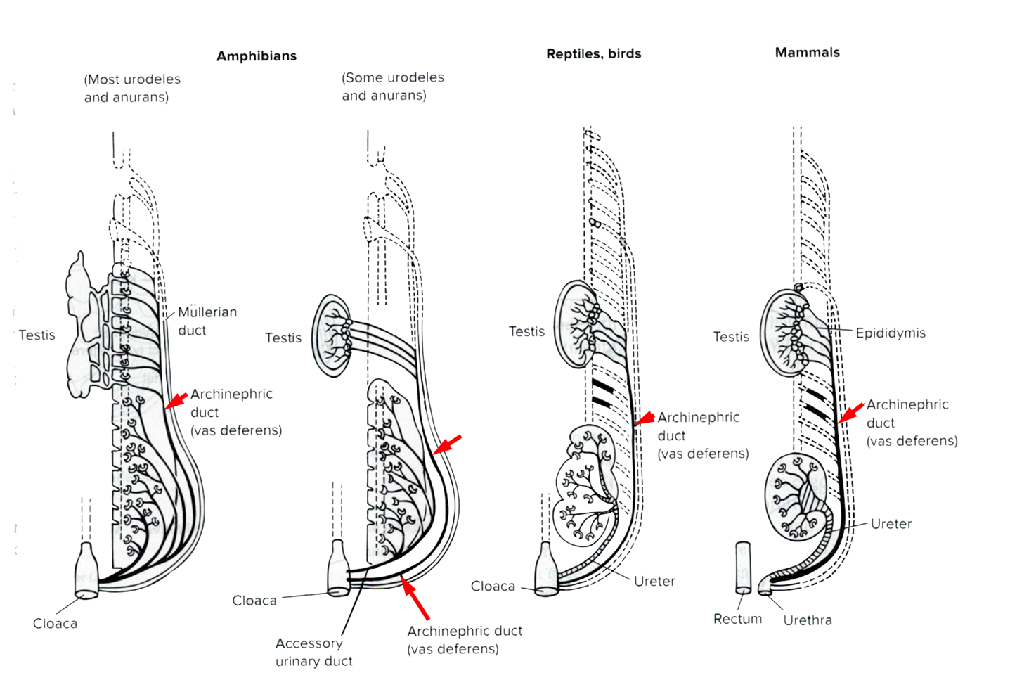
In a male, the ( ) are still draining from the anterior/cranial end of the ( ) to the ( )
renal tubules
opistonephros
archinephric duct
what is the archinephric duct also known as?
the mesonephric duct
Testes attach to the anterior/cranial end of the ( ) via ( ) and ( )
archinephric duct
rete testes
vasa efferentia
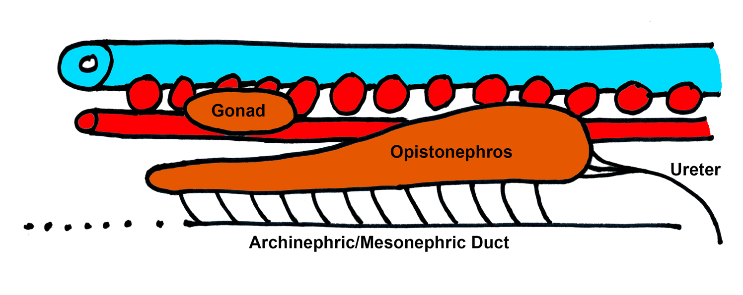
Once the testes attach to the archinephric duct via rete testes and vasa efferentia, the ( ) is essentially taken over by the ( ) for ( ) and is now known as the ( ) or the ( )
archinephric/mesonephric dust
gonad
sperm transport
ductus deferens
vas deferens
The archinephric/mesonephric duct has essentially been co-opted/taken over by the ( ). As this happens, the ( ) regresses
testes
mesonephros
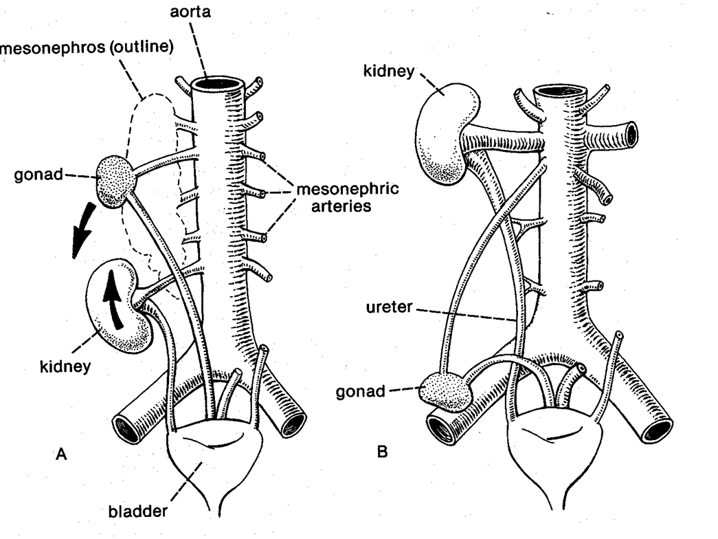
As the mesonephric duct is being taken over by the testes, the ureter is only draining the ( )
posterior/metanephric kidney
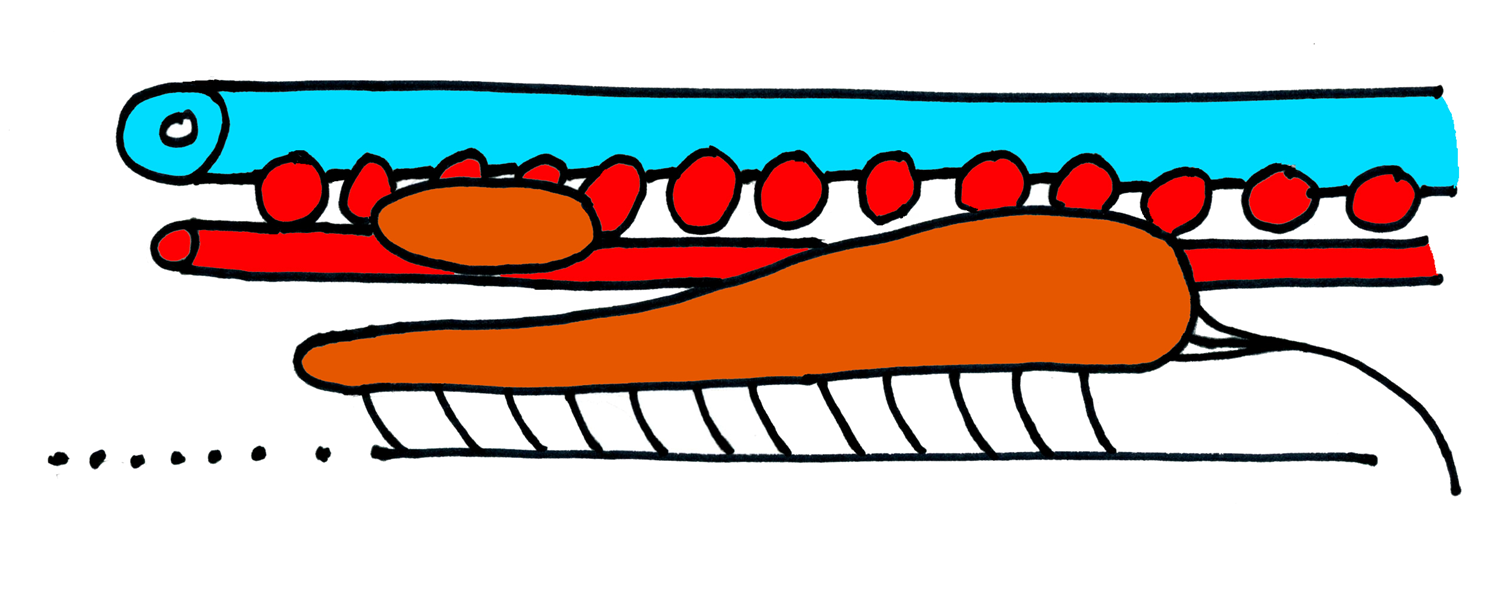
label and draw
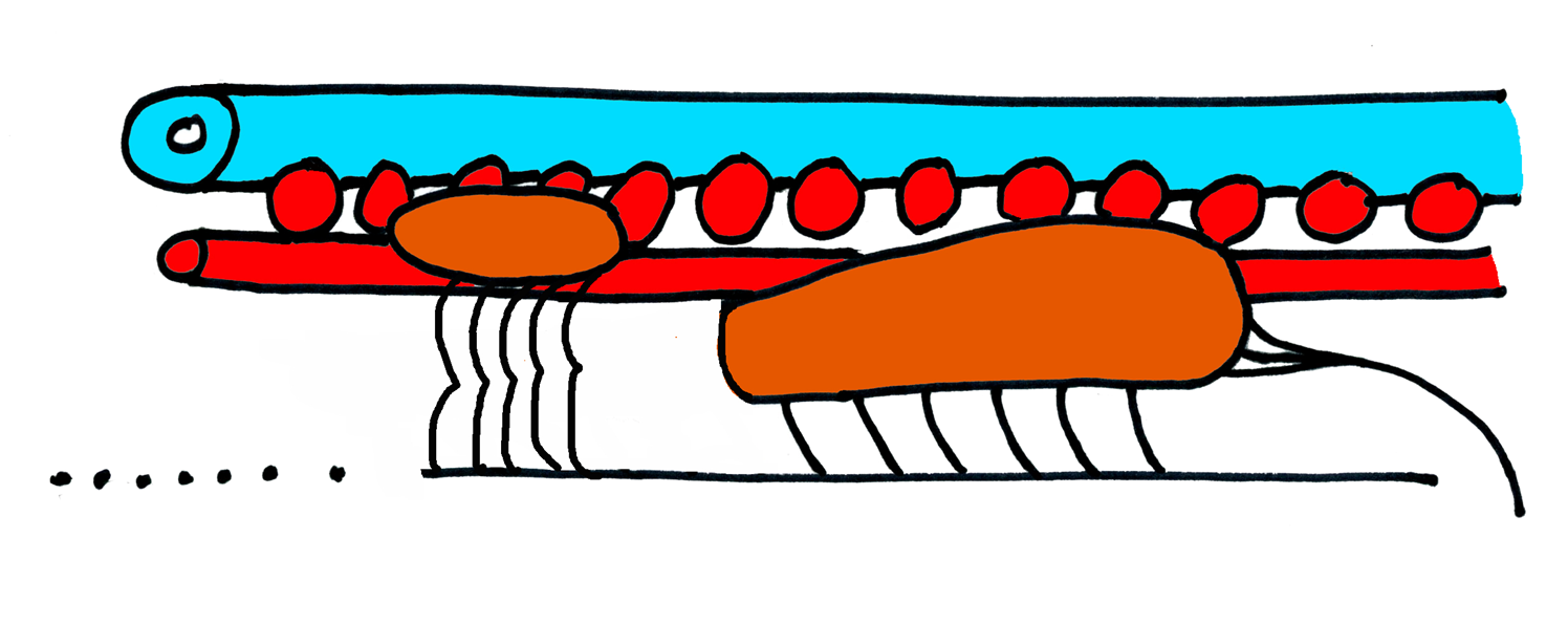
label and draw
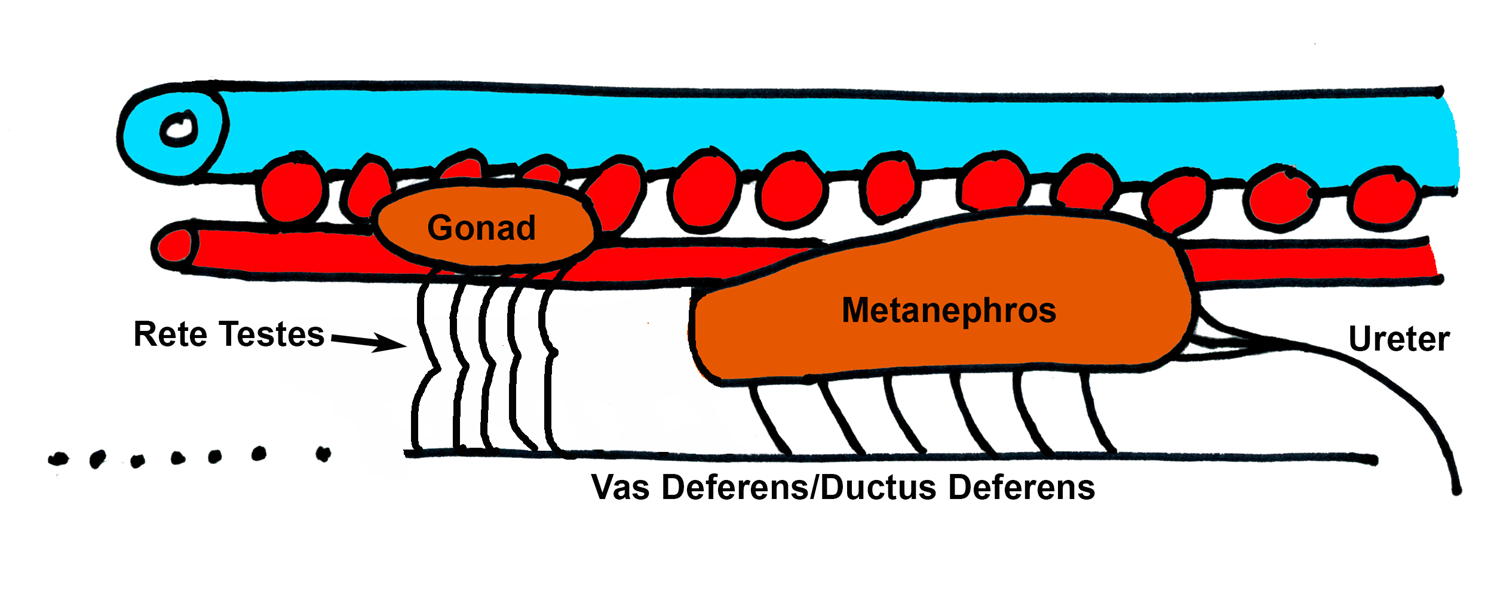
Note how testes take over mesonephric duct in males
okay
The condition in females is similar in pretty much all ( ), from ( ) to ( )
gnathostomes
fish
amniotes
In females, there is no battle for the ( ). In females, the ( ) leave the ( ) duct to the kidney, and ( )
duct
ovaries
mesonephric/archinephric
build their own duct
In females, the ( ) still grows forward toward the ( )
ureter
metanephros
females do not take over the ( ), but rather, build their own, separate ( )
mesonephric duct
oviduct
The end of the oviduct does not insert into the ( ) . It is just a ( ). Elaborated edges of the funnel are called ( )
ovary
funnel
fimbria
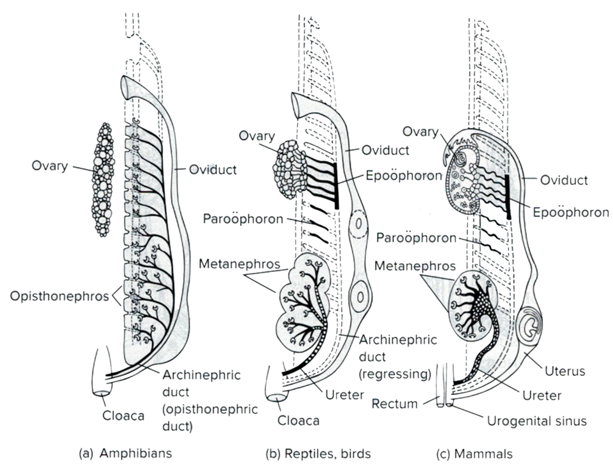
Note how ovaries do not take over mesonephric duct in females
yes
The metanephric kidney forms from the ( )-most kidney segments, but mammalian kidneys actuall move ( )
caudal
cranially (the term “ascent” is used in orthograde humans)
What is the mechanism to kidney movement in mammals?
The mechanism of this is not perfectly understood. Some researchers say that kidneys actively move cranially, whereas other suggest they stay in place and the body grows and expands past them
How do kidneys move?
They do so by attaching successively to more cranial segmental arteries, while simultaneously detaching from more caudal segmental arteries (sorta like climbing a ladder)
The final connecting arteries of the kidney are the paired
Renal arteries
As the kidneys attach to successively more cranial segmental arteries, the ureters stretch to retain their connections to the ( ), and the ( )
metanephric kidney cranially
bladder caudally
kindeys “ascend” to just caudal to ( ) and ( )
diaphram
liver
the ( ) kidney is a bit lower due to ( )
right
mass of liver
Kidneys and gonads pass one-another, so the ( ) and ( ) in males must cross one-another
ureter
ductus deferens
the ( ) and ( ) pass ( ) to the ureter
gonadal arteries
ductus deferens
ventral
The gonads start development near the ( ) but you will recall mammalian gonads are not in the ( )
mesonephric kidney
middle of the body
Mammalian gonads actually move ( )
caudally ( the term “descent” is used in orthograde humans)
The gonads follow a tract of ( ) called the ( ) into a more ( ) position. Near the ( ) of the ( ) in females, ( ) in males
connective tissue ( some liken it to a slide)
gubernaculum
caudal position
brim cranial edge
pelvic girdle
all the way out of the body
As the gonads move more caudally, they do not attach and detach from successively more ( ) . Rather, the paired ( ) stretch and remain connected ( ). Thus, ( )
caudal segmental arteries
gonadal arteries
near where they started
gonadal arteries are often quite long and sometimes connected to the nearby renal arteries
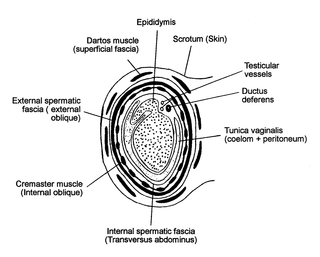
In males, the ( ) tear right through the body wall to reside in their own separate sac, the ( ). As they do this, they take with them equivalents to all the ( ) with them as the ( )
testes
scrotum
body wall layers
scrotum is built
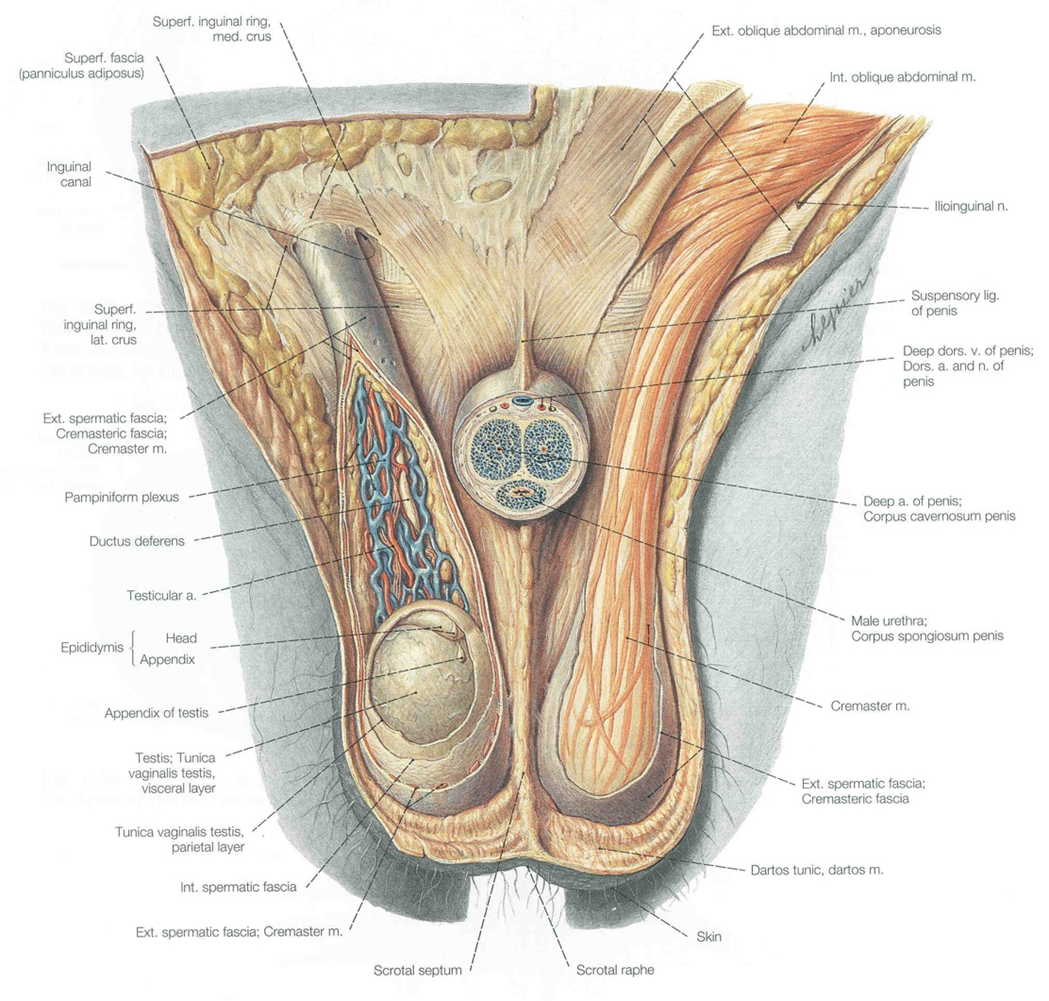
There will be a thin/tiny layer of all body wall parts equivalent to the ( ) and a tiny bit of coelom in each side of the ( ) . Called the ( )
scrotum
scrotal sac
tunica vaginalis
woah
woah
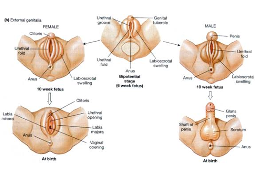
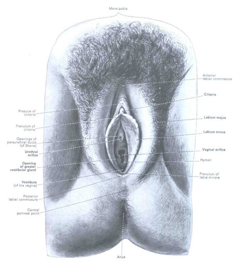
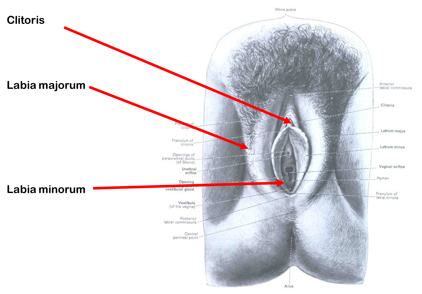
In females, erectile tissue is ( ), the ( ) do not fuse in the midline and do not enlarge as much
present
bulbs
The bulbs of the vestibule form separate masses of ( ) on either side of the ( )
erectile tissue
vaginal opening
The bulbs of ( ) will become the ( ) , singular ( )
the vestibule
labia minora
minorum
Because the bulbs of the vestibule dont fuse in the midline, the female ( ) cannot be enclosed in the midline as in the ( ) of the male
urethra
corpora spongiosa
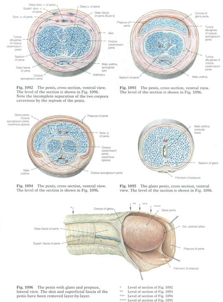
The tip end of the midline columns is the ( ). Right and left ( ) form ( ) of clitoris
clitoris ( similarly sensitive to glans of males)
crura
(much smaller) corpora cavernosa
okay
where is the clitoris, labia majorum and minorum (3 total)
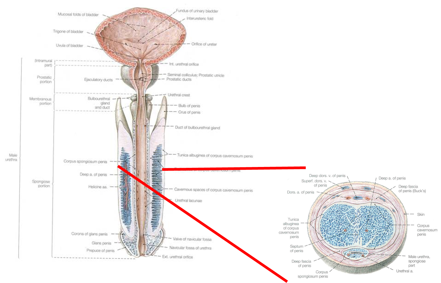
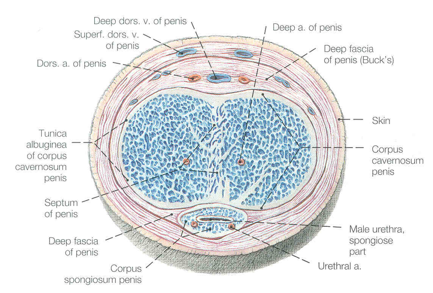
males have ( ) of erectile tissue
three columns
The right and left bulbs fuse in the midline to form the ( ) which ( )
corpora spongiosum
surrounds the urethra
In males, the urethra emerges out of tip of enlarged genital tubercle- the ( )
glans of the penis
In males, at the tip of the ( ) that is the ( )
bulbous dilation
glans of the penis
In males, right and left ( ) remain independent and form the paired ( )
crura
corpora cavernosa
The right and left sides of the penis are bound to one another by what?
tunica albuginea