Week 4: ECG Axis Hypertrophy and Enlargement
1/63
There's no tags or description
Looks like no tags are added yet.
Name | Mastery | Learn | Test | Matching | Spaced | Call with Kai |
|---|
No analytics yet
Send a link to your students to track their progress
64 Terms
What are we looking at when referring to the “Axis”?
The electrical activity of ventricular depolarization (QRS complex).
What is the axis of the heart?
Direction of the mean electrical vector
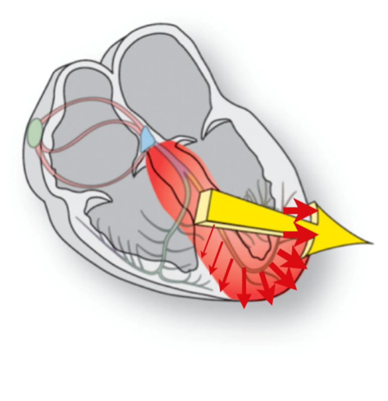
What is the first vector in ventricular depolarization?
Septal depolarization
What are the later vectors in ventricular depolarization?
Progressive depolarization of ventricles
What is the mean vector?
Average vector of all instantaneous vectors
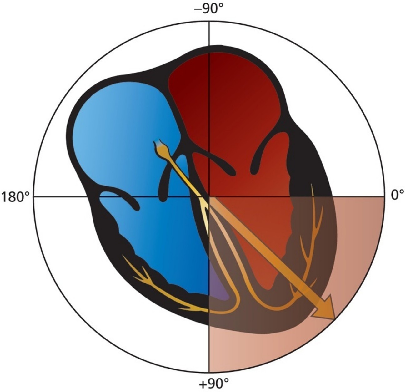
What is the mean electrical axis?
Direction of the mean vector
What direction does the normal mean QRS vector point?
Left and inferiorly
What can an abnormal axis suggest?
A change in the physical shape and orientation of the heart or a defect in its conduction system
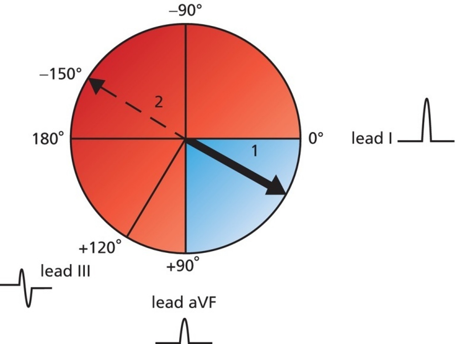
What QRS axis range is considered normal?
0° to 90°
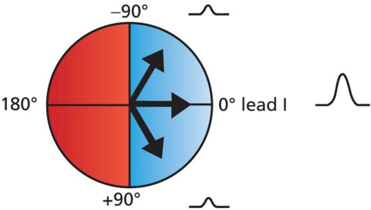
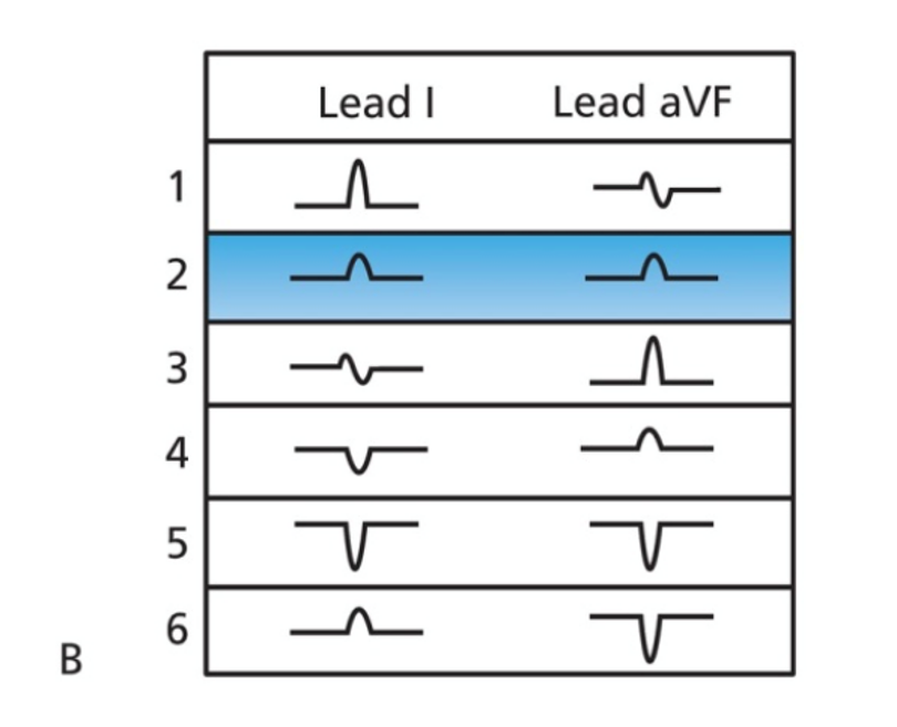
If the QRS complex is positive in leads I and aVF, what is the axis?
Normal

What does a positive QRS complex in lead I indicate about axis?
Normal if axis is between −90° and 90°
What does a positive QRS complex in lead aVF indicate about axis?
Normal if axis is between 0° and 180°
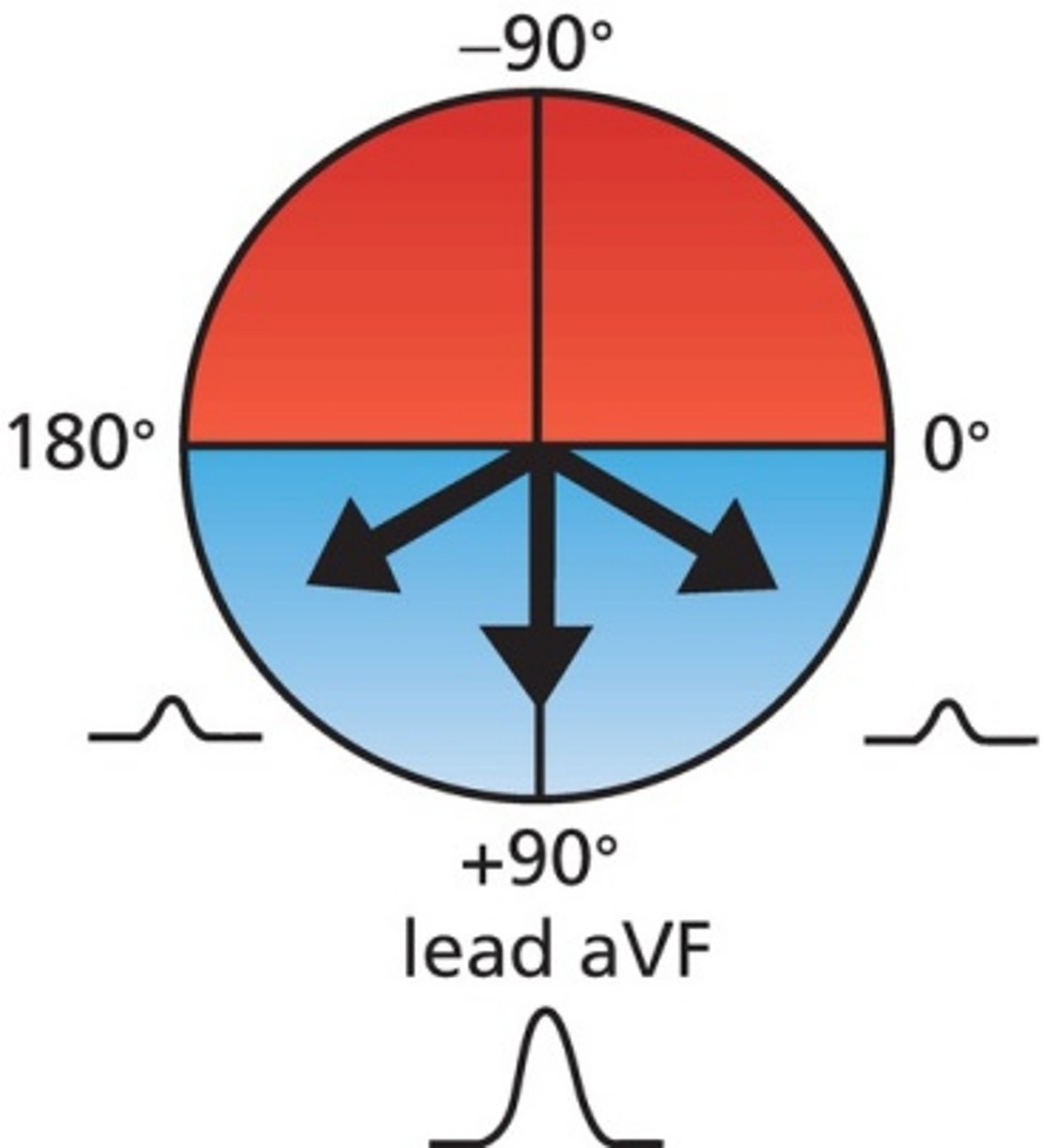
If QRS is positive in both leads I and aVF, what is the axis?
Normal

If QRS in either lead I or aVF is not positive, what does it indicate?
QRS axis is not normal
What is a biphasic QRS complex?
A QRS complex with positive and negative deflections
What is an iso-electric wave?
A biphasic wave with equal positive and negative deflections
If a lead is iso-electric
where is the axis located?
What can cause the electrical axis to change?
Rotation of the heart
How does hypertrophy affect axis?
Axis deviates toward the greater electrical activity.
How does infarcted tissue affect the QRS vector?
QRS vector turns away from the infarcted tissue.
How does RBBB affect the axis?
Axis turns to the right.
What axis range defines Right Axis Deviation (RAD)?
+90° to +180°.
What lead pattern suggests Right Axis Deviation?
Negative QRS in lead I and positive QRS in lead aVF.
What are the most common causes of Right Axis Deviation?
RVH
What axis range defines Left Axis Deviation (LAD)?
−90° to 0°.
What lead pattern suggests Left Axis Deviation?
Positive QRS in lead I and negative QRS in lead aVF.
What are the most common causes of Left Axis Deviation?
LVH
What is hypertrophy?
Increase in muscle mass.
What causes hypertrophy?
Chronic pressure overload and increased resistance.
What is enlargement?
Dilation of a heart chamber.
What causes enlargement?
Chronic volume overload.
Why does a chamber enlarge?
To accommodate more blood.
What causes Left Ventricular Hypertrophy (LVH)?
Chronic hypertension causing the LV to work harder.
How does LVH affect electrical activity?
Increased LV electrical dominance over RV.
What axis deviation is associated with LVH?
Left axis deviation.
What conditions can cause Right Ventricular Hypertrophy (RVH)?
Severe COPD and uncorrected congenital heart disease.
What axis deviation is associated with RVH?
Right axis deviation.
How can hypertrophy or enlargement increase QRS duration?
The chamber takes longer to depolarize.
How can hypertrophy or enlargement increase amplitude?
The chamber generates more current and voltage.
How can hypertrophy or enlargement shift electrical axis?
A larger percentage of electrical current moves through the expanded chamber.
What P-wave finding suggests Right Atrial Enlargement (RAE)?
Tall
What P-wave amplitude suggests RAE?
Amplitude >2.5 mm.
What happens to P-wave duration in RAE?
No change in duration.
What axis deviation may occur with RAE?
Possible RAD.
What is another name for the RAE P-wave pattern?
P pulmonale.
What causes Right Atrial Enlargement?
Increased right-sided pressures (Pulmonary HTN or pulmonary valve stenosis).
What P-wave finding suggests Left Atrial Enlargement (LAE)?
Wide
What V1 finding suggests LAE?
Terminal component ≥1 mm below the isoelectric line.
What happens to the terminal component duration in LAE?
Increased duration ≥1 small block (0.04 sec).
What axis deviation occurs with LAE?
No significant axis deviation.
What is another name for the LAE P-wave pattern?
P mitrale.
What causes Left Atrial Enlargement?
Increased left-sided pressures (Mitral valve stenosis
What are common ECG themes in RVH?
Increased R-wave amplitude in right ventricle leads and increased S-wave amplitude in left ventricle leads.
What are common causes of RVH?
COPD
What precordial lead finding suggests RVH?
R wave larger than S wave in V1.
What left-sided precordial finding suggests RVH?
S wave larger than R wave in V5–V6.
What limb lead finding suggests RVH?
RAD with QRS axis > +100°.
What lead I finding suggests RVH?
Predominately negative QRS in Lead I.
What are common ECG themes in LVH?
Increased R-wave amplitude in left ventricle leads and increased S-wave amplitude in right ventricle leads.
What are common causes of LVH?
Hypertension
What precordial lead criteria suggests LVH?
S wave in V1 plus R wave in V5 or V6 >35 mm.
What limb lead criteria suggests LVH?
R wave in aVL >11 mm.
What are secondary repolarization abnormalities of ventricular hypertrophy?
Downsloping ST-segment depression and T-wave inversion.
How do secondary repolarization abnormalities appear?
A single asymmetric wave with gradual downward slope and abrupt upward slope.