Lab Chapter 19
1/27
There's no tags or description
Looks like no tags are added yet.
Name | Mastery | Learn | Test | Matching | Spaced | Call with Kai |
|---|
No analytics yet
Send a link to your students to track their progress
28 Terms

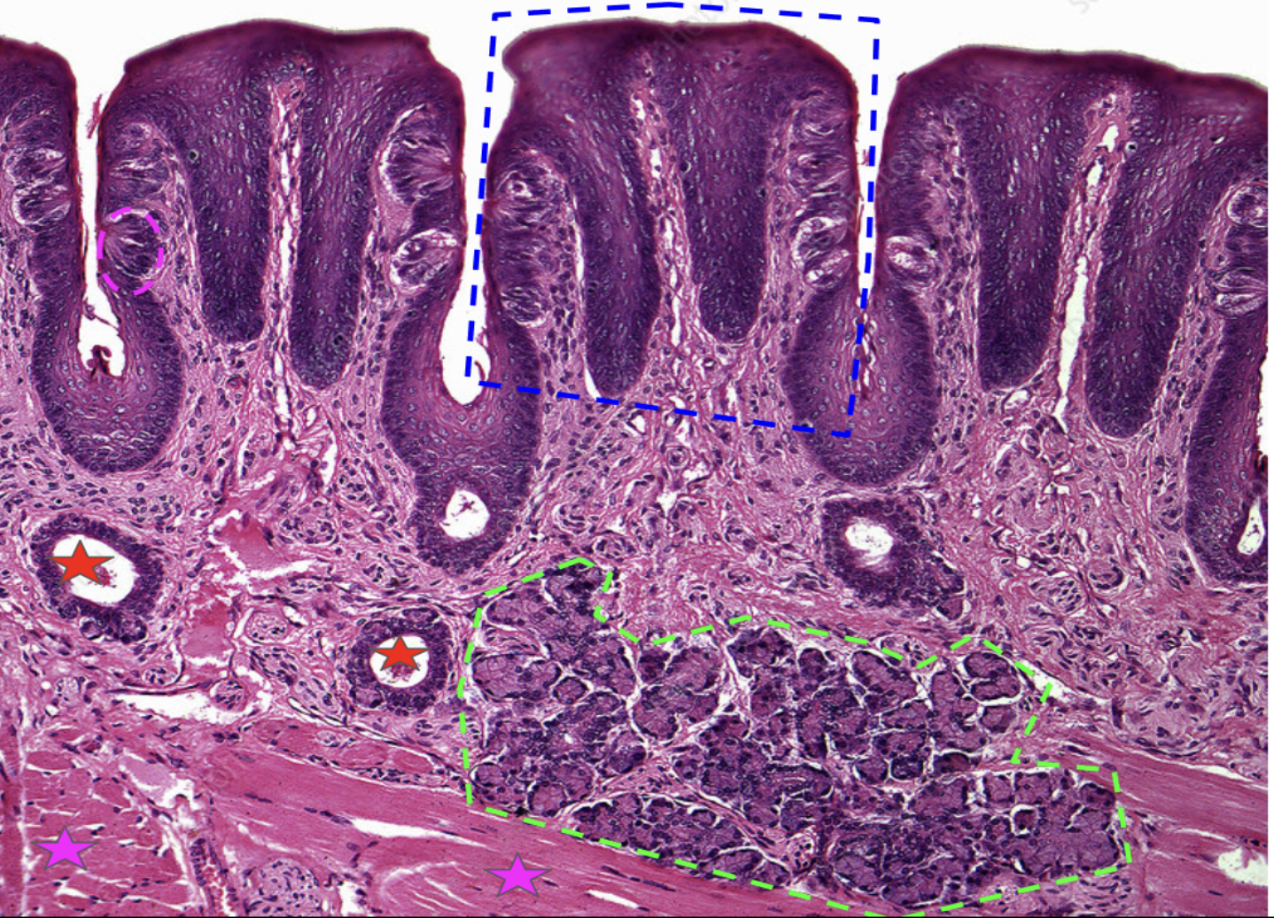
This image depicts what? Identify. Function
Tongue
Blue: Papillae - provide friction to help grab food
Pink circle: Taste buds (found along the outside of each papillae) - help us taste
Green: Lingual salivary gland (intrinsic gland) - secretes salivary amylase
Red: Lingual salivary gland ducts - carries the saliva and enzymes from lingual salivary gland
Pink star: Skeletal muscle (makes up the bulk of the tongue) - moves tongue
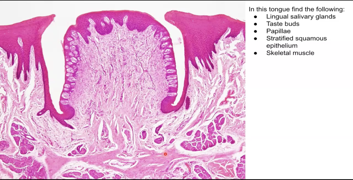
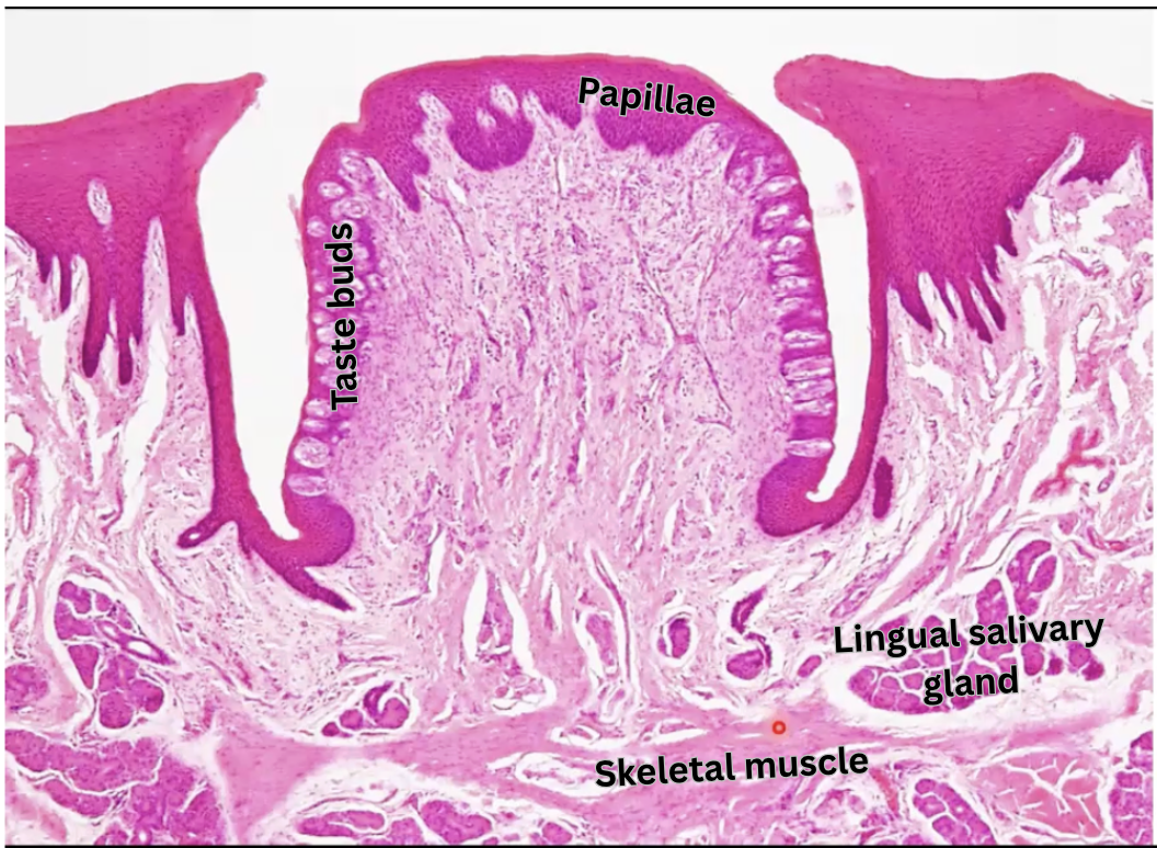
Identify the following:
Lingual salivary glands
Skeletal muscle
Taste buds
Papillae
What are the 5 types of taste
Salt
Sweet
Sour
Bitter
Umami (delicious)
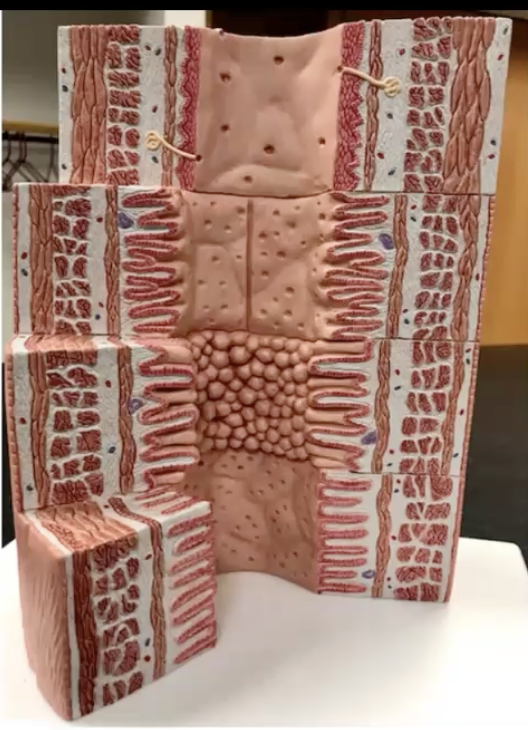
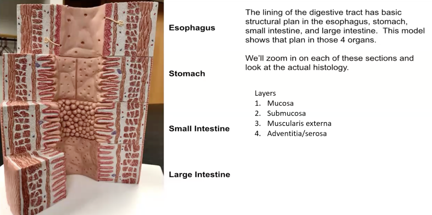
Identify each organ. What are the layers of each?
Esophagus, stomach, small intestine, large intestine (all have the same 4 layers)
- Starting from the lumen out (hole in the middle)
Mucosa
Submucosa
Muscularis externa
Adventitia/serosa
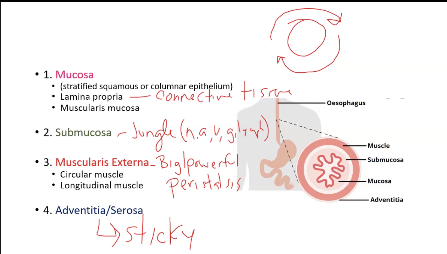
What are the individual parts of each layer? What is the function of the 3rd & 4th layer?
1. Mucosa
Stratified squamous or columnar epithelium
Lamina propria (just connective tissue)
Muscularis mucosa
2. Submucosa
Jungle (has nerves, arteries, veins, glands, etc)
3. Muscularis externa
Function is peristalsis: to move food
Circular muscle (inner layer)
Longitudinal muscle (outer layer)
Adventitia or serosa
• Adventitia: Sticky, allows organ to stick to other structures
- Serosa: Produces serous fluid to reduce friction with other organs

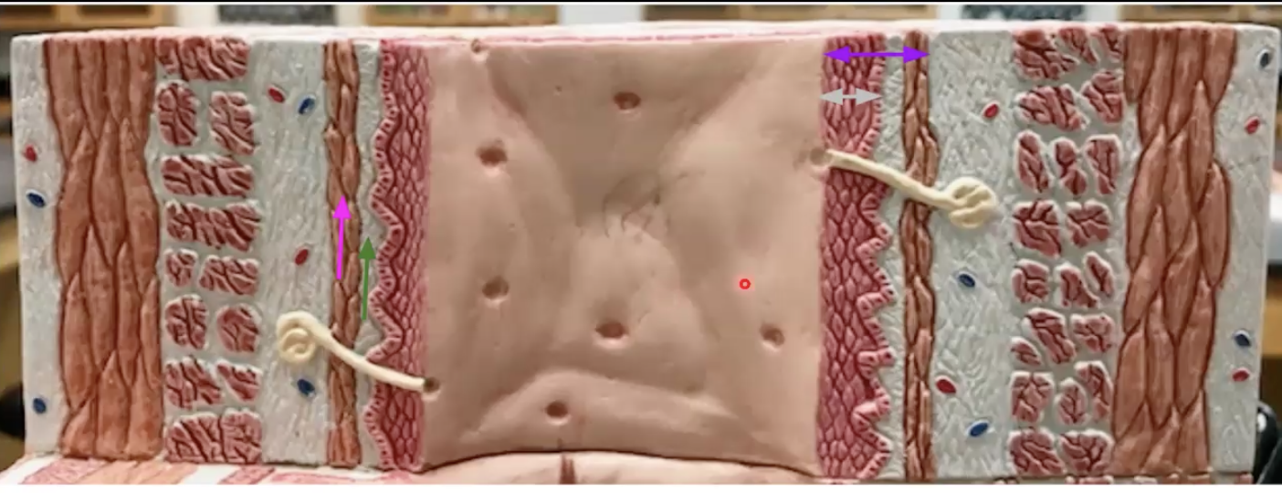
What organ is this? Identify the parts.
Function of grey and pink
Esophagus
Purple: Mucosa (first 3 parts bc mucosa has 3 layers)
Grey: Stratified squamous epithelium (notice the multiple layers of flat cells) - function is protection
Green: Lamina propria
Pink: Muscularis mucosae: shake off food that gets stuck

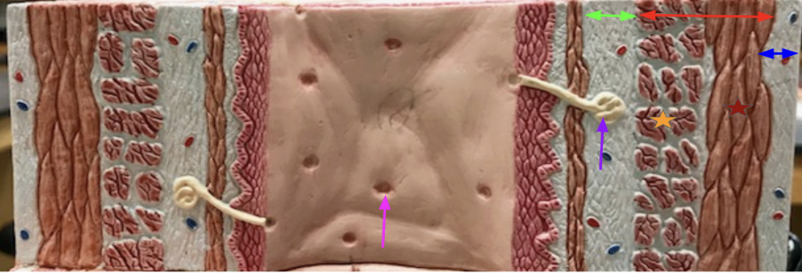
What organ is this? Identify. Function of purple
Esophagus
Green” Submucosa
Purple: Esophageal submucosal mucous gland - makes mucus so food can slide down the pipe
Pink: Opening of Esophageal submucosal mucous gland
Red: Muscularis externa
Dark red: Outer longitudinal layer
Orange: Inner circular layer
Blue: Adventitia

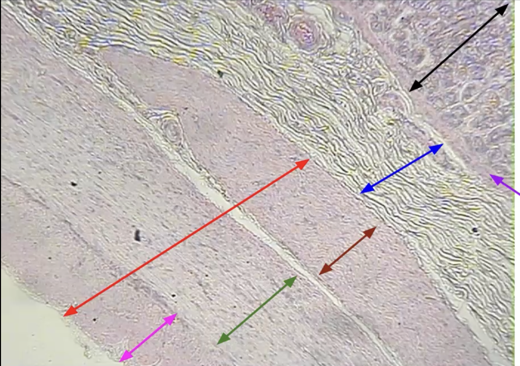
What organ is this? Identify
Esophagus
Black: Stratified squamous epithelium
Red: Lamina propria
Orange: Muscularis mucosae
Blue: Submucosa
Green: Circular muscularis externa
Light blue: Longitudinal muscularis externa

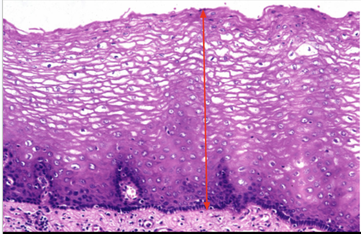
What is this image depicting? Function? Where is the only place this is located?
Stratified squamous epithelium
Protection
Only found in the esophagus/mouth

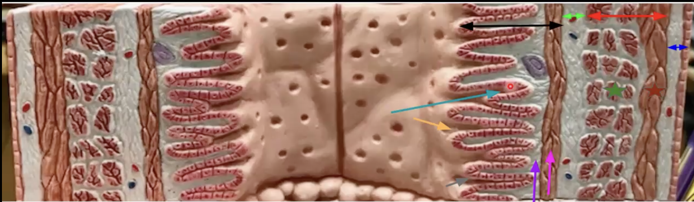
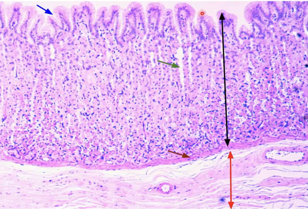
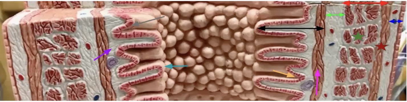
What organ is this? Identify. Function of teal and blue
Stomach
Black: Mucosa
Grey: Simple columnar epithelium
Purple: Lamina propria
Pink: Muscularis mucosae
Teal: Gastric glands - secrete gastric juice
Orange: Gastric pits (openings of gastric glands)
Neon Green: Submucosa
Dark green: Middle circular layer
Dark red: Outer longitudinal layer
Blue: Serosa - produce serous fluid to reduce friction between organs
Red: Muscularis externa
True or false: The esophagus is the only organ whos mucosa is made up of stratified squamous epithelium
What abt the Adventitia/Serosa?
True. Every other organ is simple columnar epithelium
- The esophagus is the only one that has adventitia. The other organs have serosa.
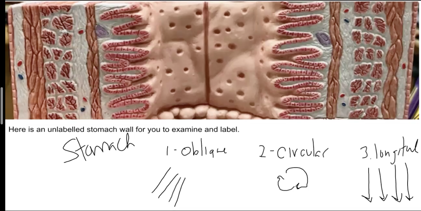
How does the muscularis externa of the stomach differ from the esophagus, small intestine, and large intestine? Why does this make sense?
- The muscularis externa of the stomach is made up of 3 layers: Inner oblique layer (not shown in the model), middle circular layer, and outer longitudinal layer. The other organs are only made up of the last two.
- The extra layer allows the stomach to perform mechanical digestion.
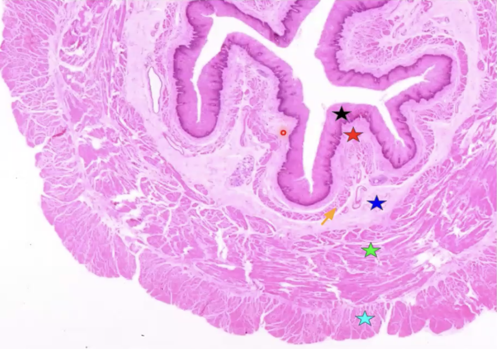
What organ is this? Identify. Function of blue
Stomach
Black: Mucosa
Blue: Gastric pit - where food is digested (holes at the top)
Green: Gastric gland (holes in the middle)
Dark red: Muscularis mucosae
Red: Submucosa


What organ is this? Identify
Stomach
Black: Mucosa
Purple: Muscularis mucosae
Blue: Submucosa
Red: Muscularis externa
Dark red: Inner oblique layer
Green: Middle circular layer
Pink: Outer longitudinal layer
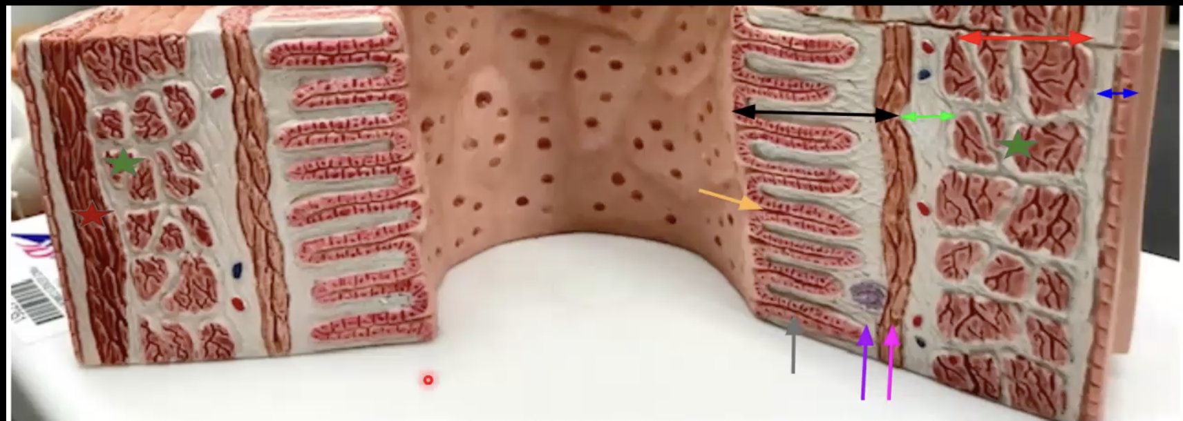
What organ is this? Identify. Function of teal and orange
Small intestine
Black: Mucosa
Grey: Simple columnar epithelium
Purple: Lamina propria
Pink: Muscularis mucosae
Orange: Intestinal crypts - secrete intestinal juice
Teal: Villi - increases surface area for absorption and digestion
Neon Green: Submucosa
Red: Muscularis externa
Green: Middle circular layer
Dark red: Outer longitudinal layer
Blue: Serosa

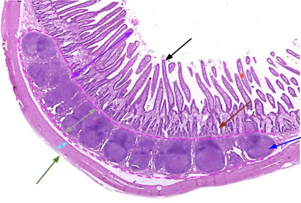
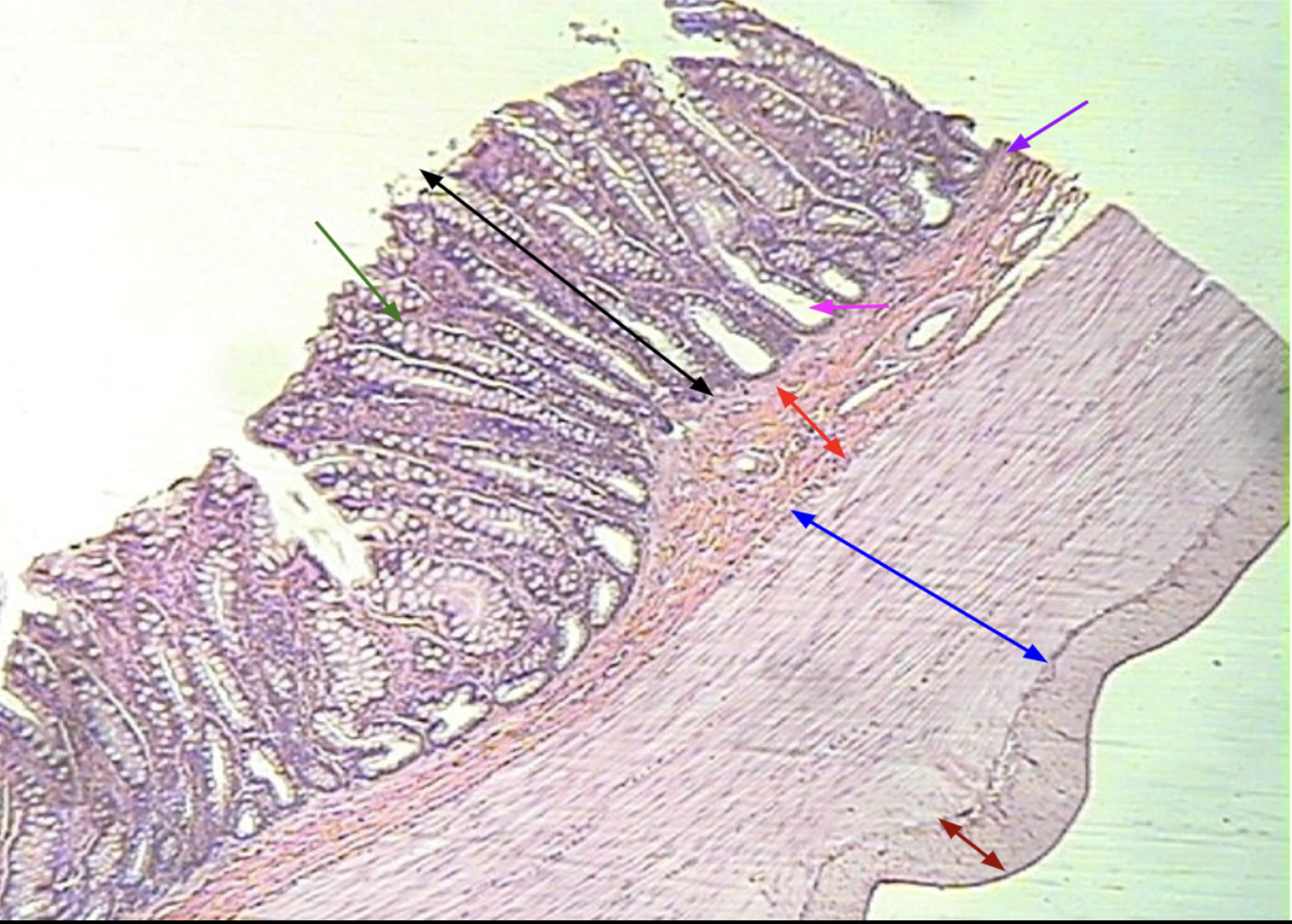
What organ is this? Identify What is function of pink?
Small intestine
Purple: Mucosa
Black: Villi
Dark red: intestinal crypt
Light green: Submucosa
Blue: Lymphatic nodule with a Peyer’s patch (Pink)
Peyer’s patch does surveillance of food, protect from pathogens (Know this he said this will definitely be on the exam)
Teal: Muscularis externa
Green: Serosa

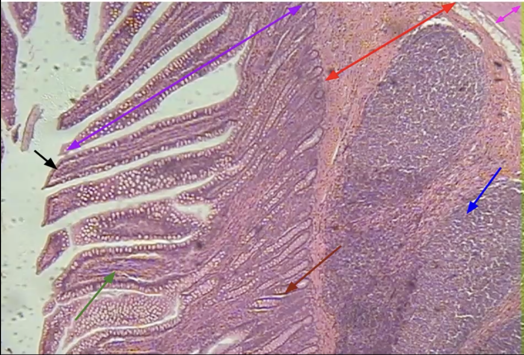
What organ is this? Identify each arrow
What are the small circles seen in structure indicated by the purple arrow?
Small intestine (know this bc it has Villus). The small circles are goblet cells, which function in producing mucous.
Purple: Mucosa
Black: Villus
Dark red: Intestinal crypt
Red: Submucosa
Blue: Lymphatic nodule (with peyer’s patch)
Pink: Muscularis externa (inner circular layer)
Green: Lamina propria

What organ is this? Identify
Small intestine
Black: Mucosa
Green: Inner circular muscularis externa
Red: Outer longitudinal muscularis externa


What organ is this? Identify. Function of organ and function of color orange
Large intestine (looks just like small intestine except there is no villi). Function: Absorb water, the pores in the middle is where water absorption takes place.
Black: Mucosa
Grey: Simple columnar epithelium
Purple: Lamina propria
Pink: Muscularis mucosae
Orange: Intestinal glands - secrete mucus. This mucus helps to smooth the passage of feces after the large intestine absorbs water from it.
Neon Green: Submucosa
Red: Muscularis externa
Green: Inner circular layer
Dark red: Outer longitudinal layer
Blue: Serosa

What organ is this? Identify.
Large intestine
Black: Mucosa
Purple: Muscularis mucosae
Red: Submucosa
Blue: Inner circular muscularis externa
Dark Red: Outer longitudinal muscularis externa
Green: Goblet cell
Pink: Intestinal gland

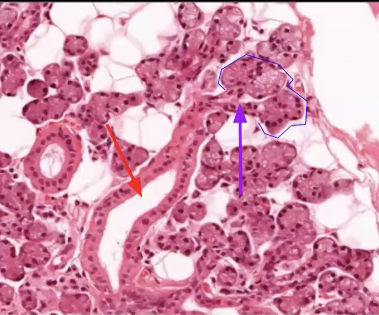
What organ is this? Identify. What is the function of the blue?
Parotid gland (Salivary gland)
Blue circle: Serous acini (cluster of cells) - secrete saliva (make amylase) into intercalated ducts
Pink: Ducts with a simple epithelium
Red: Ducts with a stratified epithelium
Purple: Connective tissue septa

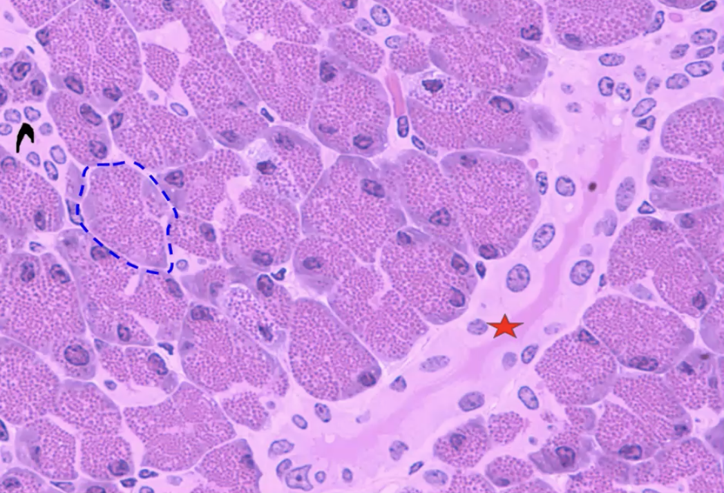
What organ is this? Identify the parts
Parotid gland
Blue: Serous acini
Purple: Intercalated duct
Red: Striated Duct
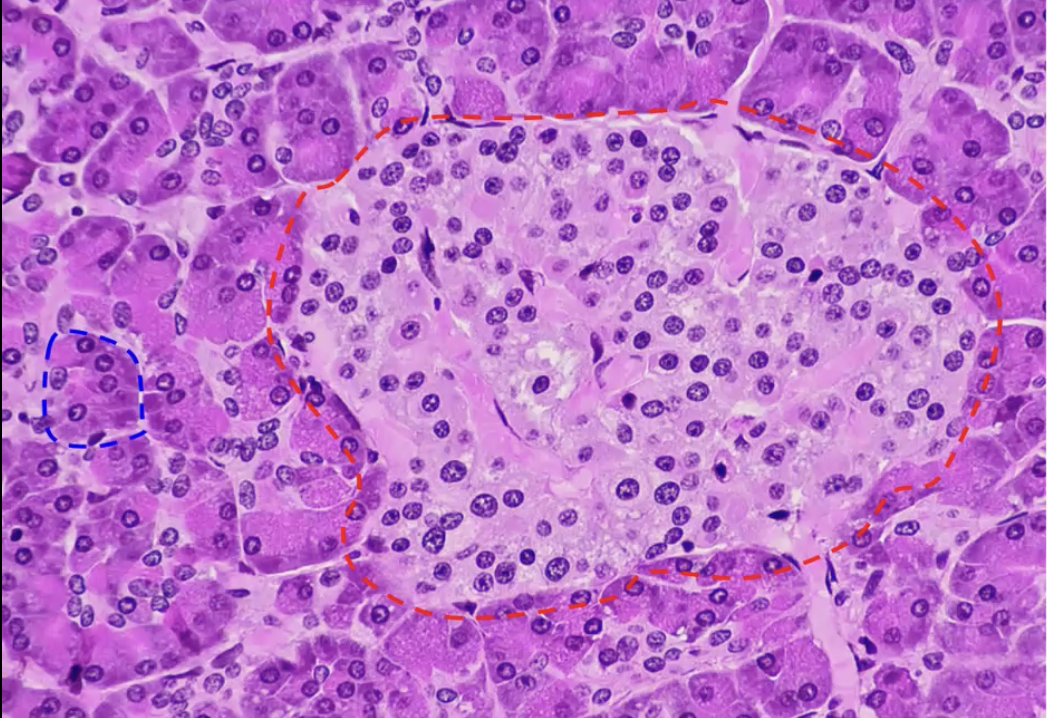
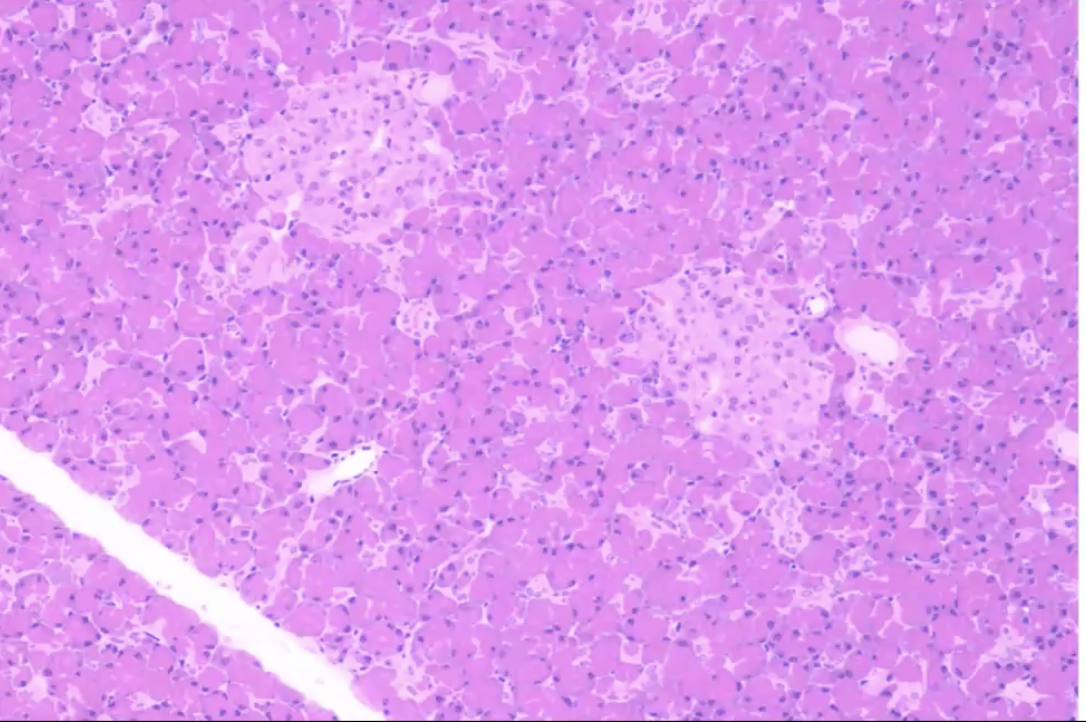
What organ is this? Identify Function of blue and red
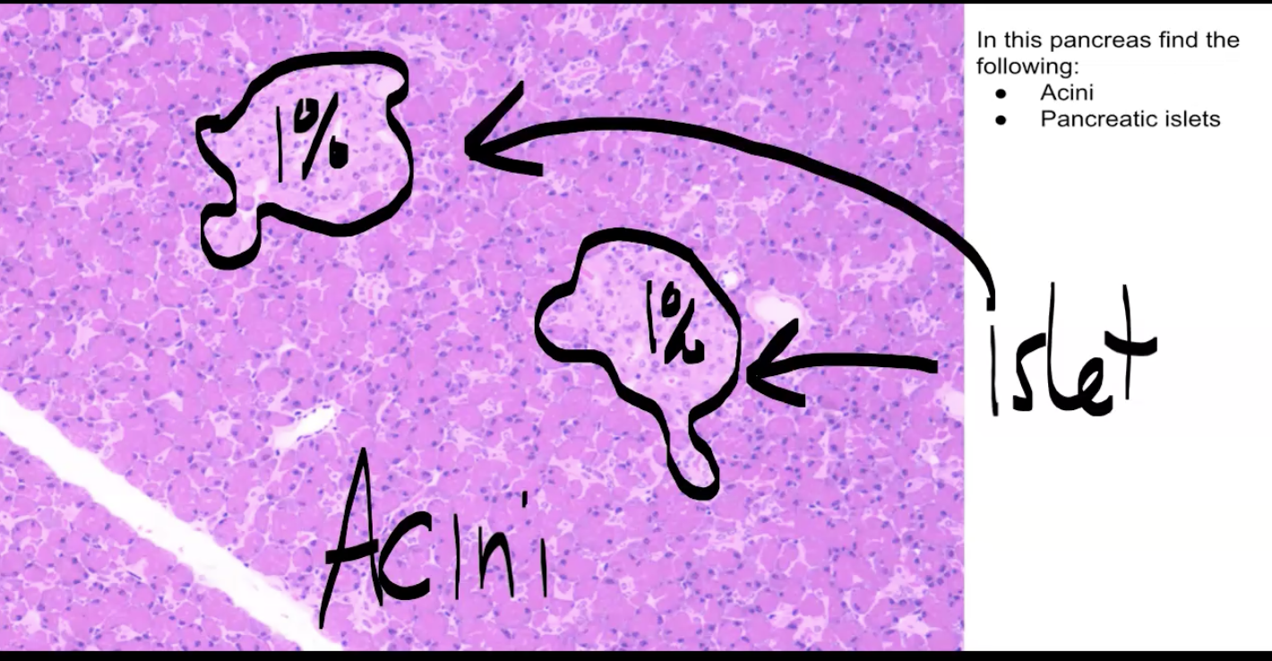
Pancreas
Blue: Pancreatic acini (majority of the cells are acini, dark purple) - they make various digestive enzymes such as amylase, protease, lipase, and nuclease
Red: Pancreatic islets (1% are are these cells, the light purple) - control sugar, make insulin and glucagon

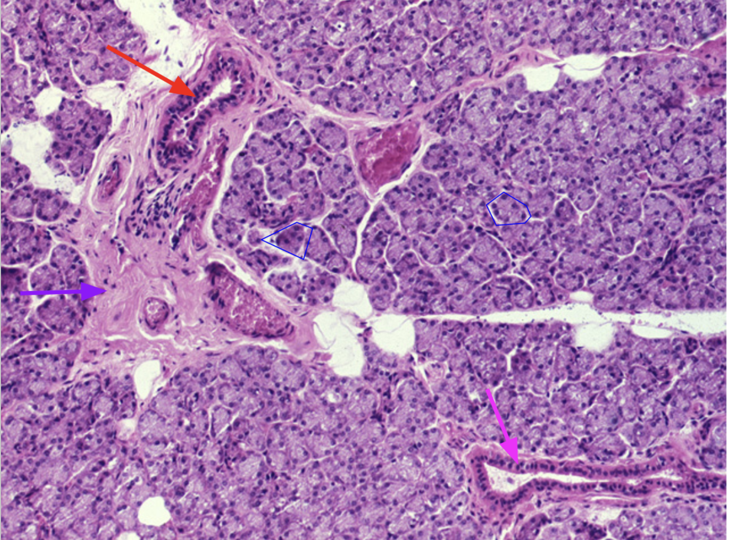
What organ is this? Identify. Function of red.
What role does the pH play?
Pancreas
Blue: Pancreatic acini
Red: Pancreatic duct - carries enzymes and alkaline fluid. The high pH of pancreatic juice helps neutralize the acidity of gastric chyme.

What organ is this? Identify the acini and islets
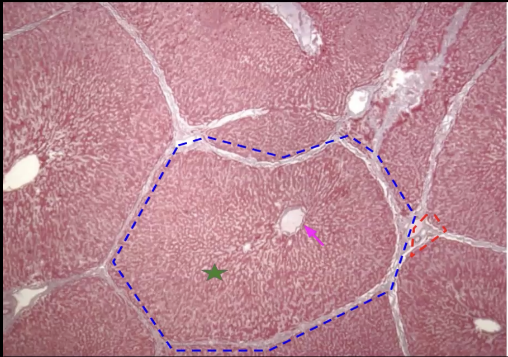
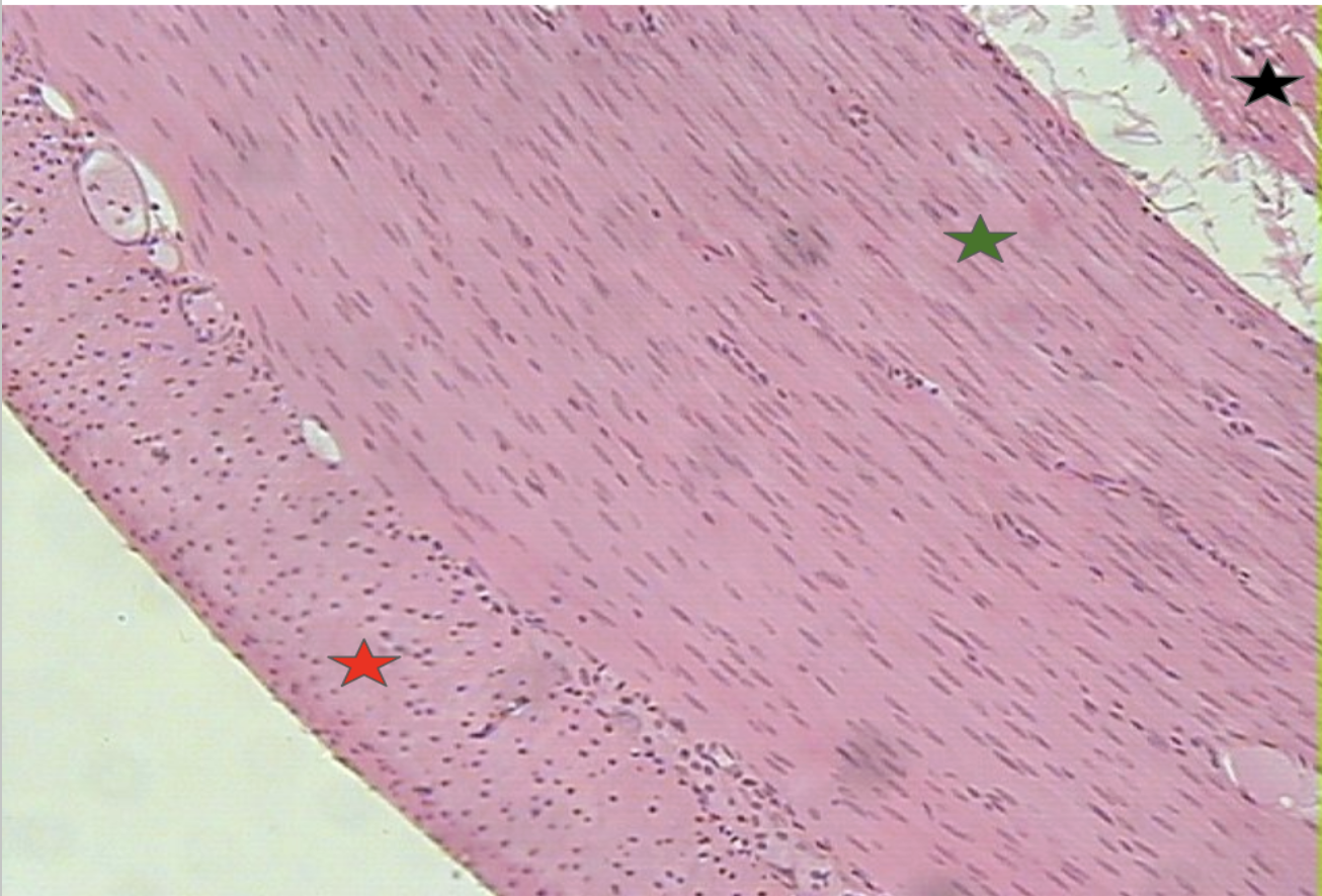
What organ is this? identify. What makes up the structure in red?
Liver (bc it is reddish brown)
Blue: Liver lobules - metabolism and break down of blood
Pink: Central vein (hole in the middle of every lobule) - carries filtered blood from the lobule
Green: Hepatocytes and sinusoidal capillaries
Red: Portal triad (made up of a vein, artery, and a duct)

What functional advantages are provided by having stratified epithelium in the esophagus and simple epithelium in the small intestine?
Stratified epithelium provides protection in the esophagus, and the simple epithelium in the small intestine allows for nutrient absorption.
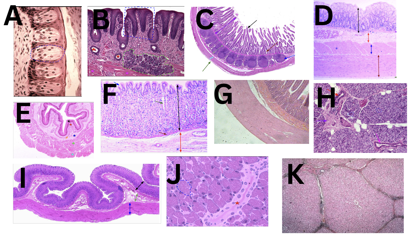
Identify the organ
A - Taste buds
B - Tongue
C - Small intestine
D - Large intestine
E - Esophagus
F - Stomach
G - Small intestine
H - Parotid gland
I - Stomach
J - Pancreas
K - Liver
