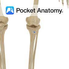
Femur, Tibia, Fibula
5.0(1)
Studied by 2 peopleCard Sorting
1/21
Earn XP
Description and Tags
Last updated 12:35 AM on 3/28/23
Name | Mastery | Learn | Test | Matching | Spaced | Call with Kai |
|---|
No analytics yet
Send a link to your students to track their progress
22 Terms
1
New cards
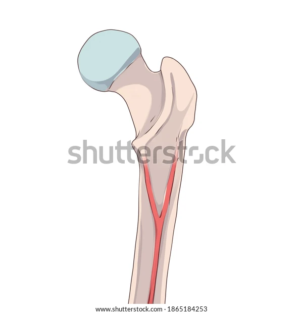
Head of femur
ball at top of femur
2
New cards
neck of femur
connects ball to femur
3
New cards
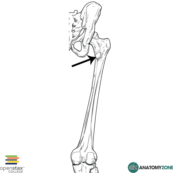
greater trochanter
top part of ridge after the neck of femur
4
New cards
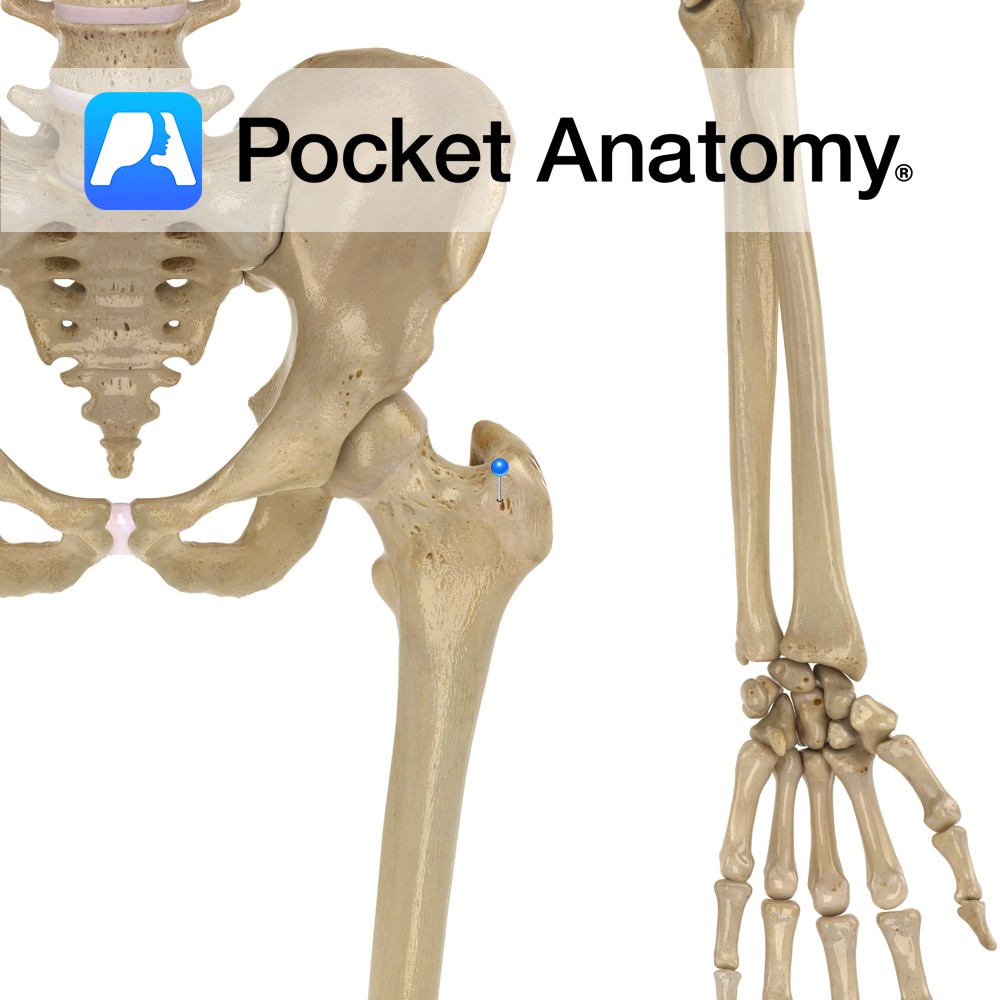
lesser trochanter
bottom part of ridge after the neck of femur
5
New cards
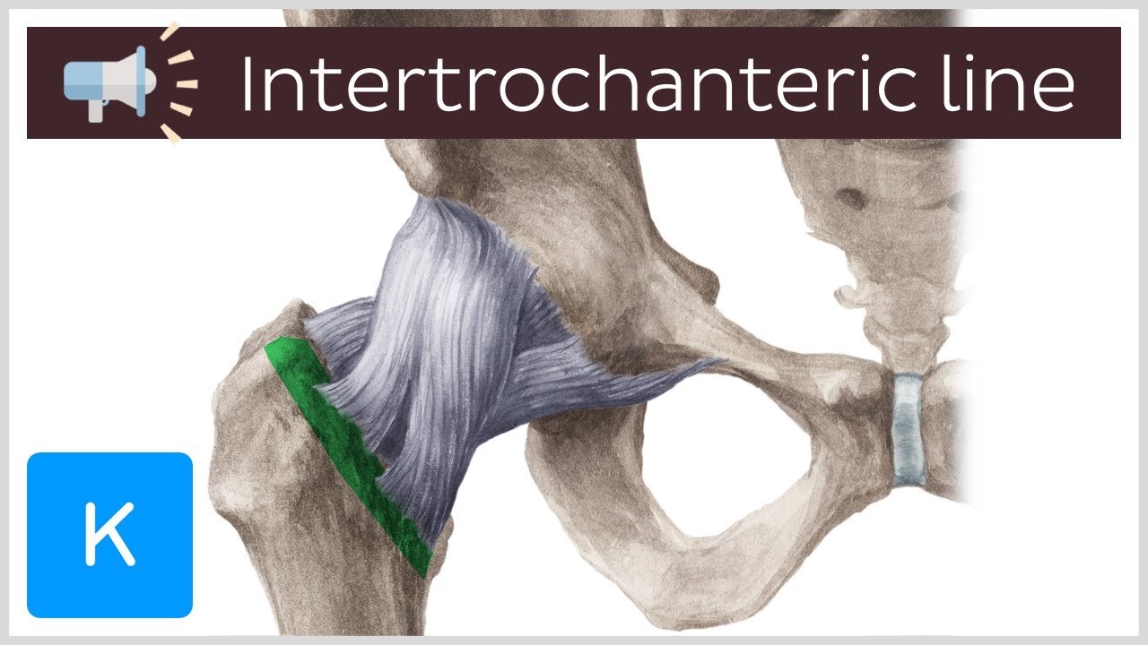
intertrochanteric line
the line after the neck on anterior side of femur
6
New cards
intertrochanteric crest
line after neck on posterior femur

7
New cards
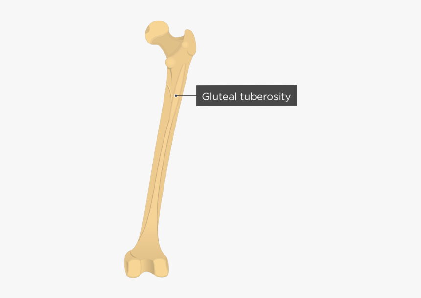
gluteal tuberosity
v on proximal posterior femur
8
New cards
linea aspera
line down posterior side of femur
9
New cards
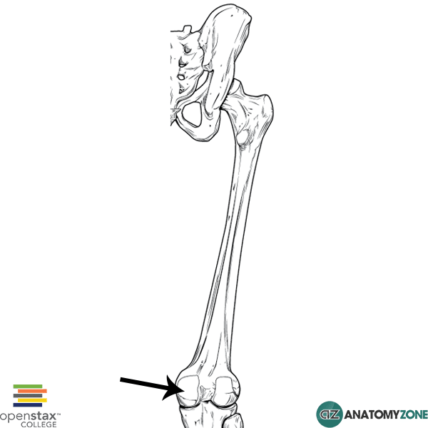
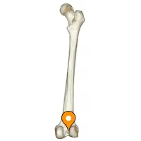
Medial condyle
medial attachment site on distal end of femur
10
New cards
lateral condyle
lateral attachment site on distal end of femur
11
New cards
intercondylar fossa
space on posterior side of femur between condyles
12
New cards
patellar surface
surface of patella
13
New cards
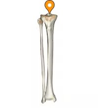
Intercondylar eminence
two dot ridges on top of tibia
14
New cards
tibial tuberosity
bump on anterior side of tibia
15
New cards
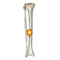
anterior crest
ridge of anterior side of tibia
16
New cards
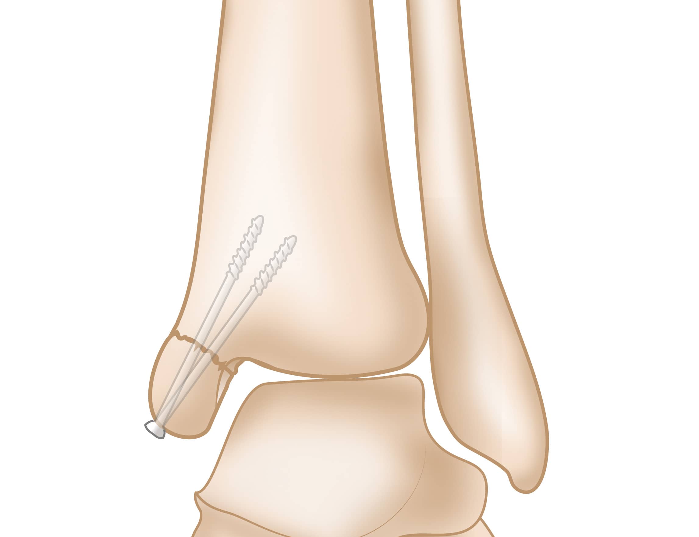
medial malleolus
point at the end of tibia
17
New cards
head
top of fibula
18
New cards
Tibialis anterior
dorsiflexes and inverts the ankle
19
New cards
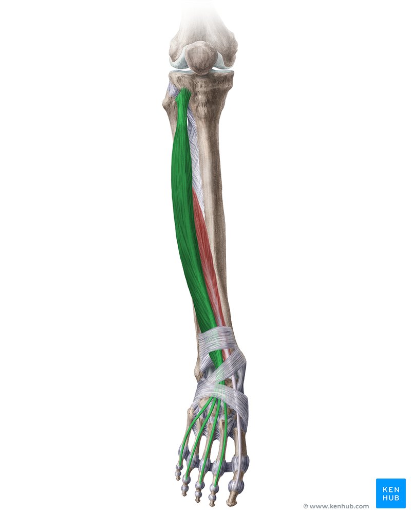
extensor digitorum longus
extends toes dorsiflexes the ankle
20
New cards
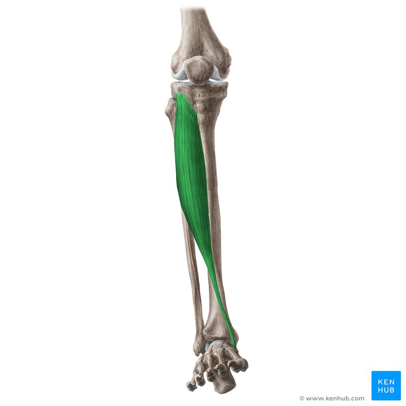
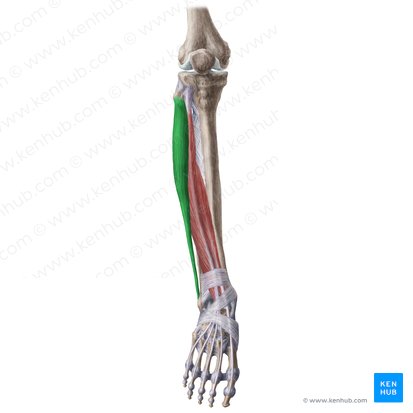
fibularis longus
plantar flexes the ankle and everts the ankle
21
New cards
Soleus
plantar flexes the ankle
O: tibia and fibula
I: calcaneus
O: tibia and fibula
I: calcaneus
22
New cards
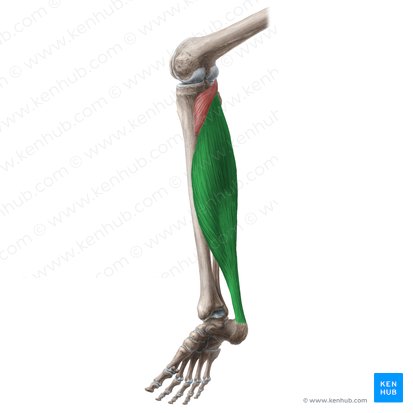
gastrocnemius
plantar flexes the ankle
O: medial and lateral condyles
I: calcaneus
O: medial and lateral condyles
I: calcaneus
