Nervous coordination and muscles
 Call Kai
 Learn
Learn Practice Test
 Spaced Repetition
 Match
Match Flashcards
 Knowt Play
1/54

There's no tags or description
Looks like no tags are added yet.
Name | Mastery | Learn | Test | Matching | Spaced | Call with Kai |
|---|
No analytics yet
Send a link to your students to track their progress
55 Terms
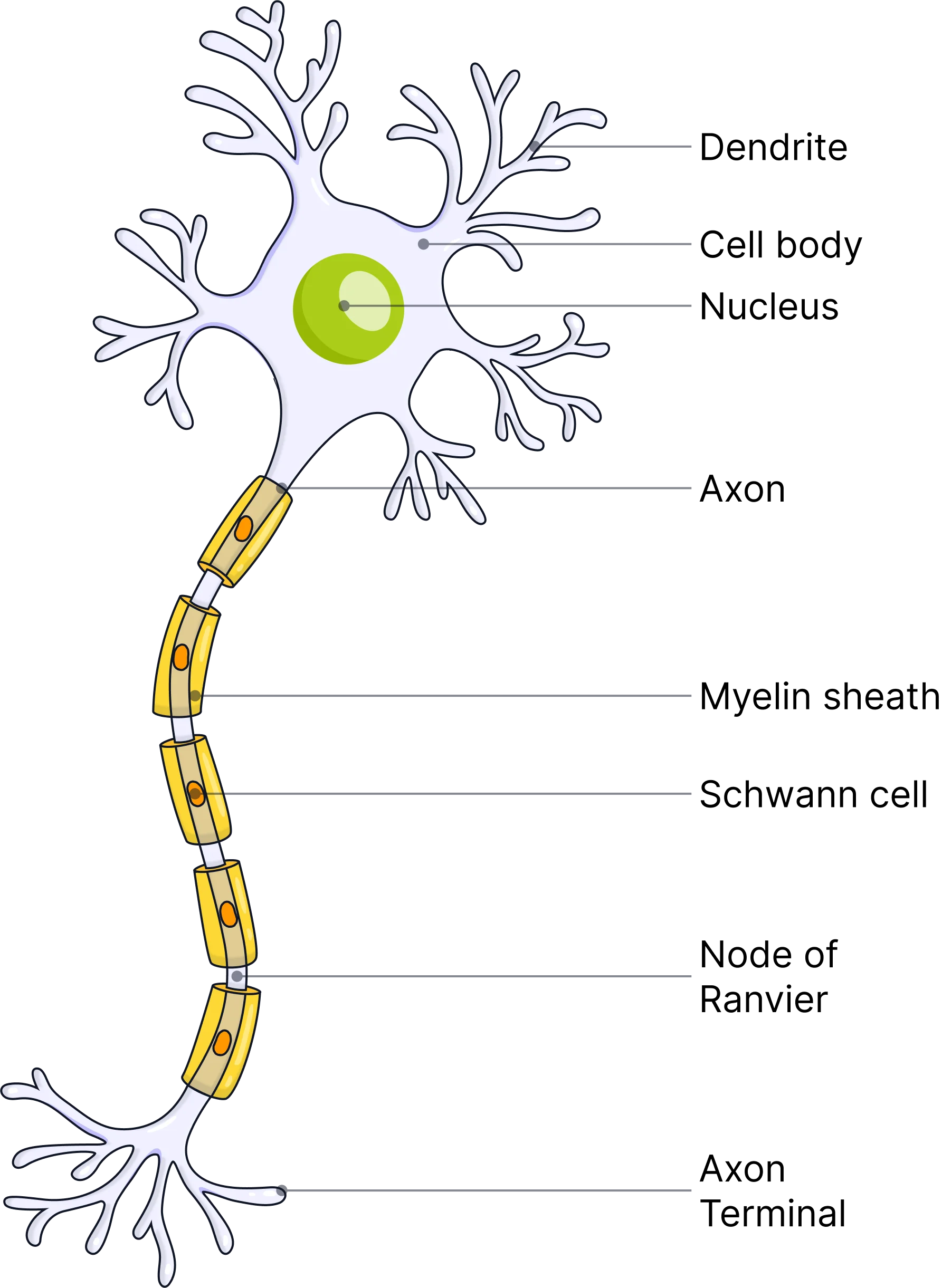
Describe the structure of a motor neurone.
What does resting potential mean?
The difference between the electrical charge inside and outside the neurone when the neurone is not conducting an impulse.
What is the value for resting potential in mV?
-70 mV
Describe and explain how resting potential is maintained in a neurone.
1) A sodium-potassium ion pump actively transports 3 sodium ions (Na+) out of the neurone.
2) While this happen, the sodium-potassium ion pump allows 2 potassium ions (K+) to move into the neurone.
3) This creates an electrochemical gradient, causing sodium ions to move in and potassium ions to move out generating -70 mV resting potential (it is more positive on the outside of the membrane than on the inside).
The cell-surface membrane of the neurone is more permeable to potassium ions as there are more potassium ion channel proteins that are always open (unlike sodium ion channels).
What is an action potential?
When the potential difference across a neurone reaches the threshold potential value therefore generating a nerve impulse.
What causes depolarisation across a neurone membrane?
The neurone membrane becoming more permeable to sodium ions (due to sodium ion channel proteins opening), therefore allowing more sodium ions to move into the neurone.
What is the threshold potential value in mV?
-55 mV.
Describe how an action potential is generated in a pacinian corpuscle (pressure sensitive).
1) Application of pressure causes stretch-mediated sodium ion channel proteins to deform and open.
2) Sodium ions move into the neurone via facilitated diffusion, causing depolarisation.
3) Once the membrane potential reaches the threshold potential an nerve impulse is generated.
What happens during repolarisation?
The potassium ion channel proteins are open causing more potassium ions to move out of the neurone which causes the outside of the neurone to be even more positive which causes the membrane potential to be even more negative than resting potential.
Why is it good that we have a threshold potential?
It means that we do not respond to every small change in our environment (as not every change causes the threshold potential to be reached).
What word do you use to describe a membrane that has a membrane potential more negative than the resting potential?
Hyperpolarised.
What is the name of the period in which resting potential is restored after hyperpolarisation?
The refractory period.
What is the all-or-nothing principle?
If the membrane potential does not exceed -55 mV an action potential and nerve impulse are not generated.
True of false? The size of a stimulus is indicated by the size of an action potential.
False. The size of the stimulus is indicated by the frequency of action potentials (larger stimulus causes more frequent action potentials).
Why can’t an action potential be generated during the refractory period?
Sodium ion channel proteins are recovering and can’t be open meaning the membrane of the neurone can’t be stimulated.
Give and explain three reasons why the refractory period is important.
The refractory period ensures that discrete impulses are produced.
The refractory period ensures that action potentials are unidirectional (if the action potential spread out in multiple directions a response would be prevented).
The refractory period limits the number of impulse transmission (this means we don’t overreact to stimuli).
What are three factors that affect the speed of conduction?
Myelination and saltatory conduction.
Axon diameter.
Temperature.
How are myelin sheaths formed?
Schwann cells wrap around the axon of the neurone (not allowing charged ions or nerve impulses to pass through them).
Describe the process of saltatory conduction and how it allows a nerve impulse to travel faster.
1) Schwann cells are wrapped around the axon of the neurone, forming the myelin sheath which does not allow ions or nerve impulses to pass through it.
2) Depolarisation that generates an action potential can only happen at nodes of Ranvier.
3) The action potential jumps from node to node (saltatory conduction).
As the action potential only has to be present at nodes of Ranvier, it skips large areas of the neurone meaning the action potential travels faster.
How does increasing the diameter of an axon increase the speed of conductance? Explain your answer.
It increases the speed of conductance because an increased axon diameter offers less resistance to the flow of ions in the axon meaning depolarisation spreads more quickly.
True or false? Increasing the temperature increases the speed of conductance (to a certain point). Explain your answer.
True. Increasing temperature gives ions more kinetic energy, causing them to diffuse faster. Increasing temperature causes enzymes involved in respiration to work faster and therefore produce more ATP for the sodium/potassium ion pump to carry out active transport.
What is a synapse?
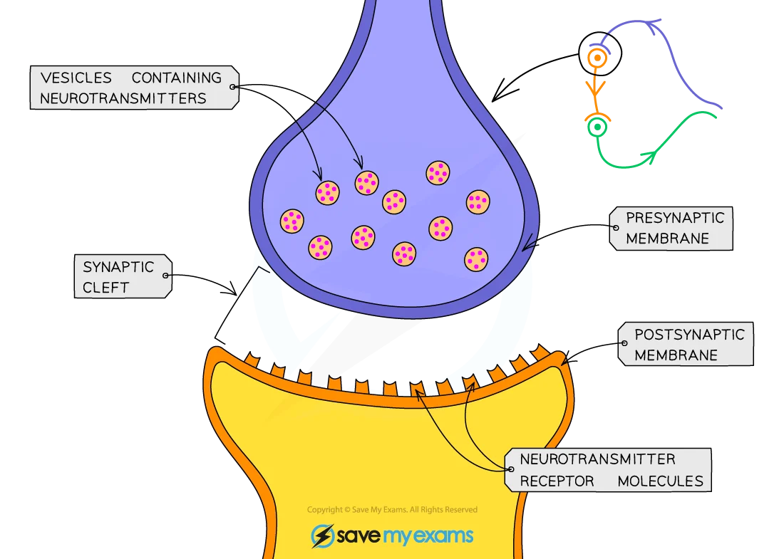
The gap between the end of an axon of one neurone and a dendrite of it’s adjacent neurone.
What is an action potential transmitted as across a synapse?
Neurotransmitters (action potentials cannot move across synapses).
Describe the process of synaptic transmission.
1) The action potential arrives at the synaptic knob.
2) The arrival of the action potential causes the synaptic knob to be depolarised.
3) Depolarisation of the synaptic knob causes calcium ion channel proteins in the cell-surface membrane to open, this causes calcium ions to diffuse into the synaptic knob.
4) The influx of calcium ions cause vesicles containing neurotransmitters to fuse with the presynaptic membrane therefore releasing the neurotransmitters into the synaptic cleft.
5) The neurotransmitters diffuse down their concentration gradient across the synaptic cleft.
6) The neurotransmitters bind to complementary receptors (on sodium ion channel proteins) on the postsynaptic membrane.
7) This binding causes sodium ion channel proteins on the postsynaptic membrane to open, allowing sodium ions to diffuse into the postsynaptic neurone, causing the postsynaptic neurone to be depolarised.
8) If the postsynaptic neurone is depolarised to the threshold potential, a new action potential will be generated in the postsynaptic neurone.
9) The neurotransmitters are then hydrolysed by an enzyme, they diffuse across the synaptic cleft where they are resynthesised at the presynaptic neurone.
Describe the structure of a synapse.
What is the neurotransmitter at a cholinergic synapse?
Acetylcholine.
What is the enzyme that degrades the neurotransmitter in a cholinergic synapse?
Acetylcholinesterase.
What does acetylcholinesterase hydrolyse acetylcholine into?
Choline, acetate.
What is summation?
Summation is the rapid build up of neurotransmitters in the synapse to help generate an action potential in the postsynaptic neurone.
What are the two types of summation?
Spatial summation, temporal summation.
Describe the process of spatial summation.
Many neurones combine all the neurotransmitter that they release, this causes the threshold potential to be reached in the postsynaptic neurone (involves multiple presynaptic neurones linked to one postsynaptic neurone).
Describe the process of temporal summation.
One neurone releases neurotransmitter frequently within a short period of time so that the threshold potential is reached in the postsynaptic neurone (involves one presynaptic neurone linked to one postsynaptic neurone).
How is a synapse adapted for unidirectional movement of action potentials?
Neurotransmitter is only released from the presynaptic neurone, there are only receptors for the neurotransmitter on the postsynaptic neurone.
What is an inhibitory synapse?
A synapse that prevents an action potential from being generated.
How do inhibitory synapses prevent action potentials?
1) Inhibitory synapses cause chloride ions to diffuse into the postsynaptic neurone and potassium ions to move out of it.
2) This movement of ions hyperpolarises the postsynaptic membrane to -80 mV.
This means that an action potential will be highly unlikely to develop as more sodium ions are required to reach the threshold potential.
Give three ways in which a drug can create more action potentials in a postsynaptic neurone.
They can mimic the neurotransmitter (causing sodium ion channel proteins to open and cause depolarisation which leads to an action potential).
They can stimulate the release of more neurotransmitter.
They can inhibit the enzyme that hydrolyses the neurotransmitter.
Give two ways in which a drug could decrease the number of action potentials generated at a postsynaptic neurone.
It could inhibit the release of neurotransmitter.
It could block receptors on sodium ion channel proteins on the postsynaptic membrane.
Describe the transmission of an action potential across a neuromuscular junction.
1) The action potential reaches the presynaptic membrane on the motor neurone.
2) This causes calcium ion channels to open, allowing calcium ions to diffuse into the motor neurone.
3) The influx of calcium ions causes vesicles containing acetylcholine to fuse with the presynaptic membrane, releasing acetylcholine into the neuromuscular junction.
4) Acetylcholine diffuses across the neuromuscular junction and binds to receptors on the sarcolemma.
5) This binding causes sodium ions to move into the sarcolemma, causing depolarisation.
6) The new action potential moves down T-tubules toward the centre of the muscle fibre.
7) The new action potential prompts voltage-gated calcium ion channels in the membranes of the sarcoplasmic reticulum to open.
8) This allows calcium ions to diffuse out of the sarcoplasmic reticulum and into the sarcoplasm.
This allows muscle contraction via the sliding filament theory.
Give four differences between a cholinergic synapse and neuromuscular junction.
Neuromuscular junctions are only excitatory, cholinergic synapses could be excitatory or inhibitory.
Neuromuscular junctions connect motor neurones to muscles, cholinergic synpases connect two neurones.
Neuromuscular junctions are the end point of an action potential, in cholinergic synapses a new action potential is generated in the next neurone.
At neuromuscular junctions acetylcholine binds to receptors on muscle fibre membranes, at cholinergic synapses acetylcholine binds to receptors on the postsynaptic membrane of a neurone.
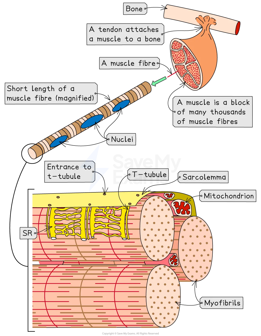
Describe a muscle fibre (muscle cell) in detail.
Myofibrils: long, cylindrical structures composed of sarcomeres.
Sarcomere: functional unit of a myofibril, the area between two Z-lines, made up of overlapping actin and myosin.
Sarcolemma: the plasma membrane of a muscle fibre.
Sarcoplasmic reticulum: a specialised form of smooth endoplasmic reticulum that surrounds myofibrils.
Sarcoplasm: the cytoplasm of a muscle fibre.
What is a sarcomere? Describe it’s structure.
The region between two Z-lines.
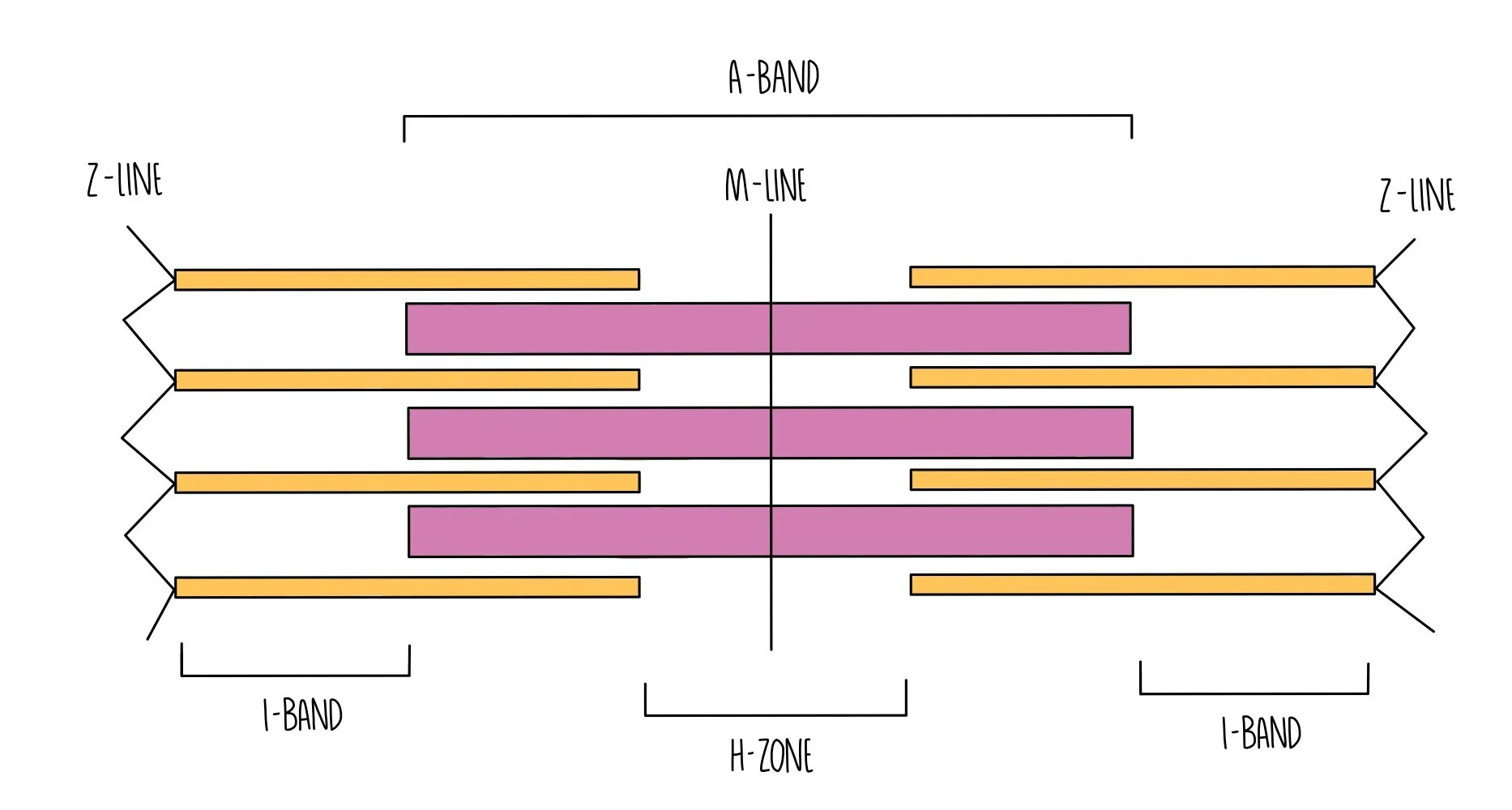
Thick = myosin, thin = actin.
I band = actin only, A band = actin and myosin overlapping, H zone = myosin only.
Describe how each band in a sarcomere changes in length when a muscle contracts.
I band shortens, A band remains the same, H zone shortens.
Describe the sliding filament theory for muscle contraction.
1) Troponin molecule on actin prevents myosin head from binding to the binding site.
2) Calcium ions (from the action potential) from the sarcoplasmic reticulum change the structure of troponin, causing troponin to pull away from the myosin binding site on actin.
3) Myosin head is now able to attach to the binding site on actin, forming an actinomyosin bridge.
4) The myosin head changes angle, pulling the actin filament along with it, this causes the ADP molecule to be released from the myosin head.
5) An ATP molecule attaches to the myosin head, this causes the myosin head to detach from the actin binding site.
6) ATPase hydrolyses ATP to ADP and Pi, this releases energy that allows the myosin head to resume its normal position.
7) The myosin head is now able to attach to the binding site behind it.
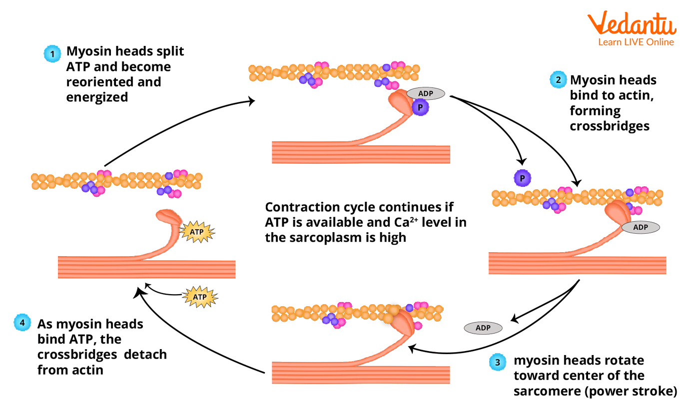
Give three things that ATP can provide energy for in muscle contraction.
Break actinomyosin bridges (detach actin and myosin).
Bend the myosin head (so it can resume it’s original position).
Active transport of calcium ions back into the sarcoplasmic reticulum when nerve stimulation stops.
What is the role of phosphocreatine?
It can give a phosphate group to ADP meaning ATP can be quickly resynthesised and used to release needed energy during short bursts of intense activity.
What is the role of glycogen granules in skeletal muscles?
They are a store of glucose that can be rapidly hydrolysed to produce glucose needed for aerobic respiration to produce ATP.
What is the role of calcium ions in the contraction of a myofibril?
1) Calcium ions diffuse into the myofibrils from the sarcoplasmic reticulum.
2) The calcium ions bind to troponin, causing it to change shape and expose the myosin head binding sites on actin.
This allows actinomyosin bridges to form.
How do fast-twitch muscle fibres and slow-twitch muscle fibres differ in structure?
Fast-twitch muscle fibres: more myosin filaments, large store of glycogen, large store of phosphocreatine, high concentration of enzymes involved in anaerobic respiration.
Slow-twitch muscle fibres: large store of myoglobin, rich blood supply, many mitochondria.
What are the differences in function of fast-twitch and slow-twitch muscle fibres?
Fast-twitch muscle fibres contract faster and with more force, slow-twitch muscle fibres contract slower and with less force but can respire aerobically for longer periods of time.
Where are slow-twitch muscle fibres commonly found?
In calf muscles.
Where are fast-twitch muscle fibres commonly found?
In biceps.
True or false? Fast-twitch fibres often respire aerobically while slow-twitch fibres often respire anaerobically.
False. FAST-TWITCH FIBRES PRIMARILY RESPIRE ANAEROBICALLY WHILE SLOW-TWITCH FIBRES PRIMARILY RESPIRE AEROBICALLY.
Compare the ATP production of fast-twitch fibres and slow-twitch fibres.
Fast-twitch fibres produce ATP faster but generate less ATP per glucose molecule, slow-twitch fibres produce ATP slower but generate more ATP per glucose molecule.
Why do fast-twitch fibres have a greater glycogen store than slow-twitch fibres?
Fast-twitch fibres need to be able to rapidly produce glucose (from hydrolysis of glycogen).
TROPOMYOSIN
TROPOMYOSIN