BIO 3.1 - GAS EXCHANGE + DIGESTION & ABSORPTION
1/42
Earn XP
Description and Tags
-SA:VOLUME ratio -gas exchange of insects, fish, humans -digestion and absorption -mass transport
Name | Mastery | Learn | Test | Matching | Spaced | Call with Kai | Chat |
|---|
No analytics yet
Send a link to your students to track their progress
43 Terms
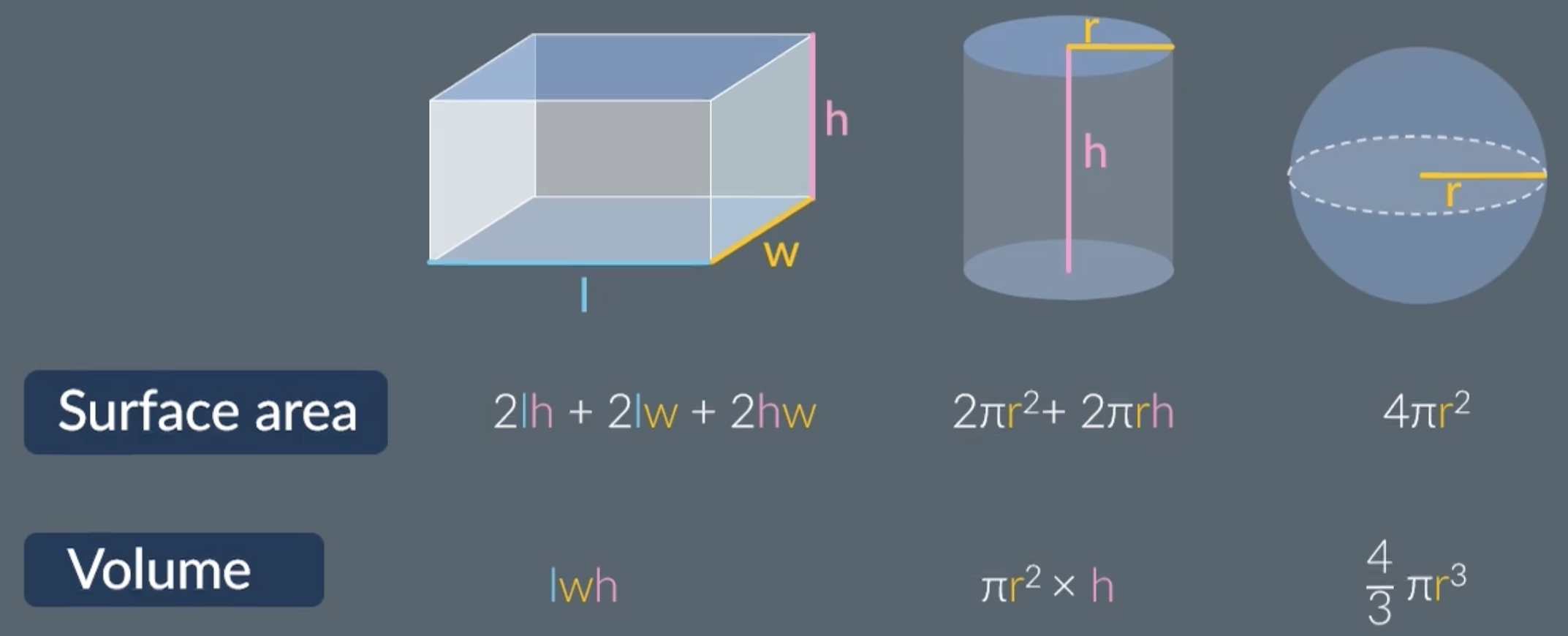
SA:VOLUME ratio
SA:V ratio = surface area / volume
smaller organisms have a larger SA:V ratio
small animals loose heat quickly because they have a larger SA:V ratio, therefore they carry out more respiration to maintain their body temperature
big animals loose heat slowly because they have a small SA:V ratio
benefits of a small/large SA:V ratio
(for small organisms) a large SA:V ratio is beneficial when the organism needs to:
lose heat quickly
absorb substances quickly
diffuse oxygen/nutrients quickly
(for large organisms) a small SA:V ratio is beneficial when the organism needs to:
conserve heat
reduce water loss
gas exchange of insects -structures
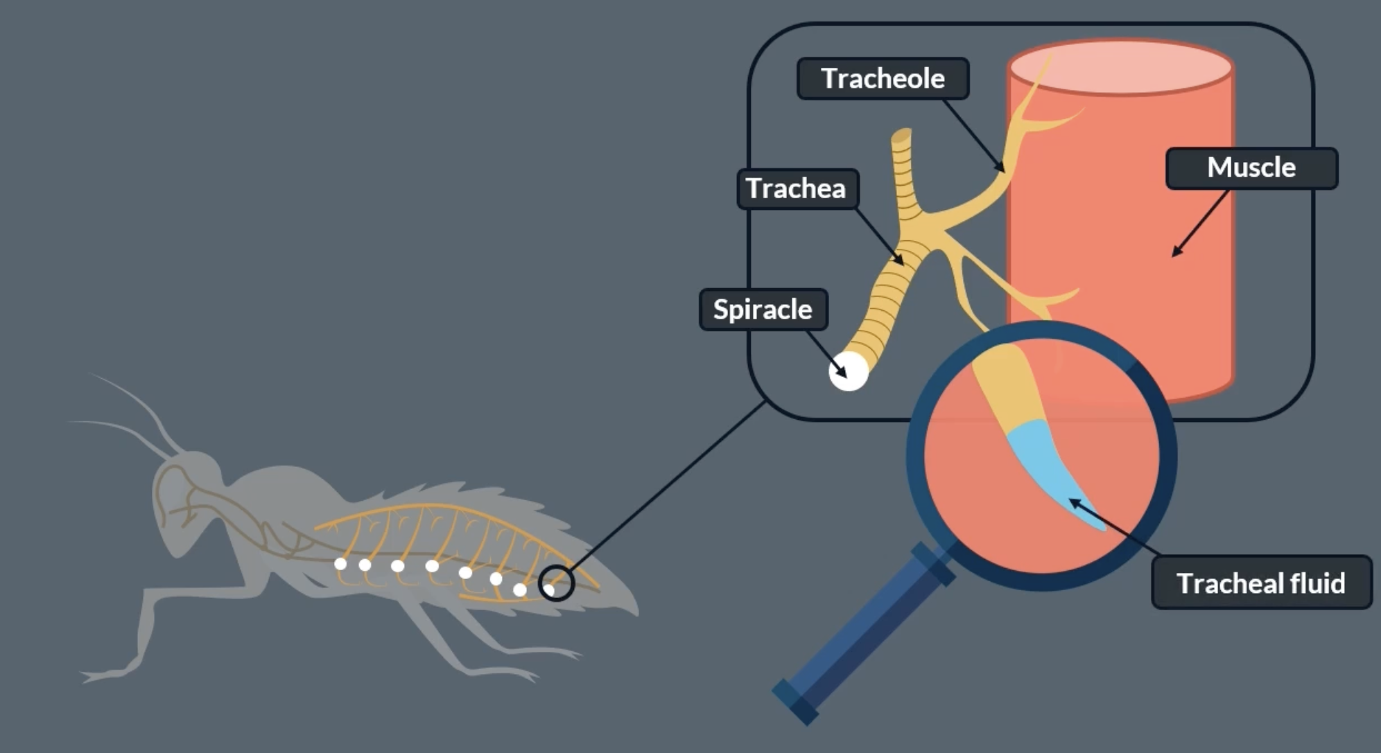
the main site of gas exchange in insects = tracheal system
spiracles on insects side extend into tracheae which then branch into tracheoles which then extend into the insects muscle tissue. allows oxygen to enter
spiracles allow air to flow in and out of the insect
trachea and tracheoles are supported by spirals of chitin, which is a strong flexible substance which help prevent the tube form collapsing as the insect moves
tracheae is the plural of trachea
have a waterproof waxy exoskeleton
difference between human and insect trachea
1. human trachea is much bigger than an insect trachea. to be precise, scientists say that a human trachea has a much bigger diameter and length than an insect trachea
2. humans only have one trachea, whereas insects have many tracheae
3. a human trachea branches into bronchi. an insect trachea branches into tracheoles
4. a human trachea is supported by cartilage, whereas an insect trachea is supported by chitin
gas exchange of insects process
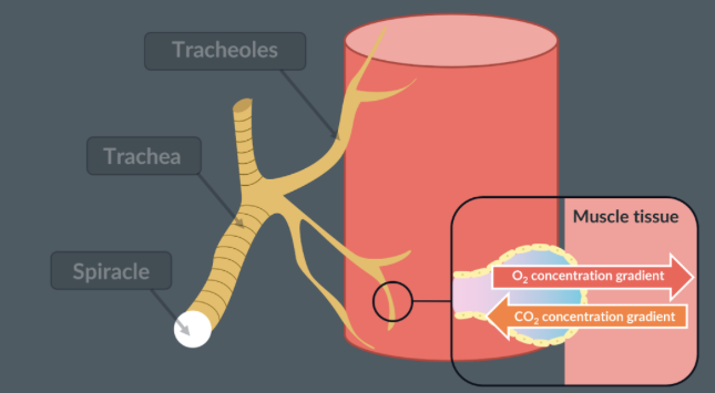
oxygen diffuses from air outside into the insects respiring muscle tissues, where the cells use up the oxygen quickly. therefore there is a lower concentration of oxygen inside the insects respiring muscle tissues than outside in the air, creating a concentration gradient in which oxygen can diffuse
oxygen enters through the spiracles, into the trachea and tracheoles, then dissolves into the tracheal fluid and diffuses into the muscle cell along its concentration gradient
aerobic respiration in muscle tissue produces carbon dioxide. therefore the concentration of carbon dioxide in the muscle tissue is greater than the concentration in the air, creating a concentration gradient in which CO2 can diffuse
CO₂ produced in the muscle cells diffuses out of the cells into the tracheal fluid, then diffuses into the air in the tracheoles and diffuses out through the spiracles along its concentration gradient
oxygen vs carbon dioxide dissolving
oxygen:
enters the tracheoles as a gas
must dissolve in the tracheal fluid before it can diffuse into muscle cells
carbon dioxide:
is already dissolved in the fluid around muscle cells so it doesn’t need to dissolve again
it simply diffuses into the tracheal fluid, then into the tracheoles and out of the spiracles
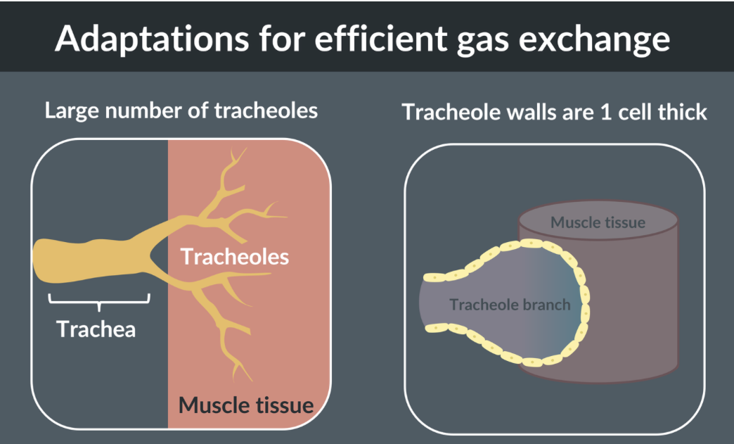
adaptations of tracheal system for gas exchange
-a large number of tracheoles that reach into the muscle tissues. this helps ensure a short diffusion pathway and maximises the surface area for gas exchange
- the walls of the tracheoles are just 1 cell thick. this also helps ensure a short diffusion pathway for gas exchange
adaptations of tracheal system for gas exchange- tracheal fluid
rate of diffusion is much faster through air rather than fluid, therefore diffusion through tracheal fluid can be slower
when rate of diffusion/ gas exchange needs to be faster (if insect is moving around more = needs more oxygen) insects can absorb the tracheal fluid into the muscle cells. the tracheal fluid is then replaced by air
this allows the final diffusion pathway to be through a gas rather than a liquid, which increases diffusion rate
limiting water loss during insect gas exchange
because insects are small they have a large SA:V ratio, meaning they experience water loss more
-insects have a waterproof lipid layer on their exoskeleton, prevent water from passing out of the cells
-have muscles around the spiracles to allow for them to be opened and closed, prevent evaporation from the spiracles. close for a short time when respiration is low
-have a small SA:V ratio of where water can actually evaporate from
methods of moving gas in the tracheal system
gas can exchange by diffusion. when cells respire, they use up oxygen and produce carbon dioxide, creating a concentration gradient between tracheoles and atmosphere
mass transport, in which an insect contracts and relaxes their abdominal muscles which creates pressure changes to help move air faster
when the insect is flying the muscle cells start to respire anaerobically to produce lactate. this lowers the water potential of the muscle cells. water moves from the tracheole fluid into muscle cells by osmosis. this reduces the volume of fluid at the tracheole ends, leaving more air space for oxygen to reach cells faster
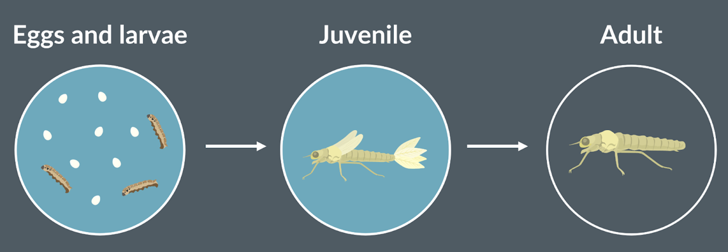
??? insects with gills
some insects live first part of their life in water where they can get oxygen from going to the surface of the water, where air can enter the spiracles
some species have gills instead of spiracles and tracheae. this adaptation is more common when the insect is a type that hunts prey underwater. hunting and chasing requires a higher rate of respiration = higher oxygen demand.
to meet this demand, the insect needs gills to extract oxygen from the surrounding water so it doesn’t have other return to surface constantly.
when these insects get older and leave the water, they lose their gills in favour of spiracles, tracheae
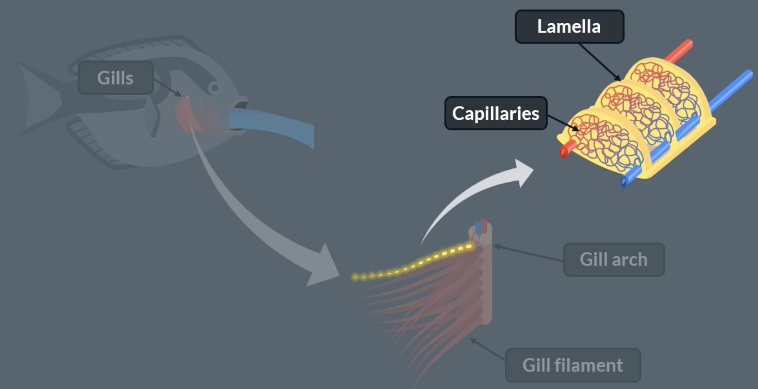
gas exchange of fish- structures
gills are the organ where gas exchange takes place
water enters the fishes mouth and is pushed put past a flap on either side of the fishes head. beneath each flap is where the gills are found. oxygen diffuses from the water into the fishes blood stream
each gill has a gill arch, which has a blood vessel running down the centre that branches off to supply each gill filament with blood
each structure in the gill contains blood vessels
lamella stick out of the gill filaments surface. all lamella contain a network of capillaries
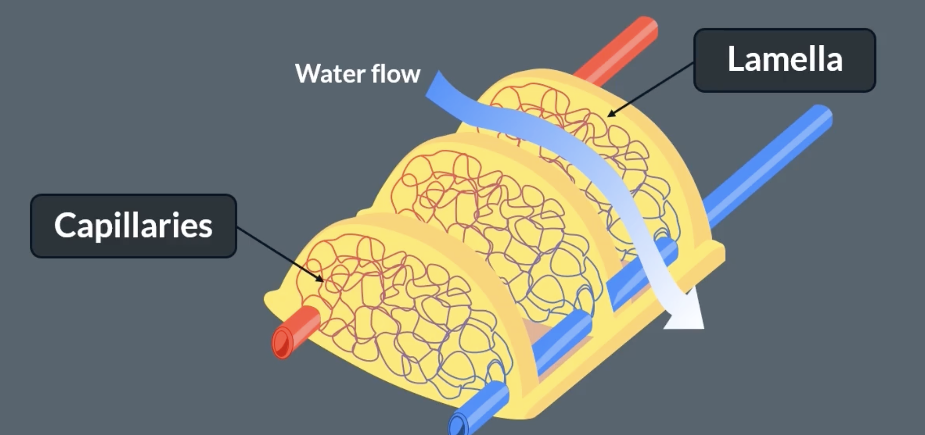
gas exchange of fish- lamellae
lamellae is where gas exchange occurs
lamellae is plural for lamella
as water crosses the gills, water runs in between the lamellae
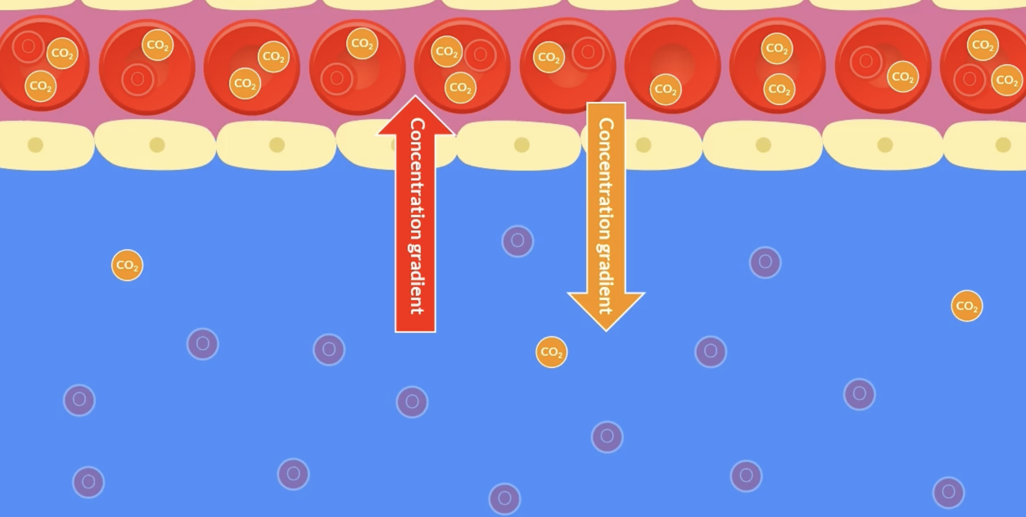
the fish uses up oxygen during respiration, therefore there is a lower concentration of oxygen in the blood than in the water, so oxygen diffuses from water into the blood stream along its concentration gradient
during respiration carbon dioxide is produced. therefore carbon dioxide concentration in the blood stream is higher than the water, so carbon dioxide diffuses from the blood into the water
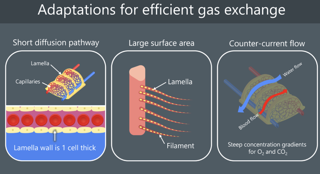
adaptations for gas exchange in fish
short diffusion pathway
there is a network of capillaries in each lamella, which brings the blood very close to the surface
the surface of each lamella is one cell thick
large surface area
there’s multiple gill arches which extend into many gill filaments. the more gill filaments = the larger the surface area for diffusion
each gill filament is covered in a large number of lamellae. the more lamellae = the larger the surface area for diffusion
counter-current flow
blood and water flow in opposite directions to maintain a steep oxygen concentration gradient along the entire length of each lamella *exact same applies for CO2
ensures equilibrium is not reached
as the oxygen diffuses into the blood stream, the now oxygenated blood flows away and brings deoxygenated blood (because blood is constantly flowing). because water flows in the opposite direction more oxygenated water can come and bring more oxygen *exact same applies for CO2
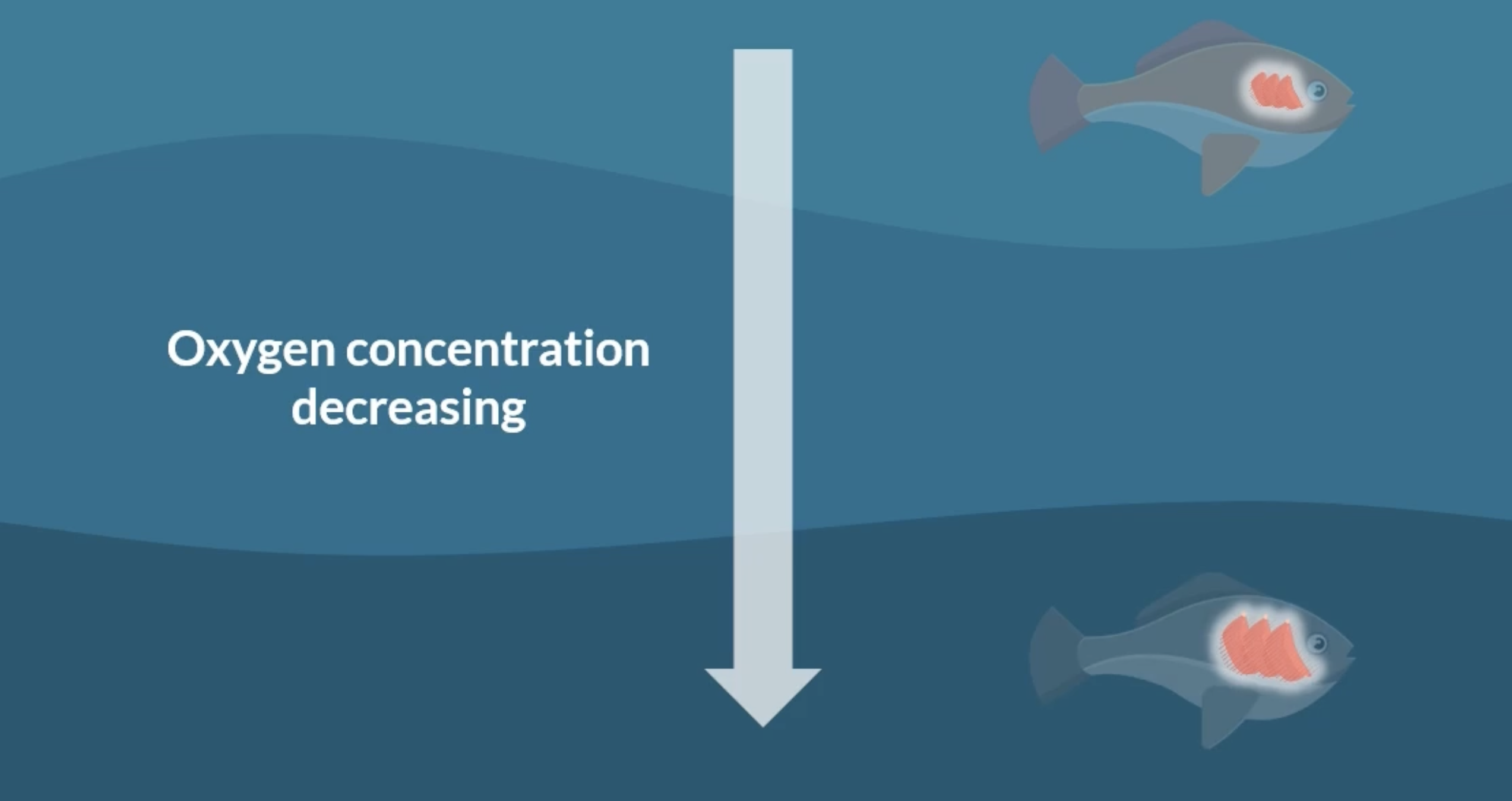
gill SA : body mass ratio
in environments where oxygen concentration is less, fish are adapted to have larger gills = larger SA. they are able to get enough oxygen
in environments where oxygen concentration is more, fish are adapted to have smaller gills = smaller SA
as the oxygen concentration of an environment decreases, the gill SA: body mass ratio increases
diffusion calculation
ficks law

gas exchange of humans- structures
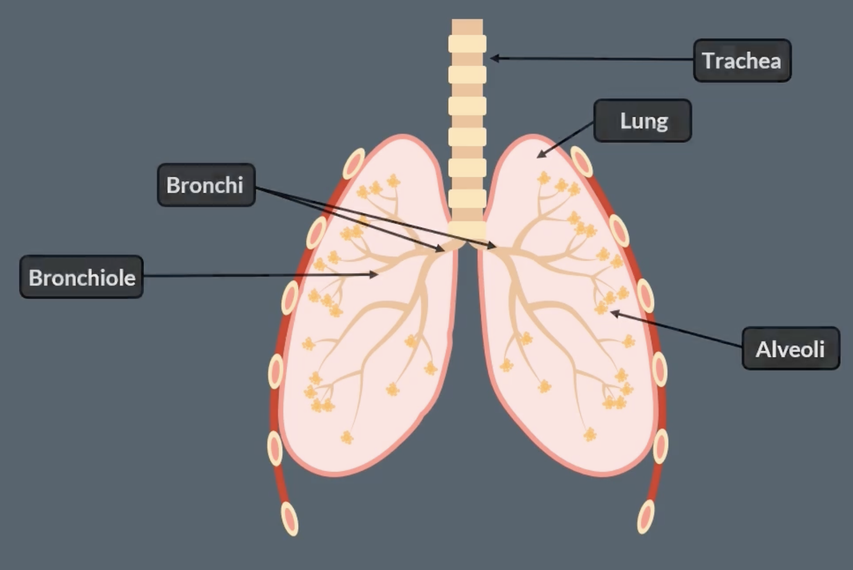
gas exchange of humans- structures 2
diaphragm is a muscle
thorax is just the chest part of your body
thoracic cavity- the internal space within the thorax that contains vital organs like the heart, lungs, trachea
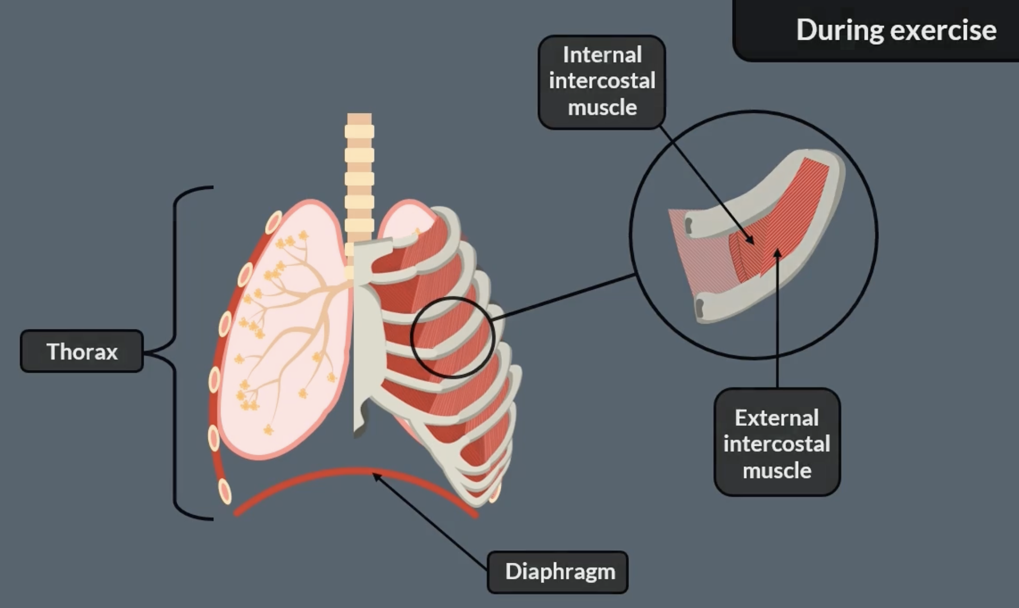
gas exchange of humans- inhalation and normal exhalation
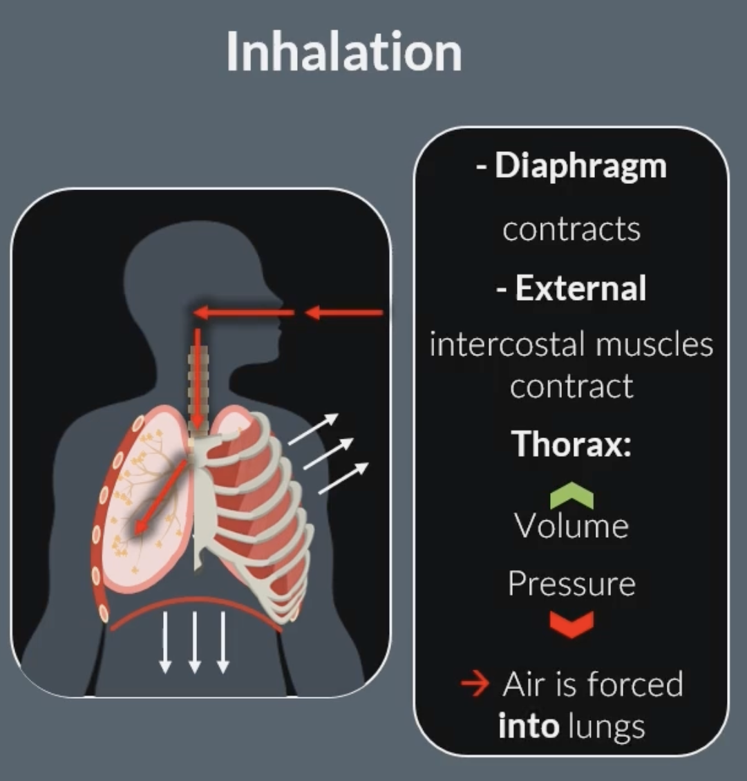
during inhalation:
external intercostal muscles contract, this pulls the ribs up and out (internal intercostal muscles relax)
diaphragm contracts and flattens, increasing space in the thorax
inside the lungs, the volume increases and the pressure decreases below atmospheric pressure
air moves into the lungs down the pressure gradient (from higher pressure outside to lower pressure inside)
-this same process occurs the same way during exercise just more strongly
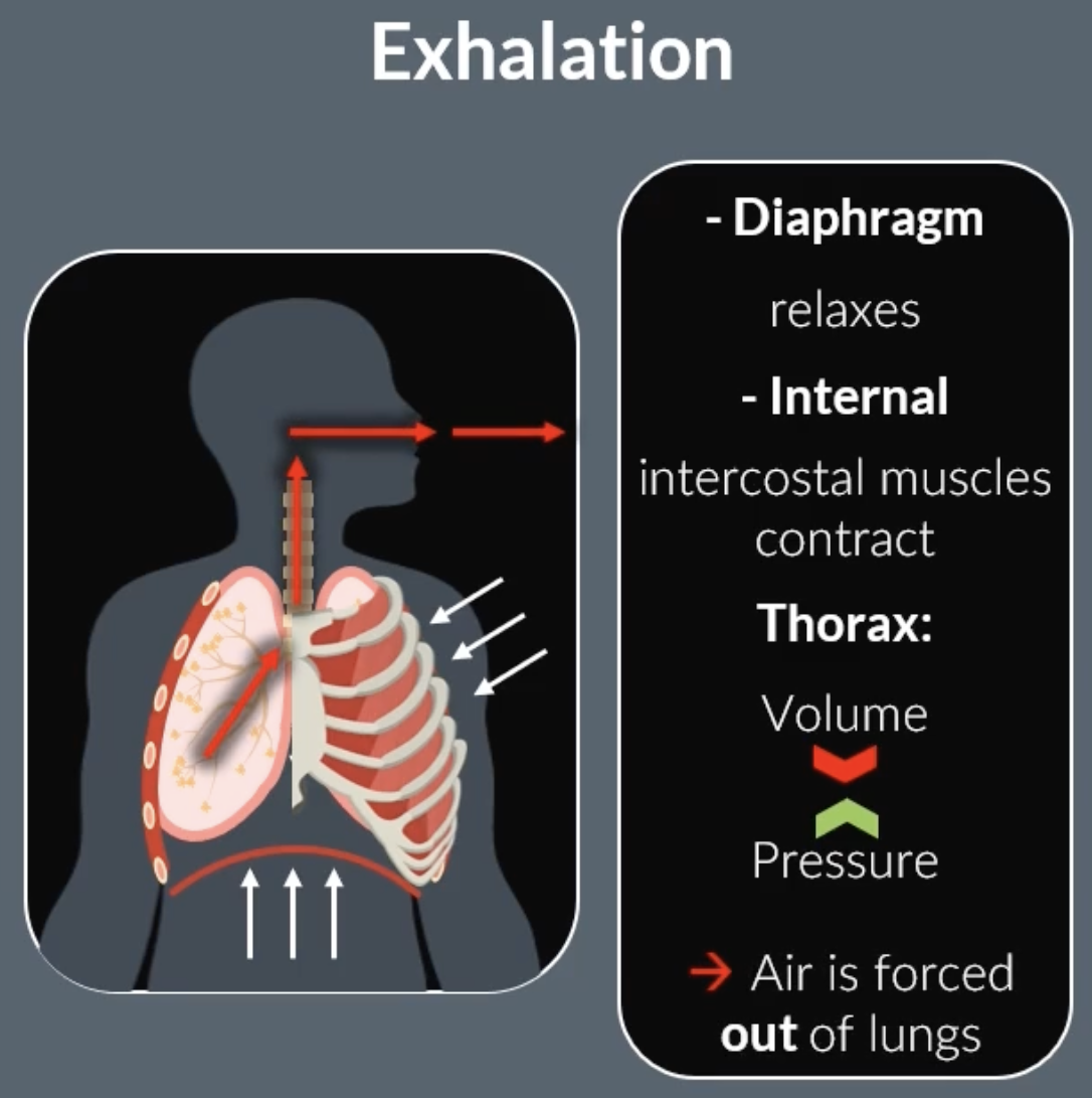
normal exhalation:
external intercostal muscles relax, pulling the ribs down and in (internal intercostal muscles relax)
diaphragm relaxes and moves up to a dome shape
this causes the volume of the lungs to decrease, so the pressure inside the lungs increases above atmospheric pressure
air leaves the lungs down the pressure gradient
-normal exhalation is a passive process
-no muscle contraction involved
-air leaves because of lung recoil and relaxation of muscles
gas exchange of humans- forced exhalation
internal intercostal muscles contract, pulling the ribs down and in (external intercostal muscles relax)
the diaphragm relaxes and moves upwards into a dome-shape
this causes the volume of the lungs to decrease and the pressure inside the lungs to rise above atmospheric pressure
air is forced out of the lungs down the pressure gradient
-forced exhalation is an active process
epithelial vs endothelial
epithelial cells- line surfaces inside and outside the body
endothelial cells- line blood vessels. forms a smooth barrier for blood flow and exchange of substances
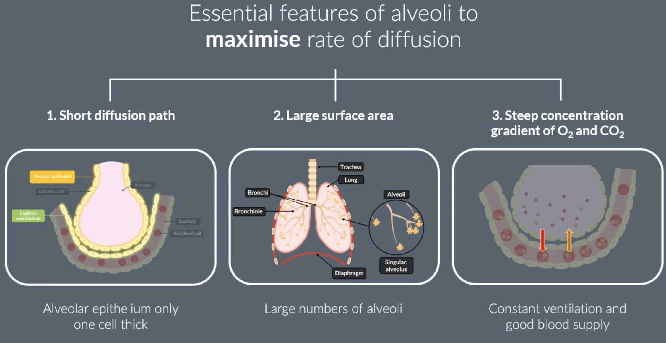
features of alveoli which maximise diffusion/ gas exchange
gas exchange occurs in the alveoli
-short diffusion path because surrounded by a single layer of epithelial cells (alveolar epithelium). the capillaries surrounding alveoli are also surrounded by a single layer of endothelial cells (capillary endothelium)
-there is many alveoli which creates a large SA
-a steep concentration gradient of oxygen and carbon dioxide is maintained because of constant ventilation and constant blood supply
breathing vs respiration
breathing= movement of air in and out of the lungs
respiration= chemical reaction to release energy in the form of ATP
antagonistic
antagonistic- two things that work in opposite ways
in terms of breathing:
muscles work in opposite pairs, one contracts while the other relaxes (eg. external and internal intercostal muscles)
digestion
large biological organisms are hydrolysed into smaller molecules that can be absorbed across cell membranes
most enzymes end in -ase
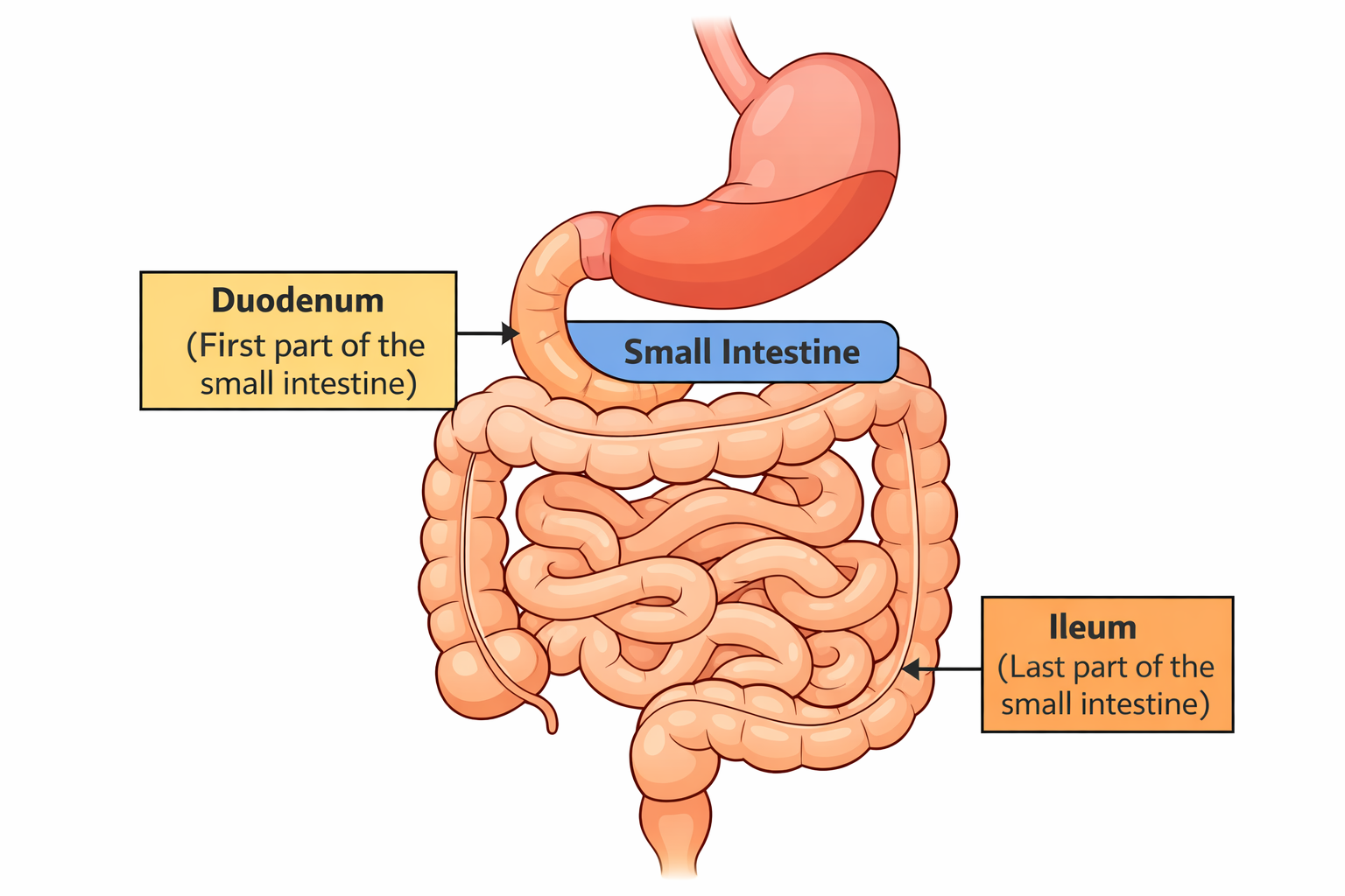
duodenum vs small intestines vs ileum
duodenum is the first part of the small intestine immediately after the stomach
ileum is the last and longest part of the small intestine
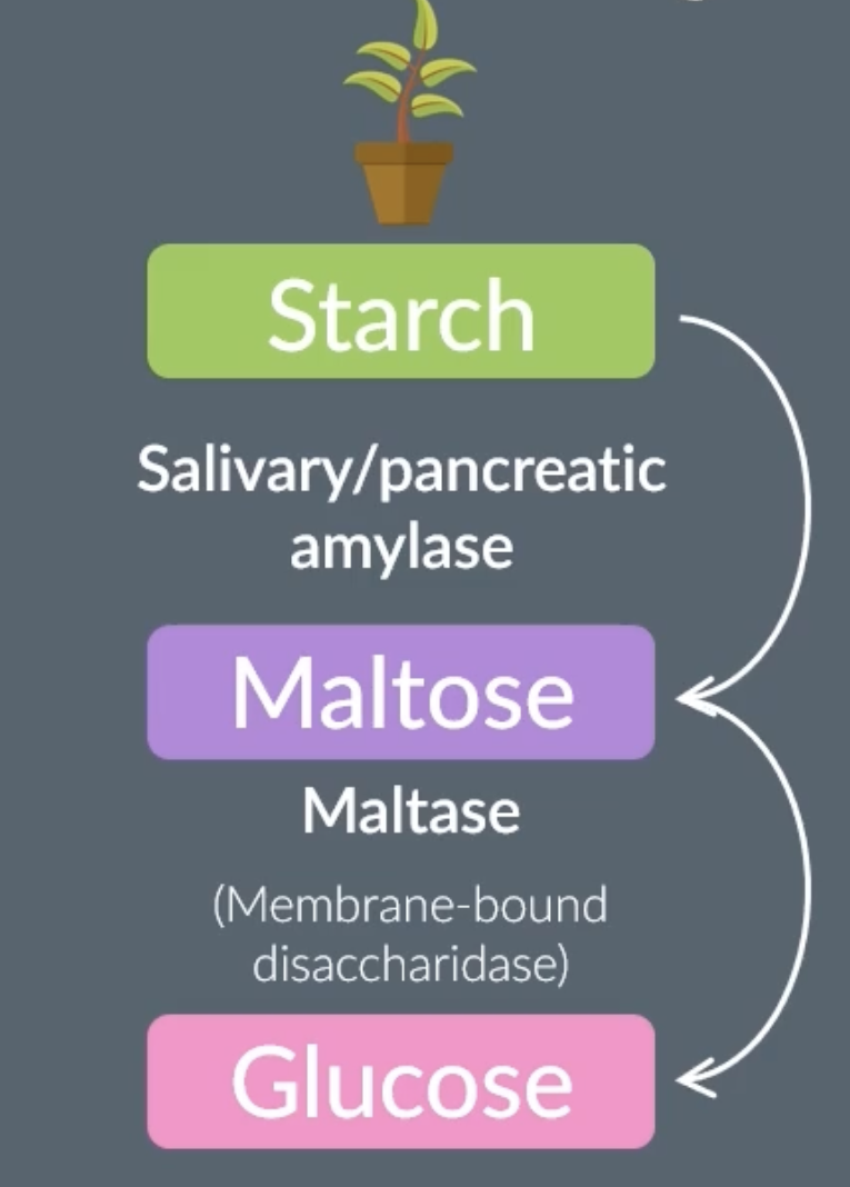
digestion of carbohydrates- starch (polysaccharide) to maltose (disaccharide)
the enzymes that digest carbohydrates are:
-amylases (produced by salivary glands and pancreas)
-membrane bound disaccharidases
digestion of carbohydrates takes place in:
-mouth. when (polysaccharide) starch enters mouth salivary glands produce amylase, which hydrolyses the glycosidic bonds of starch to produce (disaccharide) maltose.
-small intestines. any remaining starch passes the stomach and enters the small intestines. pancreas produces amylase which hydrolyses the glycosidic bonds of starch to produce (disaccharide) maltose (in the duodenum)
polysaccharide to disaccharide
in the mouth when amylase hydrolyses starch (polysaccharide) to maltose (disaccharide)
in the small intestine when amylase hydrolyses starch (polysaccharide) to maltose (disaccharide)
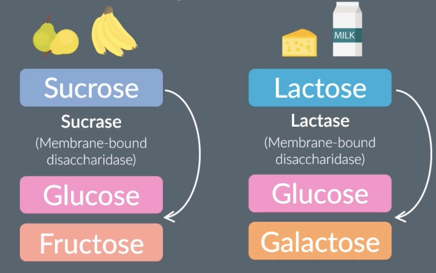
disaccharide to monosaccharide
in the ileum there is the enzyme sucrase a membrane bound disaccharidase) which is attached to the epithelial membrane. it hydrolyses the glycosidic bonds of sucrose(disaccharide) into glucose and fructose (monosaccharide)
in the ileum there is the enzyme maltase (a membrane bound disaccharidase) which is attached to the epithelial membrane. it hydrolyses the glycosidic bonds of maltose(disaccharide) into glucose (monosaccharide)
in the ileum there is the enzyme lactase (is a membrane bound disaccharidase) which is attached to the epithelial membrane. it hydrolyses the glycosidic bonds of lactose(disaccharide) into glucose and galactose(monosaccharide)
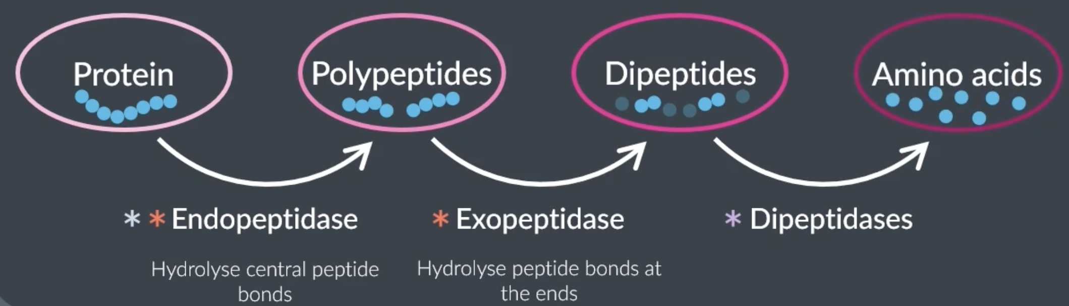
digestion of proteins
protein digestion starts in the stomach, continues in the duodenum and finishes in the ileum
3 enzymes can hydrolyse polymer proteins
-endopeptidase= hydrolyse peptide bonds between amino acids in the middle of the polymer chain to produce a shorter polypeptide chain. found in stomach and small intestine
-exopeptidase= hydrolyse peptide bonds between amino acids at the end of the polymer chain to produce dipeptides. found in small intestine
-membrane bound dipeptidase= hydrolyse peptide bonds between 2 amino acids (after the first 2 enzymes are done hydrolysing and there’s around 2 amino acids left). found on intestine epithelial cells
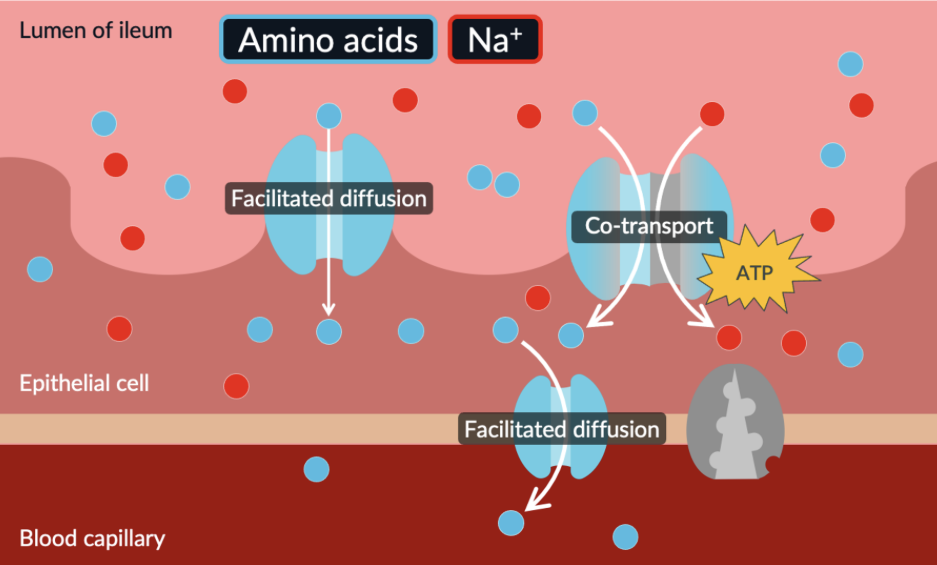
absorption of amino acids and monosaccharides
because amino acids are large and hydrophilic they cannot simply pass through the membrane so they must use proteins
there’s a high concentration of amino acids in the lumen compared to the epithelial cells. amino acids can diffuse (facilitated diffusion) into the epithelial cells. eventually the concentration of amino acids in both the lumen and epithelial cell becomes equal so there’s no concentration gradient.
therefore co transport is used to transport amino acids alongside sodium ions. sodium ions move along their concentration gradient, which provides energy for the co transporter protein to move amino acids against their concentration gradient into the epithelial
amino acids can move by facilitated diffusion from the epithelial cells into the blood. a concentration gradient is always maintained because blood is constantly flowing
absorption of monosaccharides
theyre absorbed in the exact same way as amino acids ^ same exact process
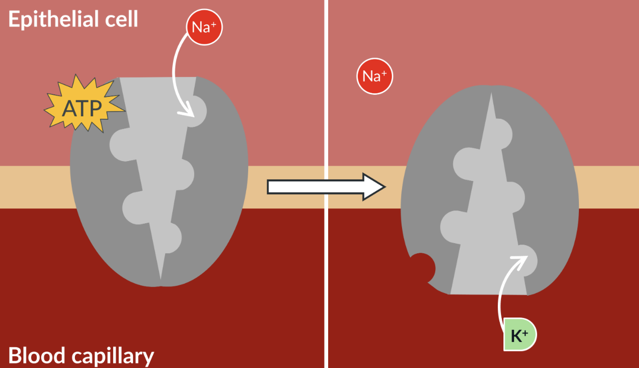
how the concentration gradient of sodium is maintained
found at the lining of the blood capillary
a concentration gradient must be maintained for sodium so it can be transported along its concentration gradient from the lumen into the epithelial cells (co transport). therefore the sodium potassium pump moves 3 sodium ions against their concentration gradient, from the epithelial cell into the blood. it also moves 2 potassium ions against their concentration gradient in the opposite direction. the sodium-potassium pump requires ATP.
ileum vs lumen
lumen is the inside of the ileum where the digested food passes through
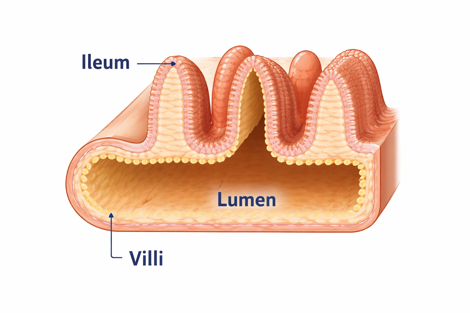
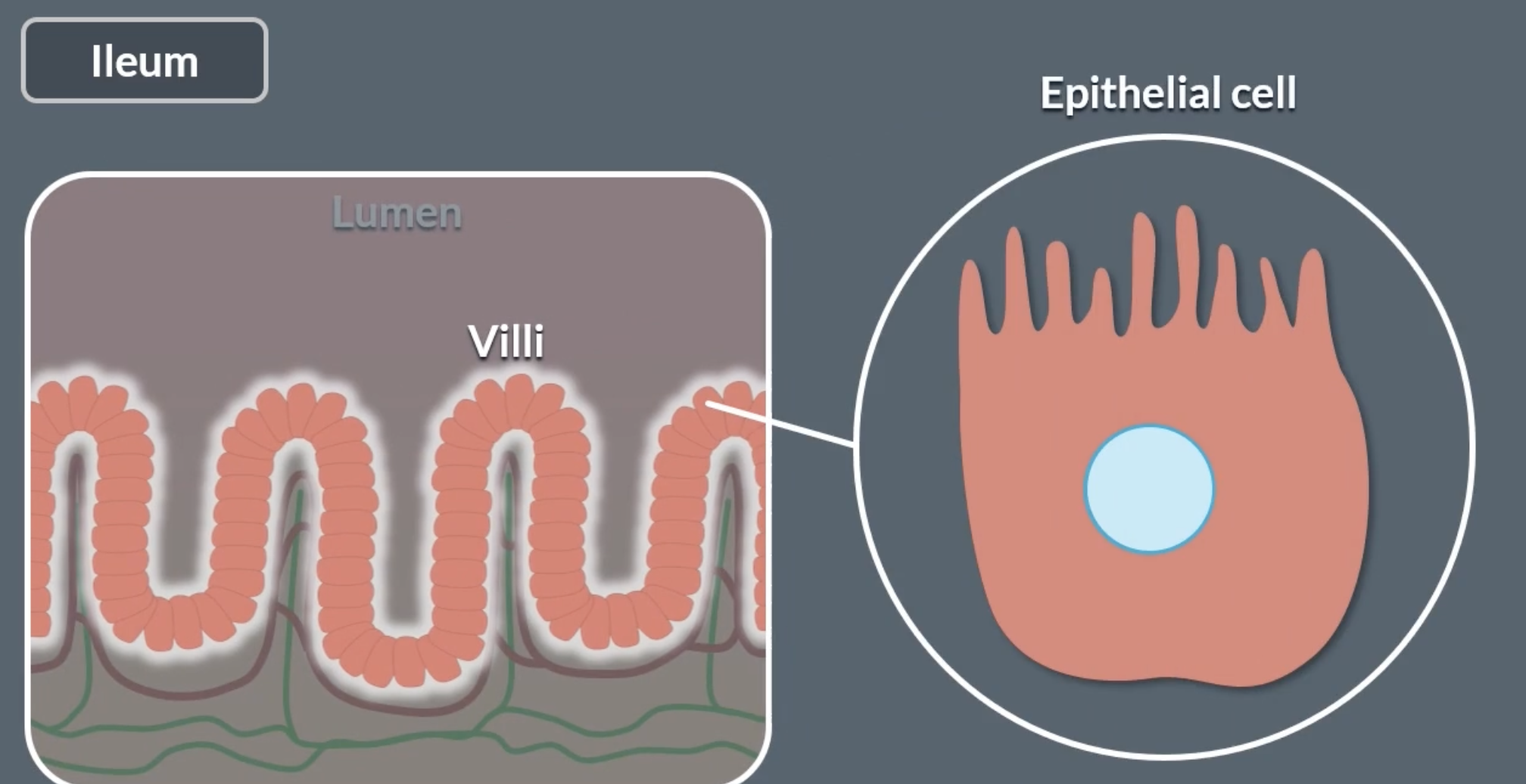
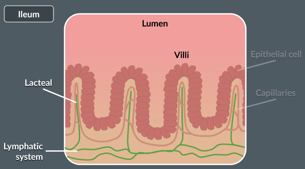
ileum more detail
ileum has bumps on the surface called villi which are lined by epithelial cells
epithelial cells have microvilli
epithelial cells separate the lumen from blood capillaries
hydrophilic and hydrophobic passing through the membrane
hydrophilic = polar, cannot simply pass through, needs proteins to cross
hydrophobic = non polar, can simply pass through
however, small polar molecules can “squeeze” through the hydrophobic core of the membrane to diffuse through even though they are hydrophilic. eg. glycerol, water, urea
ions and large polar molecules cannot diffuse through the membrane. theyre too big or charged → repelled by hydrophobic core
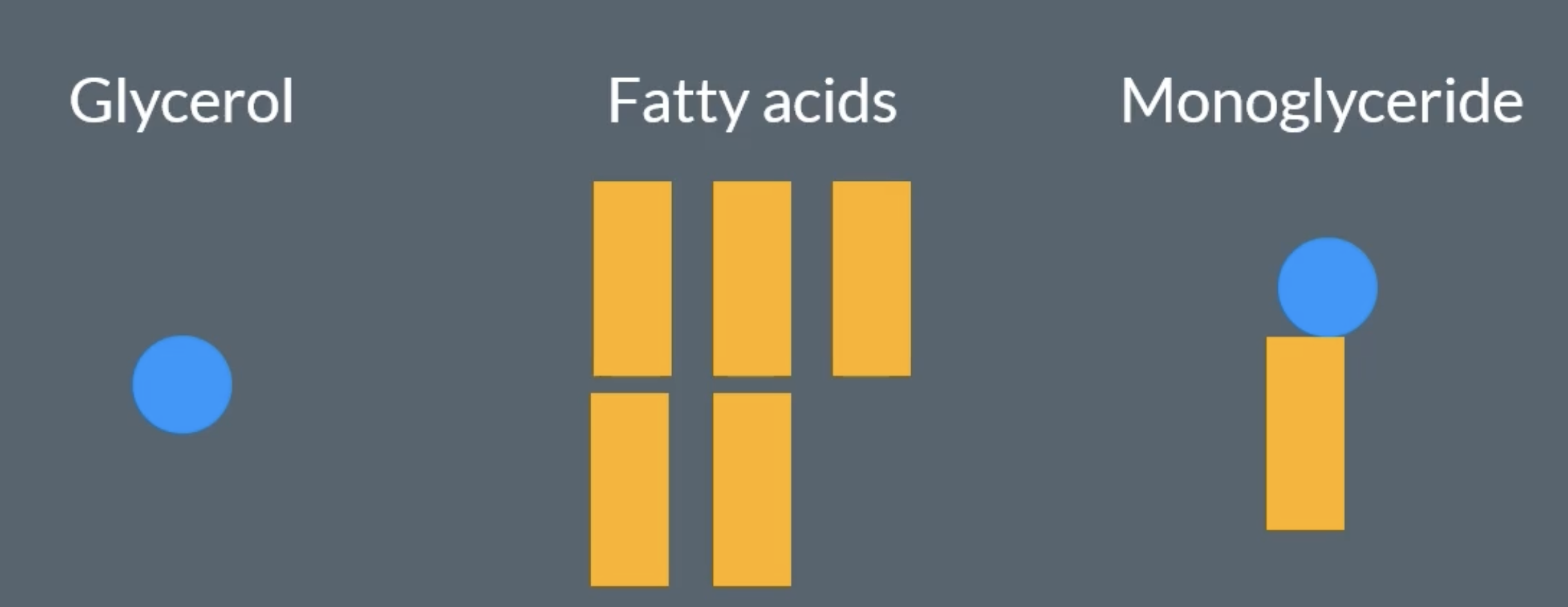
glycerol vs monoglyceride
monoglyceride is glycerol with one fatty acid still attached.
glycerol is not attached to anything, is free.
digestion of lipids
takes place in the small intestines
involves 2 stages:
-physical digestion (emulsification). bile salts are created in the liver and stored in the gal bladder. lipids get coated in bile salts to create an emulsion (emulsify the lipids), causing the lipids to split into tiny droplets. this creates a larger surface area for faster hydrolysis of lipase
-chemical (lipase). lipase is produced in the pancreas. it hydrolyses the ester bonds of lipids into monoglycerides and fatty acids
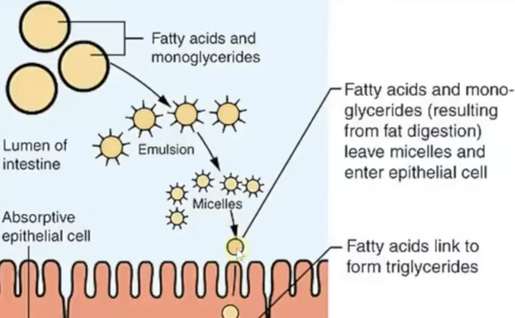
micelles and how they help with lipid absorption
-make the fatty acids and monoglycerides more soluble in water to be able to transport them into the epithelial
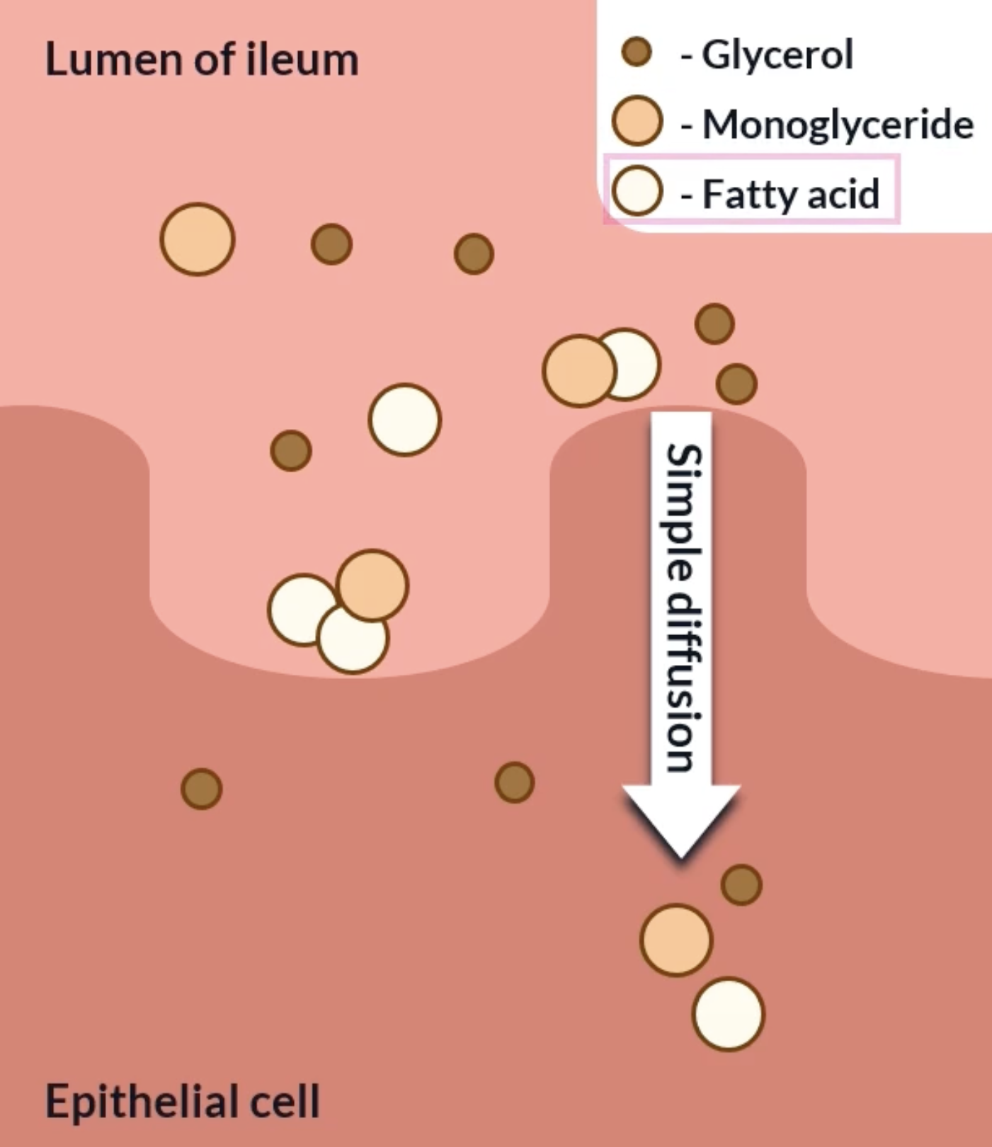
glycerol is small and polar so it is soluble in water. can easily move through the cytoplasm. because of its small size it can simply diffuse into the epithelial cells
fatty acids and monoglycerides are larger and have non polar regions so theyre are not soluble in water. therefore they are unable to easily move through the lumen to the epithelial cells. bile salts and phospholipids surround them to form micelles, micelles have a polar outer shell making them soluble in water. therefore they can move through the lumen easily. theyre released when they reach the epithelial cells where they simply diffuse in the epithelial cells (bc theyre hydrophobic and non polar)
a concentration gradient is maintained for simple diffusion because most of the monoglycerides, glycerol and fatty acids are converted back into triglycerides. this is a quick process which helps maintain the concentration gradient
absorption of lipids
short fatty acid chains can then diffuse straight into the blood
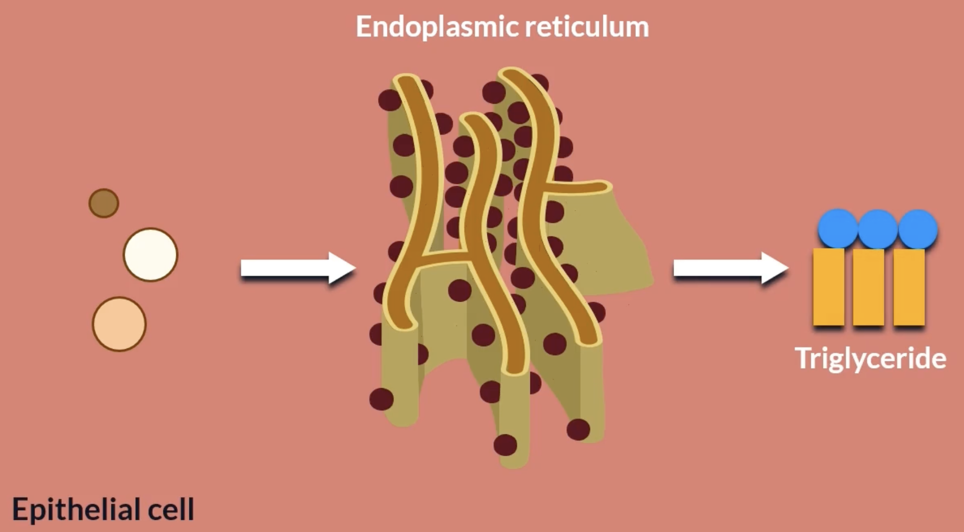
long chains cannot so they bond with monoglycerides and glycerol to form triglycerides in the endoplasmic reticulum
the triglycerides then need to exit the epithelial cells, however they aren’t soluble therefore struggle to travel through the cytoplasm. the endoplasmic recticulum combine triglycerides with cholesterol and proteins which forms chylomicrons, which are soluble.
however chylomicrons are too large to simply diffuse out of the cell membrane, so the golgi apparatus packages them into vesicles, where they fuse with the cell membrane and release the chlyomicrons by exocytosis (‘out of’ ‘cell’)
absorption of lipids 2 *lipids leaving the epithelial cells
chylomicrons don’t enter the blood because they’re too big to fit through the pores in the capillaries. instead, chylomicrons enter lymphatic capillaries, which are called lacteals and they have much bigger openings. once in the lacteals, the chylomicrons travel through the lymphatic system and eventually reach the blood.
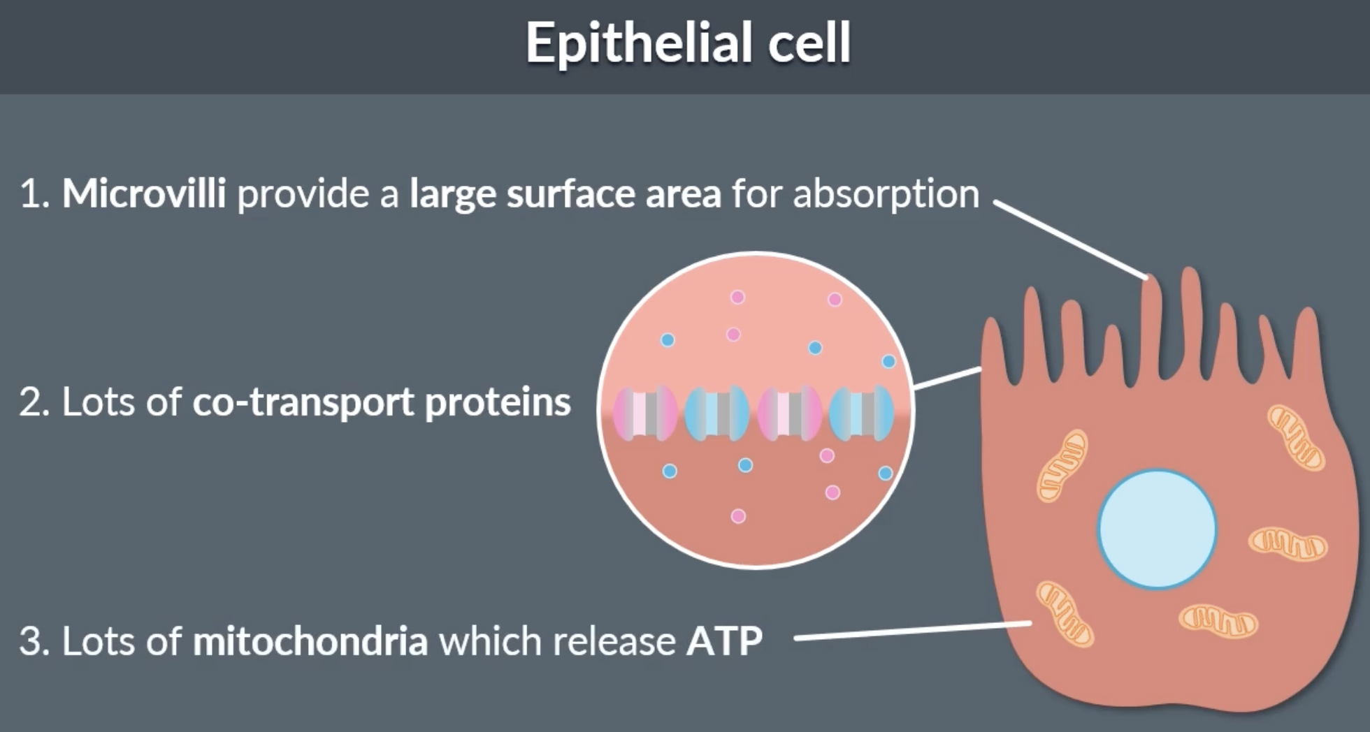
adaptations for absorption
lumen has villi and epithelial cells have microvilli, which increases SA for absorption
epithelial cells have a lot of co transport proteins to maximise the absorption of amino acids and monosaccharides
lots of mitochondria in epithelial cells because sodium potassium pump needs energy in the form of ATP to maintain a sodium concentration gradient for efficient co transport
mark scheme advice
when talking about absorption of lips always write the definition of micelles (what they contain): monoglycerides, fatty acids and bile salts