lab diagram 2
1/26
There's no tags or description
Looks like no tags are added yet.
Name | Mastery | Learn | Test | Matching | Spaced | Call with Kai |
|---|
No analytics yet
Send a link to your students to track their progress
27 Terms
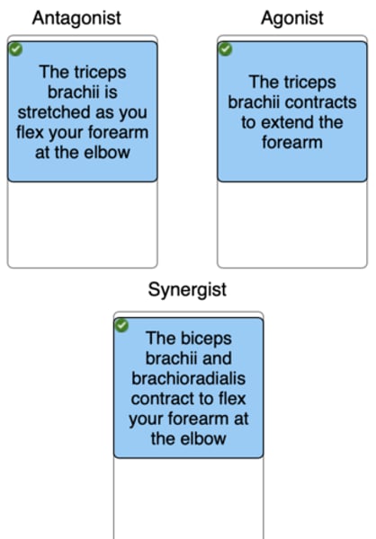
Classify the muscle actions based on whether they are acting as an agonist, antagonist, or synergist.
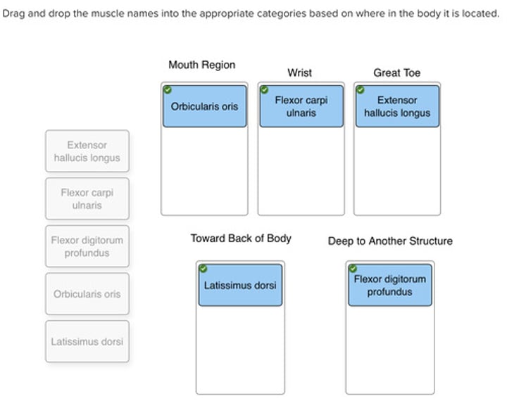
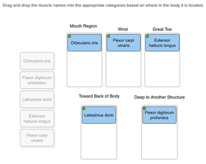
Drag and drop the muscle names into the appropriate categories based on where in the body it is located.
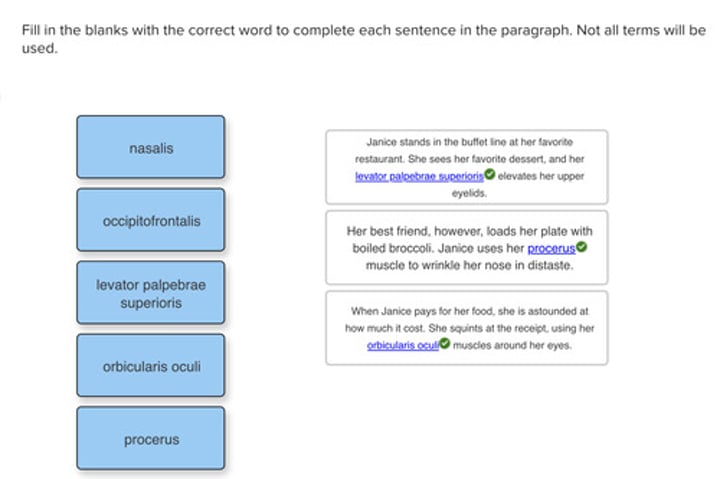
Fill in the blanks with the correct word to complete each sentence in the paragraph. Not all terms will be used.
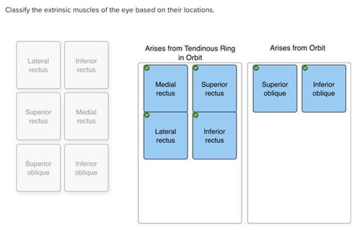
Classify the extrinsic muscles of the eye based on their locations
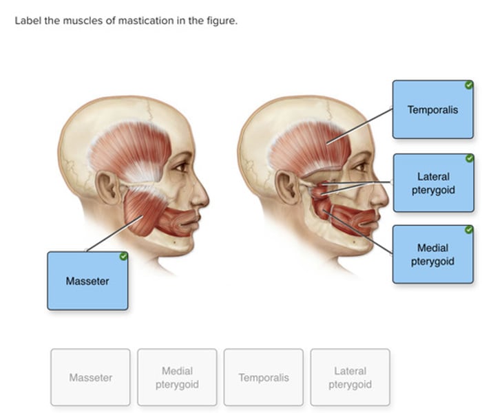
Label the muscles of mastication in the figure.
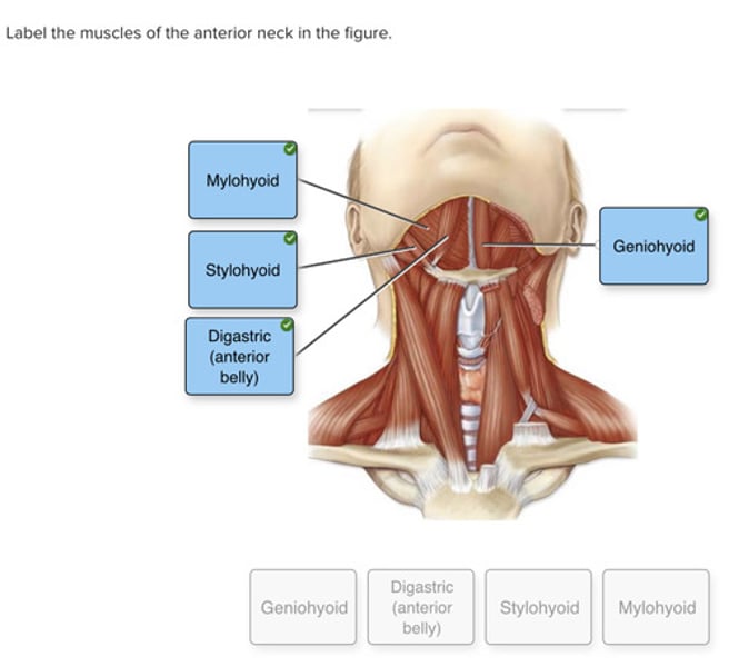
Label the muscles of the anterior neck in the figure.
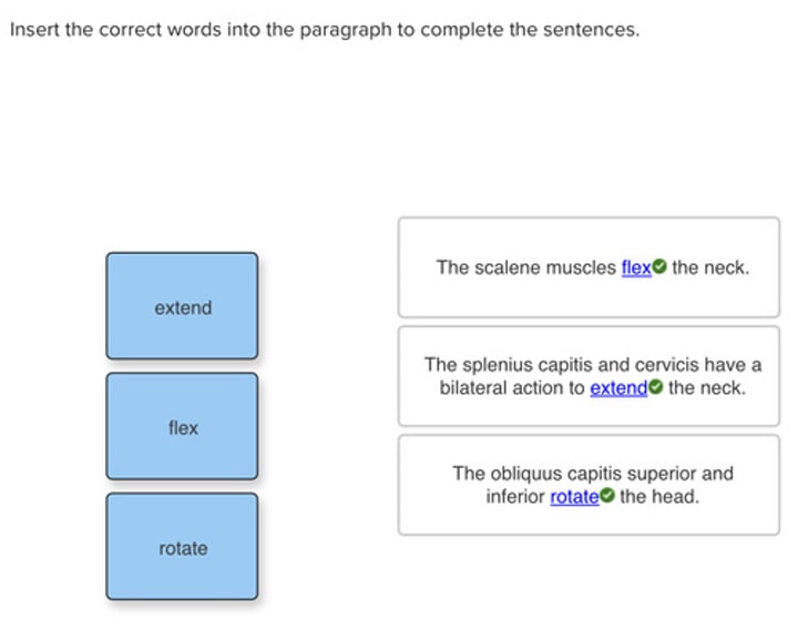
Insert the correct words into the paragraph to complete the sentences.
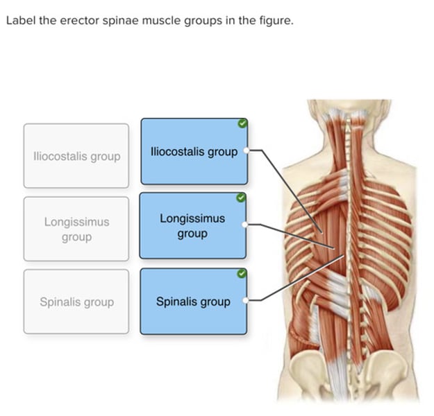
Label the erector spinae muscle groups in the figure.
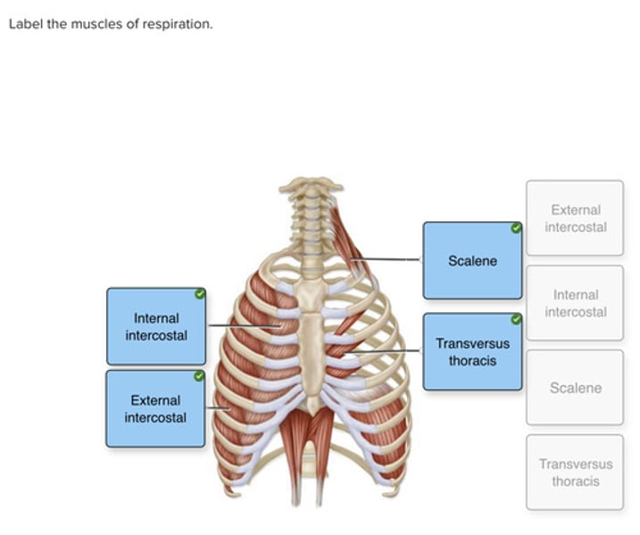
Label the muscles of respiration.
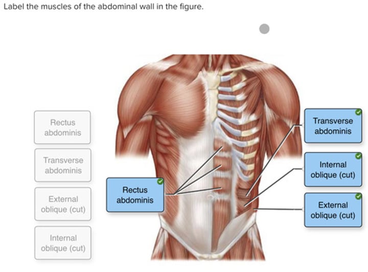
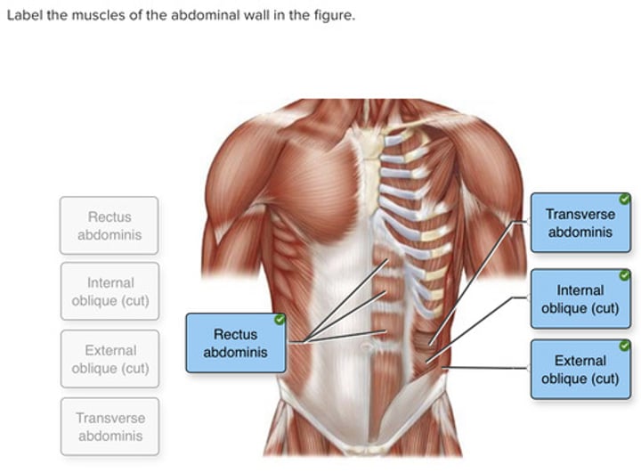
Label the muscles of the abdominal wall in the figure.
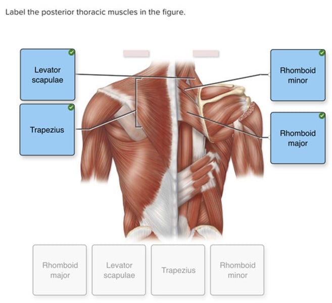
Label the posterior thoracic muscles in the figure.
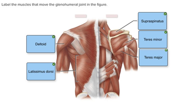
Label the muscles that move the glenohumeral joint in the figure.
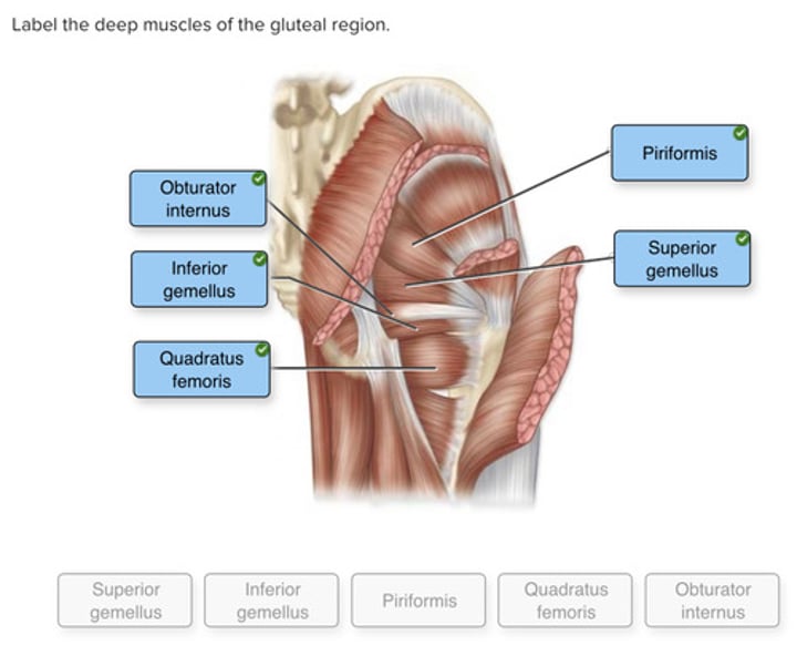
Label the deep muscles of the gluteal region.
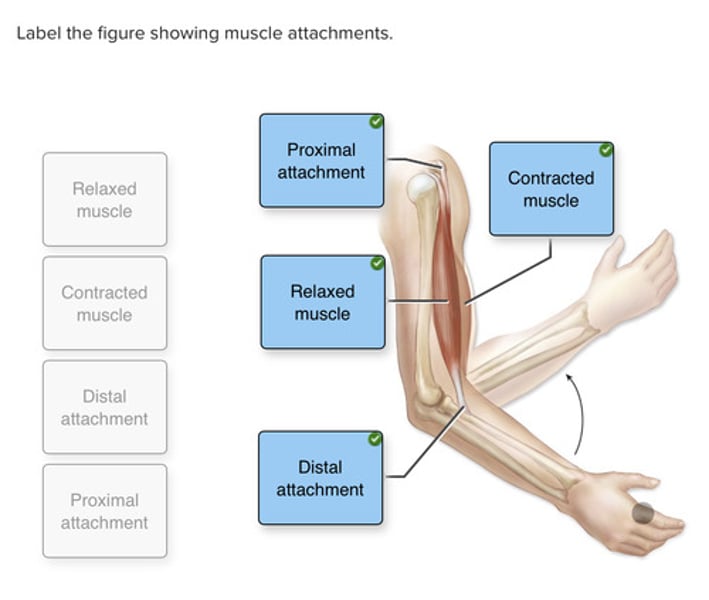
Label the figure showing muscle attachments.
Drag and drop the muscle names into the appropriate categories based on where in the body it is located.
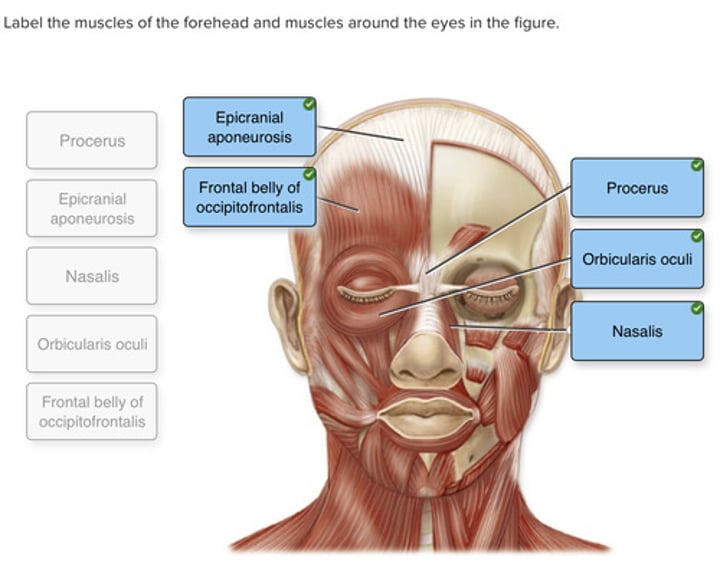
Label the muscles of the forehead and muscles around the eyes in the figure.
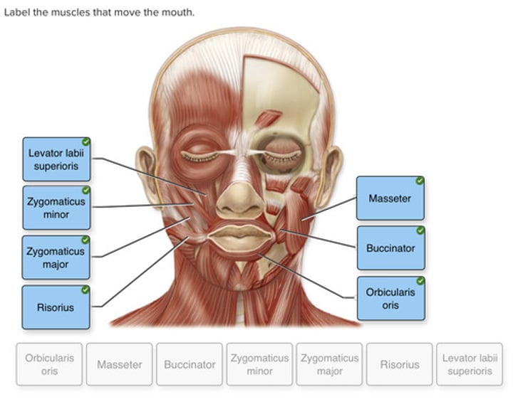
Label the muscles that move the mouth.
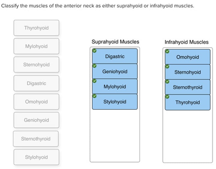
Classify the muscles of the anterior neck as either suprahyoid or infrahyoid muscles.
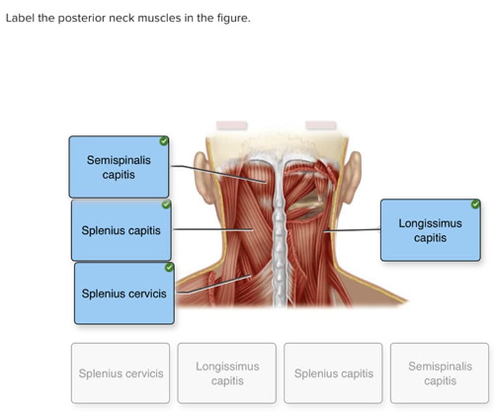
Label the posterior neck muscles in the figure
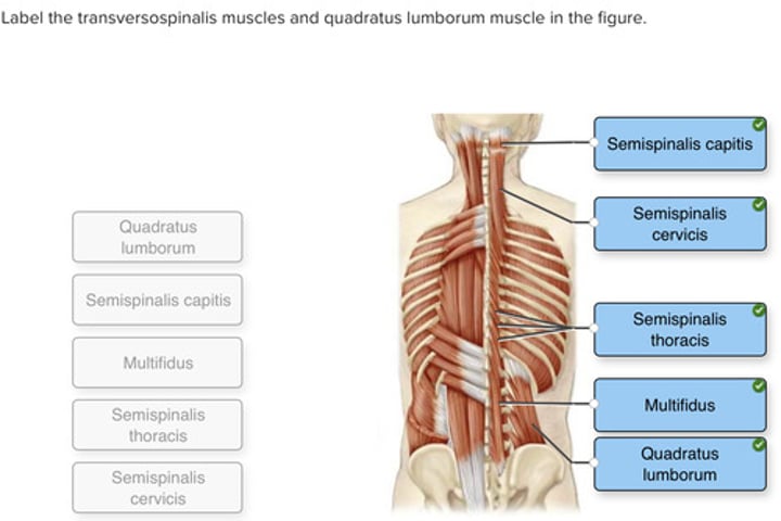
Label the transversospinalis muscles and quadratus lumborum muscle in the figure.
Label the muscles of the abdominal wall in the figure.
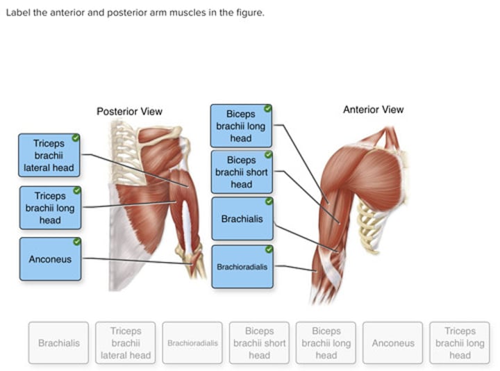
Label the anterior and posterior arm muscles in the figure.
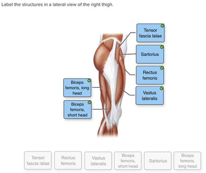
Label the structures in a lateral view of the right thigh.
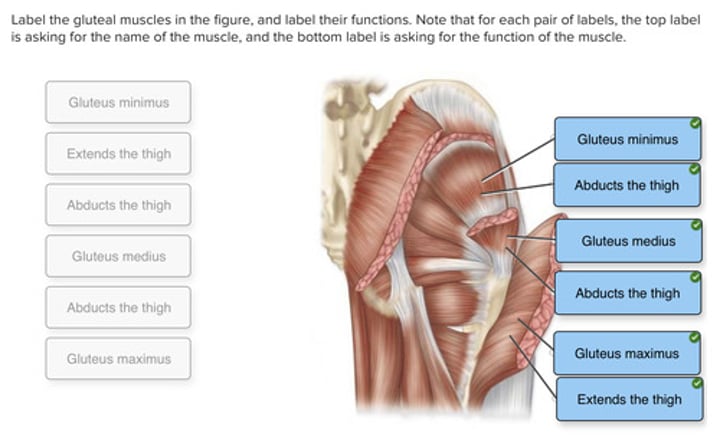
Label the gluteal muscles in the figure, and label their functions. Note that for each pair of labels, the top label is asking for the name of the muscle, and the bottom label is asking for the function of the muscle.
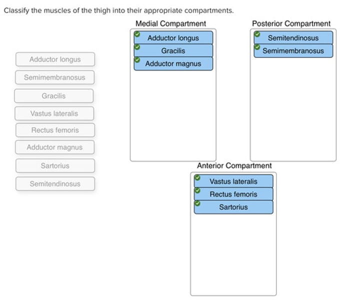
Classify the muscles of the thigh into their appropriate compartments.
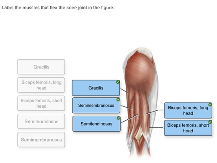
Label the muscles that flex the knee joint in the figure.
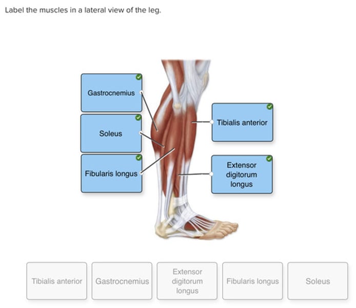
Label the muscles in a lateral view of the leg.