The Topography and Structure of the Heart
1/48
There's no tags or description
Looks like no tags are added yet.
Name | Mastery | Learn | Test | Matching | Spaced | Call with Kai |
|---|
No analytics yet
Send a link to your students to track their progress
49 Terms
THE HEART - Location
3rd to 6th Rib (3rd to 5th ICS)
More or less obliquely oriented
Apex slightly to the left side
Canine heart
Cow heart
More vertical orientation – Small and large Ruminant and Horse
Sternopericardiac ligament
Horse heart
Species differentiation of the heart location
Ruminants - 3rd to 6th Rib, Horse: 2 to 6, Pig 2 to 5 (smaller
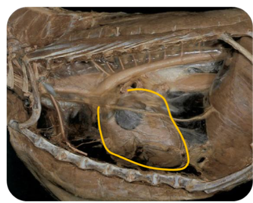
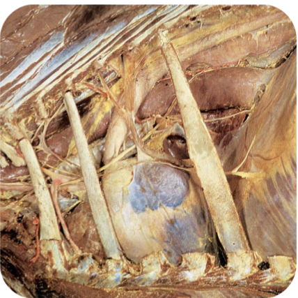
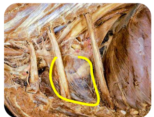
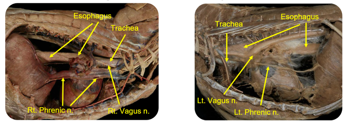
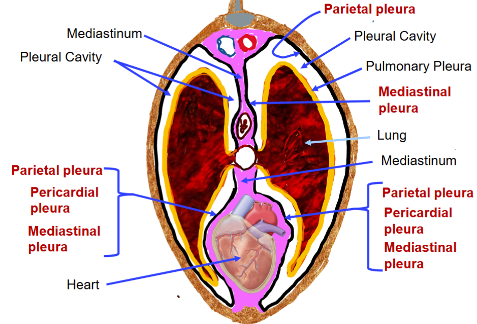
The heart: relations
The heart is located within the?
mediastinum
Any pleura that is not covering the lung is…?
is the parietal pleura
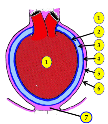
The plurae
Visceral pericardium(Epicardium)(2)
is the layer which covers the heart wall
Pericardial cavity(3)
The space between visceral and parietal pericardium, which is essentially a potential space containing small amount of serous fluid
Parietal pericardium(4)
The outer layer which is attached to the mediastinum
Fibrous pericardium(5)
is the third layer, fibrous connective tissue(Modified CT of the mediastinum),
which attaches the parietal pericardium to the mediastinal pleura (mediastinum).
Mediastinal pleura (6)
The outermost layer in this region, note that the fibrous pericardium connects the parietal
pericardium and the mediastinal pleura
Phrenicopericardiac / sternopericardiac ligament (7)
The fibrous pericardium continues as tunica adventitia of the great vessels as they pass through the mediastinum after arising from the base of the heart. It also forms Phrenicopericardiac / sternopericardiac ligament (7) at the apex of the heart
layers of the heart labeled
Function of the pericardium
provides an isolated environment to the heart
The pericardial fluid, though very little, functions as a lubricant
The pericardium prevents overdilation of the heart
The Auricle
a blind sac extended from each atrium is called auricle
The Atrium
Thin-walled chamber that receives blood. Located dorsal
The ventricle
Thick-walled chamber located ventrally that pumps the blood away from the heart
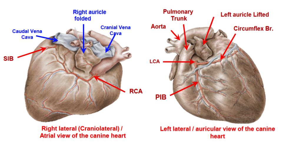
Outer anatomy of the heart
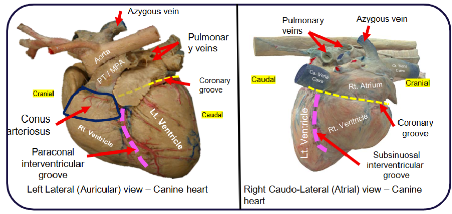
The conus arteriosus
The cone shaped part of the right ventricle where the pulmonary trunk arises from the right ventricle
The Interventricular Grooves
The grooves that demarcate ventricles from each other
Great arteries: Pulmonary trunk
Carries deoxygenated blood to the lungs
Great arteries: Aorta
Oxygenated blood supply to the body
Great veins: Cranial and Caudal vena cava
Collect deoxygenated blood from the body
Great veins: Azygos vein
Collects deoxygenated blood from most of the thoracic wall
Heart wall: Myocardium
Arrangement of the cardiac muscles
Overall reduction in the size of heart and the chambers
Pumping action: Atria → to ventricles, Rt. Ventricle → Lungs, Lt. Ventricle → Body
The cardiac cycle - Rhythmic contractions
Systole (Contraction) of the atria/ventricles.
Diastole (Dilation/ relaxation) of the atria/ventricles.
Unidirectional flow
The right AV Opening
guarded by the tricuspid valve
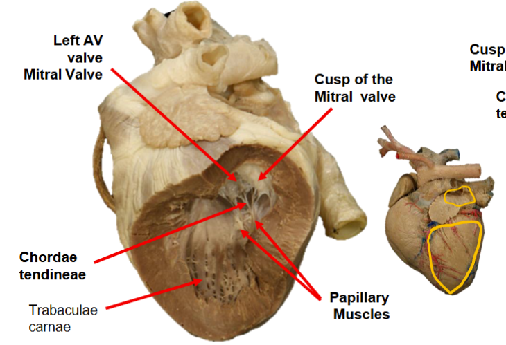
The Cusps
A roughly triangular leaf or flap of a heart valve
The left Atrium and Ventricle
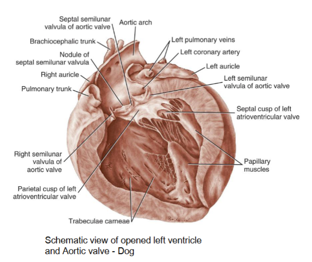
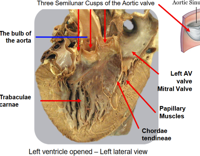
The Mitral Valve/Left AV Valve
The Aorta and Aortic Valve
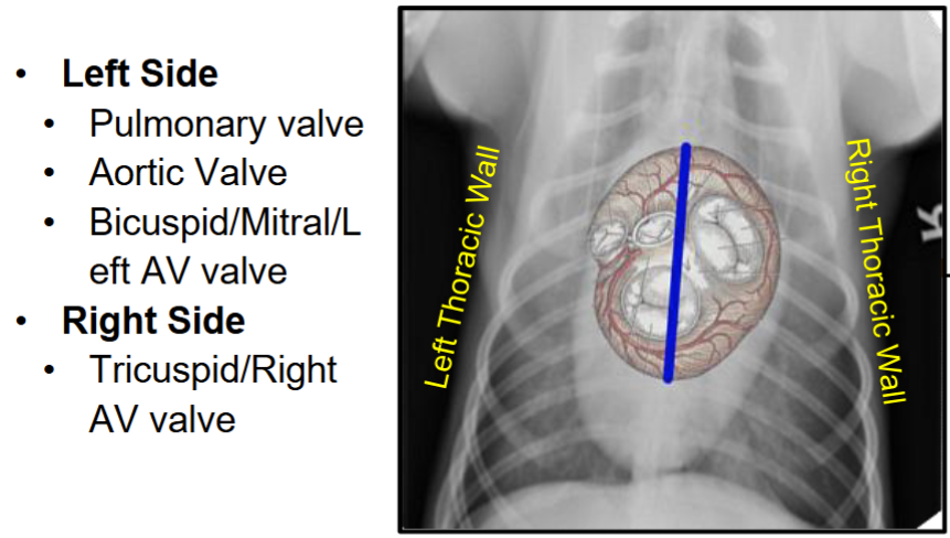
The Heart valves and their relationship with the body wall
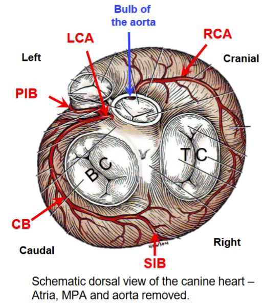
Coronary arteries - Right (Smaller) and Left
Arise from the bulb (dilated origin) of the aorta
Located in the coronary and interventricular grooves of the heart
Left coronary artery is large in what species?
Canine and ruminants
Right coronary artery is larger in what species?
Horses
Blood supply to the heart
Blood supply to the heart: Arterial supply
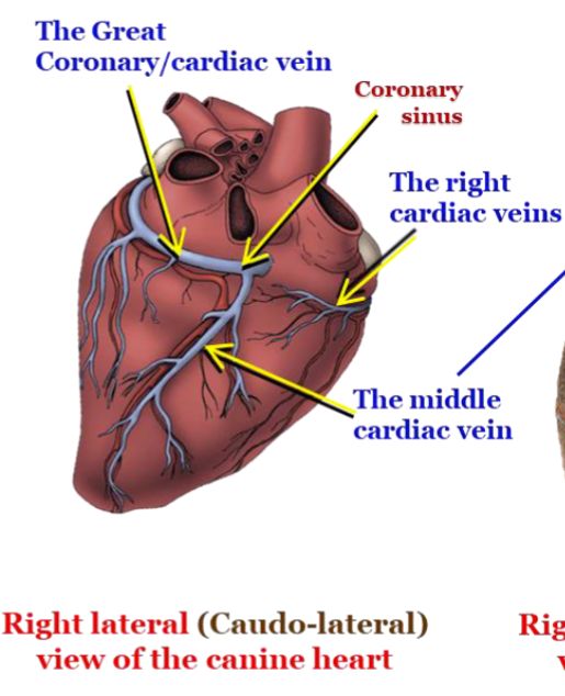
Great Coronary Vein(Cardiac Vein)
Venous return/drainage
Major Venous drainage from the heart
coronary sinus is the terminal dilated part
Opens into the right atrium
Sympathetic cardiac nerves
Sympathetic innervation
Stellate (Cervico-thoracic) and Middle cervical ganglia
Parasympathetic cardiac nerves
Parasympathetic innervation Right and left Vagus nerves
Nerve supply to the heart
Vector of heartworm disease
Mosquitoes of Aedes, Anopheles, and Culex are the most common genera acting as vectors
Common species that get heartworm?
Dogs, cats and ferrets are susceptible
Dirofilaria immitis
Where do heartworms reside in the heart?
The adult worms occlude the right side of the heart and the pulmonary trunk/arteries
General implications of heartworm infestation:
Inflammation of the pulmonary vasculature as Live, adult heartworms cause direct mechanical trauma
Dead heartworms leads to more severe vascular reactions and subsequent pathologic changes
Symptoms of heartworm disease
Coughing after exercise
Refusing to exercise or play
Seeming lethargic or weak
Loss of appetite
Unintentional weight loss