micb212 lec 1-7
1/92
There's no tags or description
Looks like no tags are added yet.
Name | Mastery | Learn | Test | Matching | Spaced | Call with Kai |
|---|
No analytics yet
Send a link to your students to track their progress
93 Terms
describe the basic properties, structure and abundance of viruses
infectious, obligate (need host cell for replication) intracellular parasite
-contains genetic material (dna/rna)
protein coat called capsid
sometimes an envelope from host cell membranes
Exists as:
Virion = inanimate virus particle outside HOST cell, extracellular
Multiplying phase = inside infected cell
abundance: 10: 1 ratio, 10³1 in ocean
explain why viruses are important in health ecology and evolution
some viruses ar beneficial
drives global cycles (kills 20-40% ocean microbes daily)
transfer genes btwn. organisms
prevent and cure diseases
compare the major hypotheses for the origin of viruses
1) virus first hypothesis - virus first. cells after (coevolved with current hosts)
2) regressive hypothesis - came from cells that got reduced
3) progressive/escape hypothesis - virus came from genes that could move between cels
outline the major steps of the viral infectious cycle
1) bind to cell receptor
2) entry and coating
3) early gene expression
4) repication of viral genome
5) late gene expresison
6) assembly of visions
7) exit
Entry:
Bacteriophages inject genome
Plant viruses enter through cell wall damage
Animal viruses enter by membrane fusion or endocytosis
Uncoating:
Capsid disintegrates
Genome is released
Early genes:
Usually help genome replication
Late genes:
Usually structural proteins
Assembly:
Structural proteins package genome
Capsid forms
Enveloped viruses insert glycoproteins into membranes
Distinguish between viral and bacterial propagation strategies
Viruses:
Not cells
Do not divide by binary fission
Must enter host cells
Use host machinery
New virions are assembled from parts
Bacteria:
Cellular organisms
Can grow and divide independently under suitable conditionsExplain how nucleic acids were established as the genetic material of viruses
Explain how nucleic acids were established as the genetic material of viruses
1950s breakthrough:
Viral genetic code is the nucleic acid genome
Not the protein coat
Examples:
Bacteriophage T4 = DNA genome
Tobacco Mosaic Virus = RNA genome
Hershey-Chase experiment:
protein contains sulfur, dna contains phosphorus
Viral protein labeled with radioactive sulfur
Viral DNA labeled with radioactive phosphorus
Protein mostly stayed outside, didn’t enter the bacteria, but phosphorus was able to get in the bacteria
Conclusion:
DNA, not protein, carried genetic information for bacteriophage
Interpret viral nomenclature and classification conventions
Viruses classified by:
Nature/sequence of nucleic acid
Capsid symmetry
Presence/absence of envelope/lipid membrane
Dimensions of virion and capsid
Hierarchy:
Realm
Kingdom
Phylum
Class
Order
Family
Genus
Species
Naming endings:
Order = -virales
Family = -viridae
Genus = -virus
Describe the Baltimore classification scheme
Baltimore system classifies viruses by:
Genome type
How genome makes mRNA
Core rule:
All viruses must make mRNA that host ribosomes can translate
Viruses do not encode complete protein synthesis machinery
Classes:
I: dsDNA (double strand)
DNA → mRNA
II: ssDNA (single strand)
ssDNA → dsDNA → mRNA
III: dsRNA
dsRNA → mRNA using RdRp
IV: (+)ssRNA
Genome can be directly translated
V: (-)ssRNA
Must carry RdRp to make mRNA
VI: (+)ssRNA-RT
RNA → DNA by reverse transcriptase → provirus → mRNA
VII: dsDNA-RT
Gapped/partial dsDNA → full dsDNA → mRNA
Must know:
dsRNA and (-)ssRNA need RdRp in the virion
(+)ssRNA can be translated immediately
All viruses must reach mRNA
how many numbers od viral genomes
only 7. they must make mRNA
Identify the types of biological information encoded by viral genomes
Viral genomes encode information for:
Genome replication
Genome assembly and packaging
Timing/regulation of replication cycle
Modulation of host defenses
Spread to other cells and hosts
Viral genomes do not encode:
Complete protein synthesis machinery
Energy production proteins
Membrane biosynthesis proteins
Classical centromeres
Classical telomeres
L2: Describe how approaches for propagating viruses have evolved over time
animal to animal experimental infection → embryonate chicken egg → cultured cells
primary cell culture: from animal cells w limited life span
continuous cell lines;: single cell type that can be propagated indefinitely (immortizlaied)
recognize morphological changes in cultured ells that indicate viral infection
cytopathic effects (CPE): rounding, detachment, lysis, synctium(fused cells), shrinking, membrane alterations, accumulation of viral proteins
explain the purpose and principles of a viral plaque assay and be able to calculate a viral tier from plaque counts and dilution factors
virus titer: conc of infectious particles in a sample
measured by inoculating (introducing pathogen) serial dilutions to host cell cultures, lab animals, chicken embryo
list and compare methods used to measure infectious and physical viral particles
wuantaitve assay: measure amt of infection n include plaque assay, fluorescent, infectious centre assay,
all or nothing: measure presence or absence of infection (end point dilution)
biological vs physical assay
biological: such as plaque assay and end point titration do not detect noninfectious particles (only infectious)
physical: all particles accounted for example electron microscopy or immunological methods
plaque essay
A plaque assay measures infectious virus particles. Serially diluted virus is added to cells, plaques form where infected cells are damaged/killed, and each plaque is counted as one PFU.
calc viral titer
PFU/mL = #plaques ÷ volume plated × dilution factor.
Example: 17 plaques from 0.1 mL at 10^-6 = 17/0.1 × 10^6 = 1.7 × 10^8 PFU/mL.
define multiply of infection (MOI) and interpret its practical meaning
#Number of infectious particles ADDED per susceptible cell
ratio of infectious agents (like viral particles) to infection targets (like host cells) in an experimen
Important: MOI does not mean each cell receives exactly that many virions. Infection depends on random collisions between viruses and cells, so some cells get no virus, some get 1 virus, and some get multiple viruses. This follows a Poisson distribution. For example, at 106 cells infected at MOI = 1, only about 63.2% of cells are infected, while 36.8% remain uninfected.
identify high level approaches used to detect viral protein and nucleic acids
Viral proteins are detected mainly by antibody-based methods like immunostaining, Western blotting, immunoprecipitation, and ELISA. Viral nucleic acids are detected by PCR, qPCR, RT-qPCR, DNA microarrays, and sequencing.
understand key term used in viral reverse genetics that describe how engineered nucleic acids are used to recover or modify viral genomes
particle to plaque forming unit (PFU)
# number of physical particles / # of infectious particles
a sinle particle CAN initiate an infection
focus forming unit assay FFU
modification of plaque assay
-determines the titers of viruses that DONT form plaques (bc not all viruses kill the cells they infect)
-after virus infects cell, cell is treated to permeabilized (small holes in membrane) so antibodies enter
-antibody recognizes viral protein is added to stain infected cell
expressed in FFU/mL
endpoint dilution assay
-develope before plaque assay but still used for viruses that dont form plaques+meausring virulence in animals
infection is detected in cell culture by cytopathic effect (cell damage) or eggs and animals (virus growth, disease, death)
P(k) = e^-m * m^k /k
physical methods/assays
serological : immunostaining : antibodies an be used to VISUALIZE viral proteins in infected cells //tissues
immunoblotting or western blotting: antibodies used to IDENTIFY viral proteins in protein mixture
immunoprecipitation: antibodies used to ISOLATE specific viral protein
enzyme linked immunosorbent assay (ELISA)
can detect viral antigens and antiviral antibodies
-uses solid surface, plastic coated w antibody or viral protein
detecting viral antigens: antiviral capture antibodies attached to plastic surface. sample added and antigens in the sample bind to the antibodies. second indicator antibody binds to antigen thats bund to the first antibody (called capture antibody)
detecting antiviral antibodies: viral protein attached to surface. sample added. antiviral antibodies in sample bind to viral proteins
polymerase chain reaction (pcr) and 2 subtypes
oligonucleotides are used to amplify viral dna sequences
real time/quantiative PCR (qPCR) can quantify viral DNA in sample
reverse transcription (RT)-qPCR can quanift viral RNA
dna microarrays
unique viral dna sequences fixed to solid support are incubated with complementary sequences amplified from sample
what is Sanger sequencing
method to identify the essence of a dna fragment (one fragment at a time)
high throughput deep sequencing
massively parallel sequencing reactions that allow scientists to sequence dna and RNA in rapid manner
define, distinguish and describe functions and relationships of viral structural proteins
function is to 1) protect genome: assembly of stable protective protein shell, specific recogization and packaging of nucleic acid genome, interaction with hoste cell membranes to form envelop
2) deliver genome
bind to host cell receptors, uncut genome, fusion w cell membrane, transport genome to aprioriate site of cell
explain concept of metastability in viral particles
A: Virus particles are stable enough to protect their genome outside the cell, but unstable enough to disassemble after infection. During assembly, energy is stored in the virus like a spring-loaded particle. When the host cell provides the correct signal, such as receptor binding or low pH, that stored energy helps the virus change shape or come apart so the genome can be released. Metastability means the virus has not reached its minimum free-energy conformation.
reasons : to protect viral genome,e to come apart and release gnome, spring loaded
identify methods used to visualize viral particles and analyze their structure
electron microscopy, x ray crystallography,
cry-electron microscopy (cryoEM) and tomography, = freezes viral particles in water
NMR nuclear magnetic resonance spectroscopy
describe the two major categories and general features of viral capsid architecture
The two major capsid architectures are helical symmetry and icosahedral symmetry. A capsid is the protein shell surrounding the viral genome, and its main job is to protect the genome. Rod-shaped viruses usually use helical symmetry, while round viruses usually use icosahedral symmetry.
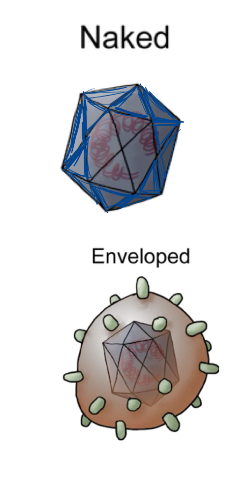
describe viral envelopes and how they are acuiqred
lipid bilayer derived from host cell
acquired fly budding of nucleocapsid through cellular membrane
nucleocapsids inside envelope may be helical OR icosahedral
identify the basic structural components and recognize additional non structural elements that may be incorporated into virions
The basic structural components of a virion are the viral genome, the capsid, and sometimes a viral envelope. Some virions also include extra components such as matrix/tegument proteins, cores, enzymes : (polymerases, integrases, proteases), transcription activators, cellular components ( tRNAs, histones, lipids, and host proteins.) These extra elements are packaged because they help the virus begin infection efficiently after entering the host cell.
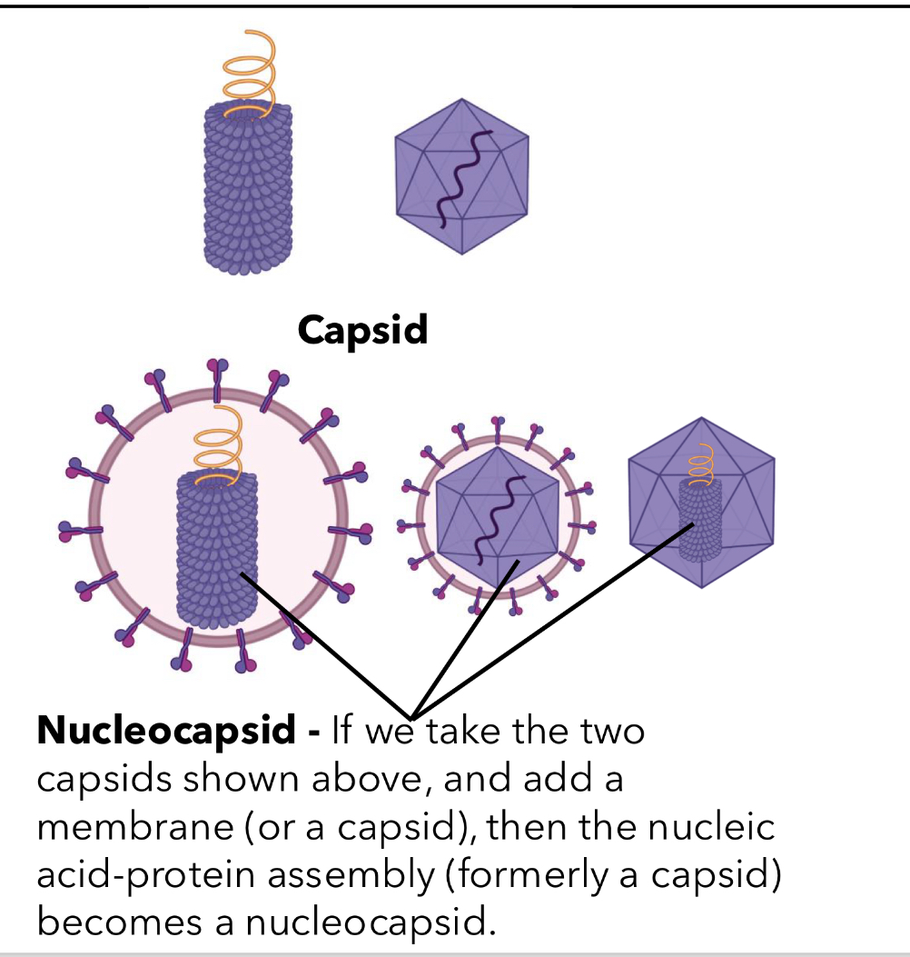
capsid
: protein shell surrounding genome
nucleocapsid (core)
nucleic acid,, protein assembly within vision
virion
infectious viral particle outside host . extracellular
envelope
viral membrane, host cel derived lipid bilayer
capsid vs nucleocapsid
capsid is outer protein shell surrounding viral nucleic acid genome protecting genome
can hv helical or icosahedral symmetry
how is metastability achieved
stable: created by symmetrical arrangement of many identical proteins to provide maximal contact
unstable: structure not oermantenly bonded tat
riles for self assembly
each subunit has identical bonding contracts w its neighbours and usually NON covalent
general features of helical capsids
: In helical capsids, coat protein subunits repeat around the viral genome in a spiral/helix. The coat proteins make identical interactions with each other and with the genome, allowing a large stable structure to be built from one type of protein subunit.
general features of icosahedral capsids
A: In icosahedral capsids, the capsid forms a closed shell with 20 triangular faces. It has 2-fold, 3-fold, and 5-fold axes of symmetry and can form a closed shell with as few as 60 identical subunits
interactions w neighbours r identical
when a capsid contains more than 60 subunits, each occupies a ___ position
quasiequivalent
what is T triangulation number
number of subunits comprising the structural unit , multiply by 60?
viral envelope glycoproteins and what do they do
A: Viral envelope glycoproteins are integral membrane proteins in the viral envelope. Their outside ectodomain helps with attachment, antigenic recognition, and fusion, while the internal domain helps with assembly. They often form oligomeric spikes on the virion surface
transfection
transformation + infection
production of infectious virus from the transofmraiton of cells by viral dna/rna
first done wth bacteriophage lambda
describe the requirements for a sucesful infection
sufficient quantity of virus
accessible, susceptible and permissive
no antiviral response
pathogenesis
process of producing a disease
e.x how does virus enter host? host response? where does rep occur? how doesinfection spread? what tissues are infected?
describe how viral particles move within the host cell after entry
skin, mucosal surfaces, eye, fetus (from mother placenta to kid e.x zekavirus)
describe factors that influence viral transmission
Viral transmission depends on whether the virus can access the host through entry sites like skin, mucosal surfaces, respiratory tract, alimentary tract, urogenital tract, eye, or fetus. It also depends on whether the virus reaches susceptible/permissive cells, how much virus is shed, how stable the virus is outside the host, and whether transmission is direct, vector-borne, horizontal, vertical, or healthcare-associated.
describe mechanisms of viral attachment and entry
1) adhere to cell surface w no specificity to look for receptor
2) attach to specific receptor on cell surface
3) penetration
4) transport and uncoating of genome
distinguish between class 1 and 2 viral fusion proteins
class 1: perpendicular to membrane spikes, mostly alpha helical, form trimers
class 2: parallel to membrane , mostly beta sheets and form dimers
infection
spread
transmission
spread from 1 susceptible host to another . required to maintain chain of infection
direct is human → human
vector borne is mosquito → human
identify routes of viral shedding
viruses can be shed through respiratory secretions/aerosols, nasal secretions, mucosal shedding, skin lesions, blood or blood supply, urine, semen, feces, insect vectors, and germline/vertical transmission. Shedding matters because it is how virus exits one host and reaches another.
susceptible cell
has functional receptor for given virus '
but doesnt mean cell can support viral REPLICATION
resistatnnt clel
has no receptor
may or may not be competent to support viral replication
permissive cell
capacity to replicate virus
doesnt mean its susceptible
isolation vs quarantine
isolation: separates sick ppl with contagious disease FROM people who are not sick
quarantine: separate and restricts movement of people who were EXPOSED to disease in CASE they become sick
nosocomial and iatrogenic
when Individual is infected while in hospital or health care facility
iatrogenic: activatey of healthcare worker leads to infection
germ line transmission
transmitted as part of genome
exceptions for cellular receptors for viruses
essental for all viruses except fungi (no extracellular phase) and plants (enterr cells by mechanical damage)
facts abt viral receptors (4)
diff viruses can hv same receptor
viruses in same family may hv diff receptors
1 virus can bind multiple receptors
a virus can use diff receptors for diff cell types
how do non enveloped viruses bind
via capsid surface or protrusions or transmembrane glycoproteins
e.x poliovirus, adenovirus, influenza
what are co receptors
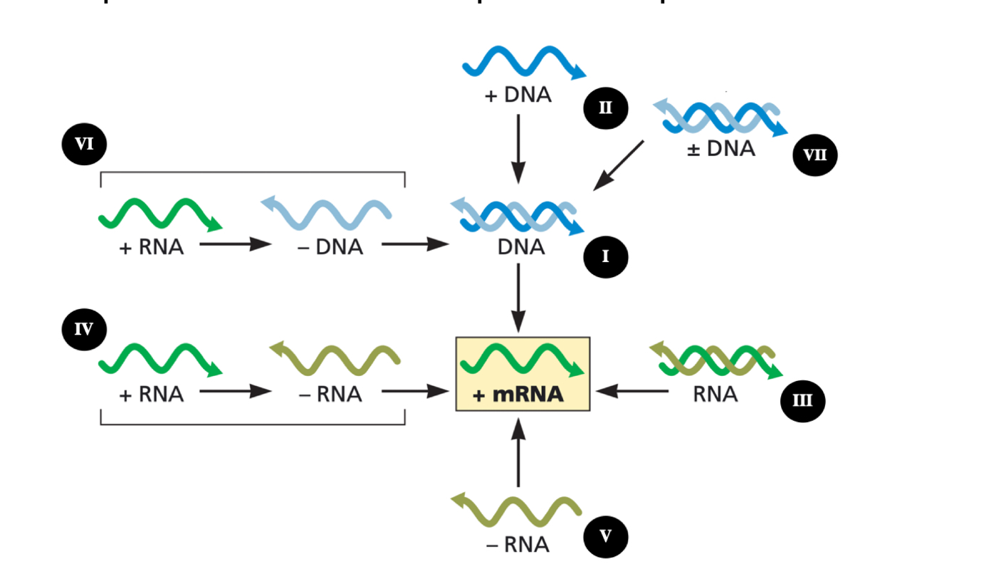
explain the central goal of viral mRNA synthesis
he central goal is to make viral mRNAs that can be translated by the host cell’s protein synthesis machinery. Viral genomes must ultimately produce mRNA because ribosomes translate mRNA, not every type of viral genome directly. Sometimes the genome itself can act as mRNA, like poliovirus, but +RNA does not always automatically mean functional mRNA because mRNA may need modifications to be recognized by ribosome
describe how diff classes of viral genomes generate mRNA
Different viral genome types reach mRNA in different ways. Positive-sense RNA viruses can sometimes use their genome directly as mRNA. Negative-sense RNA viruses must copy their genome into positive-sense mRNA using viral RdRp. dsRNA viruses must use RdRp to make positive-sense mRNA from the RNA genome. DNA viruses make mRNA using DNA-dependent RNA synthesis, usually using host transcription machinery if they replicate in the nucleus, but cytoplasmic DNA viruses need their own transcription machinery. The main idea is that every Baltimore class has a different route, but all must produce mRNA.
describe the role of viral and host enzymes in mrna synthesis
A: Viral and host enzymes copy viral genomes into mRNA or new genomes. RNA viruses use RNA-dependent RNA polymerase/RdRp to copy RNA from RNA templates. DNA viruses use DNA-dependent RNA polymerase to make mRNA from DNA. Nuclear DNA viruses can often use host enzymes, while cytoplasmic DNA viruses need to encode more of their own enzymes.
explain key modifications of viral mRNAs
: Viral mRNAs may need modifications like a 5′ cap, poly(A) tail, and splicing so they can be stable, processed, exported, and translated efficiently. Viruses can get a 5′ cap by cap-snatching, encode viral capping enzymes, use viral proteins like VPg, or use non-canonical/uncapped 5′ ends.
summarize the general strategies viruses use to regulate gene expression
hey can use viral or host DNA-binding proteins, viral co-activators, early versus late gene expression, feedback loops, cascade regulation, and mRNA processing like splicing.
distinguish viral mRNA synthesis from genome replication
: mRNA synthesis makes viral messages that can be translated into proteins. Genome replication makes complete new copies of the viral genome to package into new virions. mRNA synthesis does not always copy the whole genome, but genome replication must copy the genome end-to-end without losing sequence.
describe the major replication strategies and why they differ across the Baltimore classes
Replication strategies differ because each Baltimore class starts with a different genome type. RNA viruses need RNA-directed RNA synthesis using RdRp. DNA viruses use DNA-dependent DNA synthesis. Reverse-transcribing viruses use reverse transcription. The strategy depends on how the virus copies its genome and how it reaches mRNA.
explain the 5’ end problem and the strategies viruses use to solve it
The 5′ end problem happens with linear DNA genomes. DNA polymerase needs a primer and extends from a 3′OH. After the RNA primer at the end is removed, the very end of the DNA may not be copied. This can cause shortening of the genome ends over repeated rounds of replicatio
Circular genomes solve the problem because there are no free ends. The slide says “Circles solve the 5′ end problem.”
Parvoviruses use DNA priming. The slides label this as “DNA priming: Parvoviruses.”
Adenoviruses use protein priming. or replicating in cytoplasm
autoregularoty loop vs cascade regulation
Autoregulatory loop: a viral protein regulates its own expression.
Cascade regulation: one viral gene product turns on the next stage of viral gene expression.
t/f dna rep need primer
true always primer dependent
L6 Explain why viruses depend on host translation machinery.
Viruses depend on host translation machinery because they must make proteins, but they do not encode full ribosomes. All viruses make mRNA that can be translated by host ribosomes. Even if viruses encode their own polymerases or replication proteins, they still rely on host ribosomes, tRNAs, and translation factors to synthesize viral proteins.
Describe how viral mRNAs engage host ribosomes and the major mechanisms of translation initiation.
A: Viral mRNAs can engage ribosomes through normal 5′ cap-dependent initiation, where a cap-binding protein binds the 5′ cap, recruits the 40S ribosomal subunit, the 40S scans to the first AUG, and then the 60S subunit joins. Viruses can also use alternative mechanisms, including ribosome shunting, internal initiation/IRES, and methionine-independent initiation. Ribosome shunting lets ribosomes bypass structured RNA regions. IRES-mediated initiation lets ribosomes bind internally without a 5′ cap, as in poliovirus. Methionine-independent initiation allows initiation without normal initiator Met-tRNA.
Explain how viruses maximize coding potential.
viruses maximize coding potential by making more than one protein from limited genome space. Strategies include polyproteins, where one large protein is cleaved into multiple mature proteins; leaky scanning, where ribosomes skip weak start codons and initiate downstream; re-initiation, where the 40S subunit starts again after translating an upstream ORF; stop codon readthrough, where ribosomes continue past a stop codon; and ribosomal frameshifting, where the ribosome shifts reading frames to make a different or longer protein.
Describe ways viruses can manipulate the host translation environment.
Viruses can manipulate host translation by suppressing cellular protein synthesis and redirecting translation toward viral proteins. Poliovirus is an example: host protein synthesis drops after infection, and later protein synthesis mainly reflects viral protein production. Some viruses block host cap-dependent translation by cutting translation factors such as eIF4G, while viral RNAs can continue translating through IRES-mediated initiation.
Describe the common assembly sites and major strategies for making viral sub-assembl
Viral assembly often happens in specific cellular locations where viral components are concentrated, such as viral “factories,” inclusions, vesicles, the nucleus, or cellular membranes. Assembly depends on host cell machinery, including chaperones, transport systems, secretory pathways, and nuclear import/export machinery. Viral particles are complex, so they often assemble in steps. Sub-assemblies form first, then combine to make the complete virion. Sub-assemblies can form from individual proteins made from different mRNAs, from a polyprotein that folds and is then cleaved, or with help from viral/cellular chaperone
Explain how viruses ensure specificity for packaging their genomes.
Viruses use packaging signals in their viral genome to distinguish viral genomes from cellular DNA or RNA. Packaging signals are specific genome sequences that direct the viral genome into the assembling virion. For DNA virus examples, adenovirus has packaging signals near the inverted terminal repeat, recognized by viral protein IVa2. SV40 polyomavirus has packaging signals near the origin of replication, and mutations in this region can cause loss of encapsidation.
Q: Explain how enveloped viruses acquire their membrane.
: Enveloped viruses acquire their membrane by budding through host cellular membranes. This usually happens after internal viral structures have assembled. The envelope can come from different membranes depending on the virus, including the nuclear membrane, ER, Golgi, or plasma membrane. Budding can be driven by different viral components, such as envelope glycoproteins, internal matrix/capsid proteins, or matrix proteins. For example, influenza assembles and buds at the plasma membrane, while herpesvirus can acquire its mature envelope from the Golgi membrane
Describe how enveloped and non-enveloped viruses exit cells and why maturation might be important for infectivity.
Enveloped viruses usually exit by budding through a cellular membrane or by moving through the secretory pathway and being released by exocytosis. Some enveloped viruses use host machinery like the ESCRT pathway, which performs the final membrane-cutting step so the virus can detach from the cell. Non-enveloped viruses often exit by cell lysis, including apoptosis, necroptosis, viroporin-induced membrane rupture, or loss of membrane integrity. Some non-enveloped viruses can also exit non-lytically through vesicular release or exosomes. Maturation is important because some viruses are not fully infectious immediately after assembly. pH changes or proteolytic processing can cause conformational changes that make the viral particle infectious, as shown for Dengue and Zika viruses.
Explain the influenza replication cycle, including the host cell receptor used and the cell types expressing that receptor.
nfluenza is an enveloped, segmented, negative-sense ssRNA virus. It attaches when HA/hemagglutinin binds sialic acidon host cells. Human influenza mainly infects ciliated columnar epithelial cells in the respiratory tract, including the sinuses, nasopharynx, larynx, trachea, bronchi, and bronchioles. Even though sialic acid is common, influenza tropism is limited because HA must be cleaved by Tryptase Clara, which is produced by Clara/club cells in the bronchiolar epithelium.
Explain the two modifications of HA that are necessary for the influenza genome to enter the host cell.
A: First, HA0 must be cleaved into HA1 and HA2. HA1 binds the receptor, while HA2 contains the fusion peptide. Second, low pH in the endosome triggers a conformational change in HA, exposing the fusion peptide so the viral envelope can fuse with the endosomal membrane and release the genome into the cell.
: Explain how influenza steals the 5′-methylated cap from cellular pre-mRNA during synthesis of its own mRNA by RdRp.
A: Influenza uses cap-snatching. The viral RNA-dependent RNA polymerase binds the 5′ methylated cap of a host pre-mRNA, cuts the host RNA about 10–13 nucleotides downstream, and uses that capped fragment as a primer. The viral RdRp then extends the primer using the negative-sense viral RNA genome as the template to make viral mRNA.
Explain how influenza synthesizes full-length positive RNA to use as a template for synthesis of progeny genomes.
Influenza first makes shorter positive-sense viral mRNAs for translation. Later, when NP protein accumulates, NP binds the growing positive RNA and allows the polymerase to copy the entire negative-sense genome segment. This creates a full-length positive-sense RNA intermediate, which is then used as the template to make new negative-sense genome segments for progeny virions.
Describe how influenza progeny assemble and are released from the host cell.
nfluenza vRNPs assemble in the nucleus, then are exported and transported to the plasma membrane. The M1 matrix protein localizes to the membrane and interacts with vRNPs and viral membrane proteins. Final assembly and budding occur at the plasma membrane. NA/neuraminidase cleaves sialic acid so new virions can detach from the host cell instead of sticking to the cell surface.