Psych 106 Final
1/221
There's no tags or description
Looks like no tags are added yet.
Name | Mastery | Learn | Test | Matching | Spaced | Call with Kai |
|---|
No analytics yet
Send a link to your students to track their progress
222 Terms
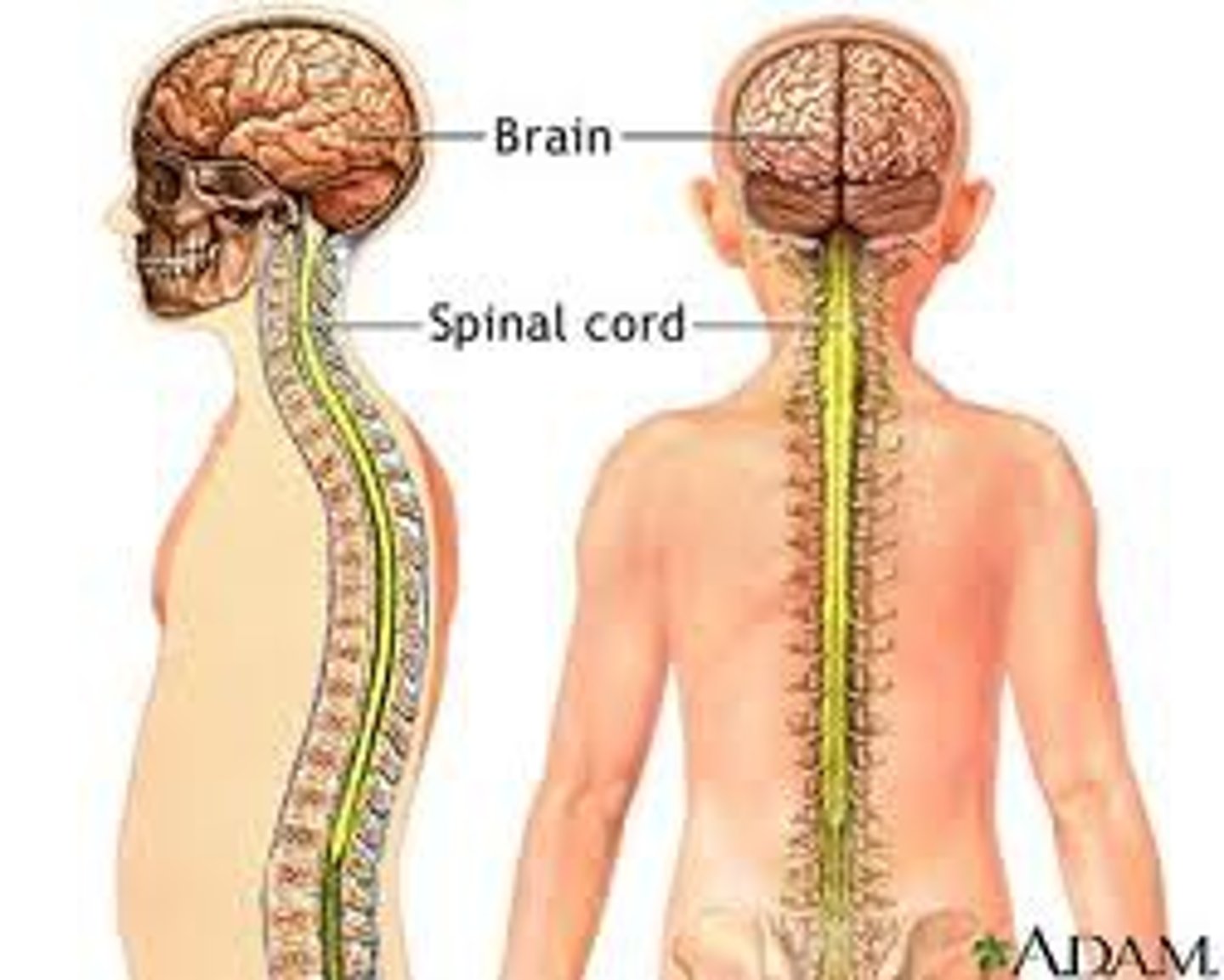
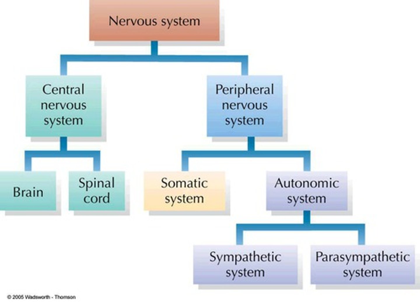
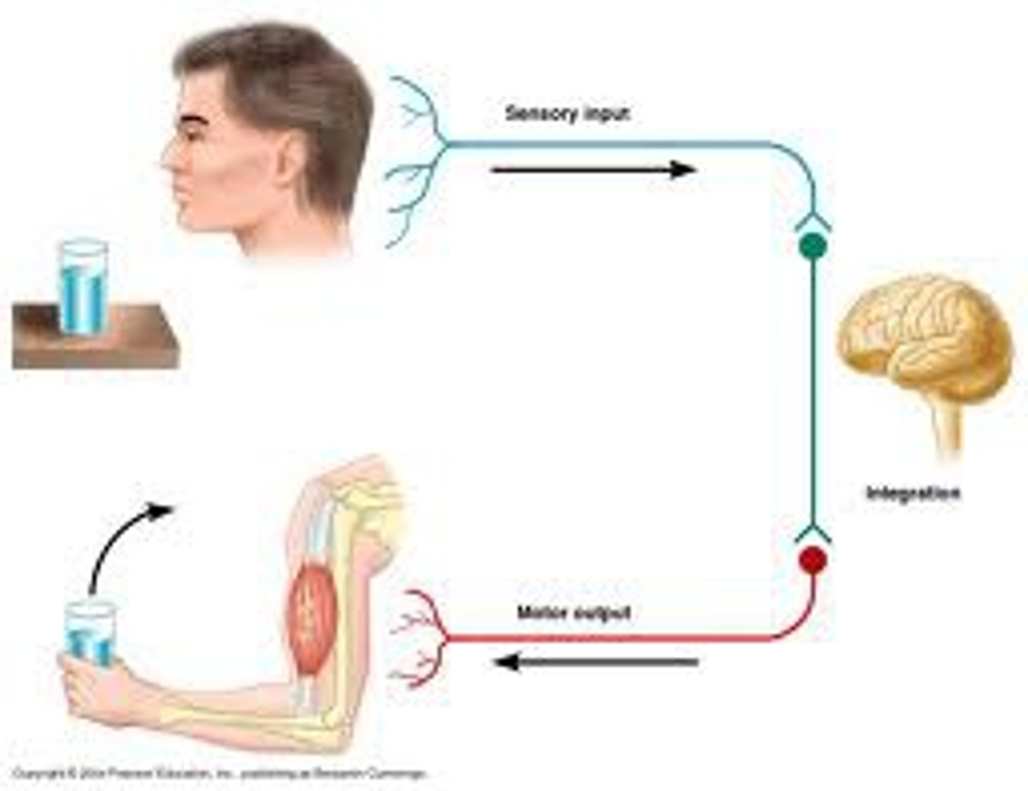
What does the central nervous system consist of?
The Brain and the Spinal Cord
What is the difference between the central and peripheral nervous system?
CNS: works at processing information and sends out responses based off of sensory input
PNS: works more as a communication between the body and the CNS
What is the somatic nervous system?
The Somatic nervous system is a part of the PNS (peripheral nervous system), that controls voluntary muscles and sends sensory info to CNS.
What is the autonomic nervous system?
The autonomic nervous system is part of the PNS, in which it controls involuntary muscles. In involves the sympathetic and parasympathetic nervous system.
What is the difference between the sympathetic and parasympathetic nervous systems?
The sympathetic nervous system is focused on expending energy (higher heart rate, faster breathing, fight-or-flight), while the parasympathetic is focused on conserving energy (lower heart rate, increase digestion, slower breathing)
What do the terms dorsal, ventral anterior, and posterior refer to?
Dorsal*: More towards top
Ventral**: More towards the bottom
Anterior: More towards the front
Posterior: More towards the back
*Dorsal can also mean more towards the back
**Ventral can also mean more towards the stomach
What are sagittal, coronal, and horizontal cuts?
Sagittal: Vertical cut through brain dividing it from left to right
Coronal: Vertical cut through brain dividing it from front to back
Horizontal: cut through brain that is parallel to base dividing it from top to bottom
What do ipsilateral, contralateral, unilateral, and bilateral mean?
Ipsilateral: on the same side
Contralateral: on opposing sides
Unilateral: one side
Bilateral: two sides
What is the difference between white matter and gray matter?
Gray Matter: holds cell bodies and synapses and is included in cortex and subcortical nuclei (appears darker)
White Matter: holds myelinated axons and includes white matter tracts that connects gray matter to other itself (appears whiter because of myelin)
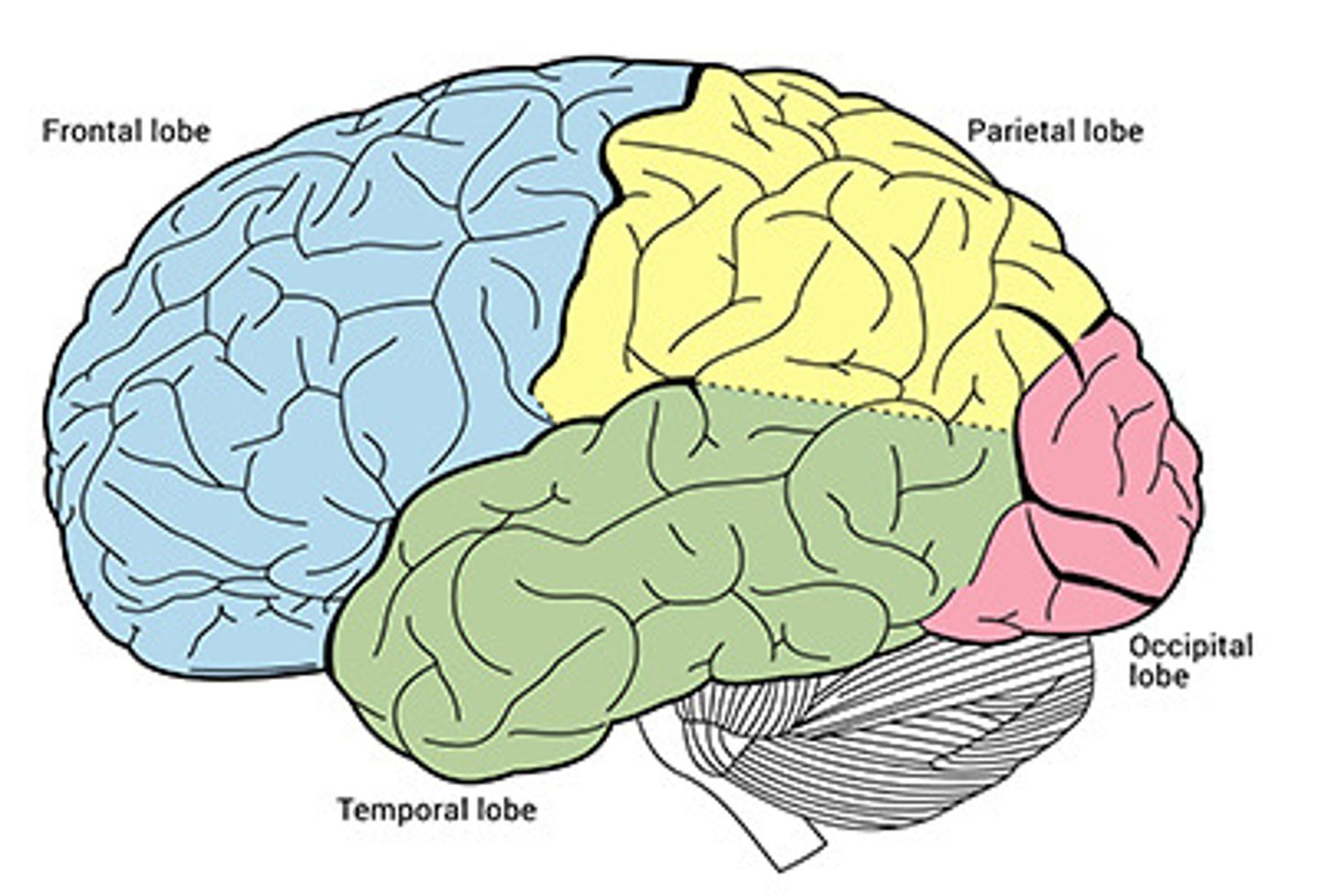
What are the four lobes of the brain?
Frontal lobe
Parietal lobe
Temporal lobe
Occipital lobe
What is the major function of the ventricles?
Protection of the brain (acts as a buffer)
Maintaining Homeostasis and intracranial pressure (maintaining temperature)
Removing toxins and waste products
What is the major function of the cerebral cortex?
The outermost part of the brain (3mm thick) which is intensely folded (the more folding = the more cortical surface without increasing head size) that deals with perception, cognition, decision-making
What is the major function of the cerebellum?
Maintain posture
Dexterity
Smooth execution of movement
Integration of action planning, movement, and sensory feedback
What is the major function of the basal ganglia?
Regulate motor activity
Start/stops movement
Action planning
Learning reward, skills, habits
What is the major function of the thalamus?
"Relay center" that takes in sensory information from sensory organs and will direct it to the cortex
What is the major function of the hypothalamus and pituitary gland?
Controls body regulation like (body temp, hunger/thirst, sexual activity)
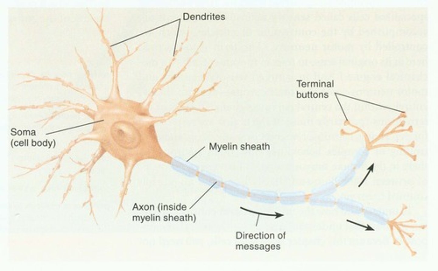
What are the main structures of a neuron?
Dendrites (take in info/input)
Soma (Cell Body)
Axon
Terminals (release info/output)
What is the difference between afferent and efferent neurons?
Afferent Neurons send info from sense organs to the brain (sensory neurons)
Efferent Neurons carry info back from Brain to the muscles (motor neurons)
What is the structure and permeability of the neuronal cell membrane?
The membrane is a lipid bilayer, uncharged molecules (H2O, O2, CO2) can move through easily, but larger molecules or ions cannot pass through easily (Na+, K+, Cl-, Ca2+)
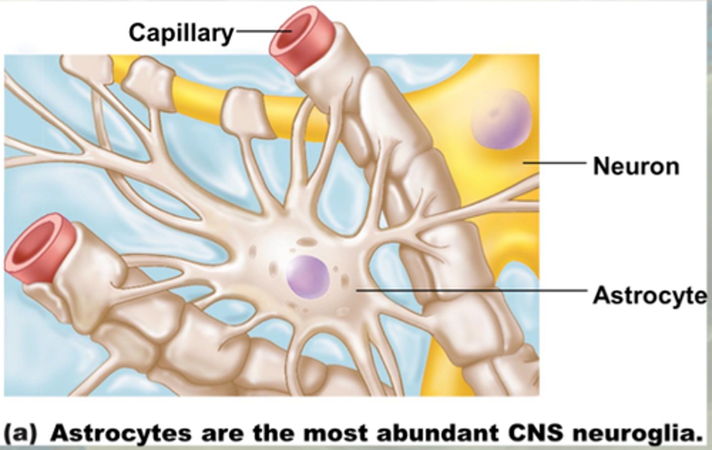
What are the three major types of glial cells in the CNS and what are their functions?
Astrocytes: Create Blood Brain Barrier and modulate synaptic strength
Oligodendrocytes: form myelin around axon (myelin sheath)
Microglia: Immune cells and Phagocytes (can eat other cells)
What are astrocytes and what is their role in the blood-brain barrier?
Astrocytes are glial cells that wrap around blood vessels in the brain and protects the brain from viruses, pathogens, and toxins
What are oligodendrocytes and Schwann cells?
Oligodendrocytes and Schwann Cells both have the main function to produce myelin. The difference is that Oligodendrocytes do it for the CNS, where as Schwann Cells do it for the PNS
What is the concentration of Na+, K+, Cl-, and Ca2+ ions in the extracellular and intracellular space?
High Concentration of Na+/Cl-/Ca2+ on outside/extracellular, low on inside
High Concentration of K+ on inside/intracellular, low on outside
What are chemical and electrical gradients?
Chemical/Concentration Gradient: force in which ions move to a side where there is less concentration to balance it out on either side
Electrical Gradient: force in which ions move so there is no charge difference on either side
Based on electrochemical gradient, which ions are driven into or out of the cell?
Na+/Ca2+/Cl- wants to move inside (because the intracellular of a resting neuron is more negative)
K+ wants to move in because of electrical gradient, but the concentration gradient doesn't want to bring it in as there is a high concentration inside the cell already
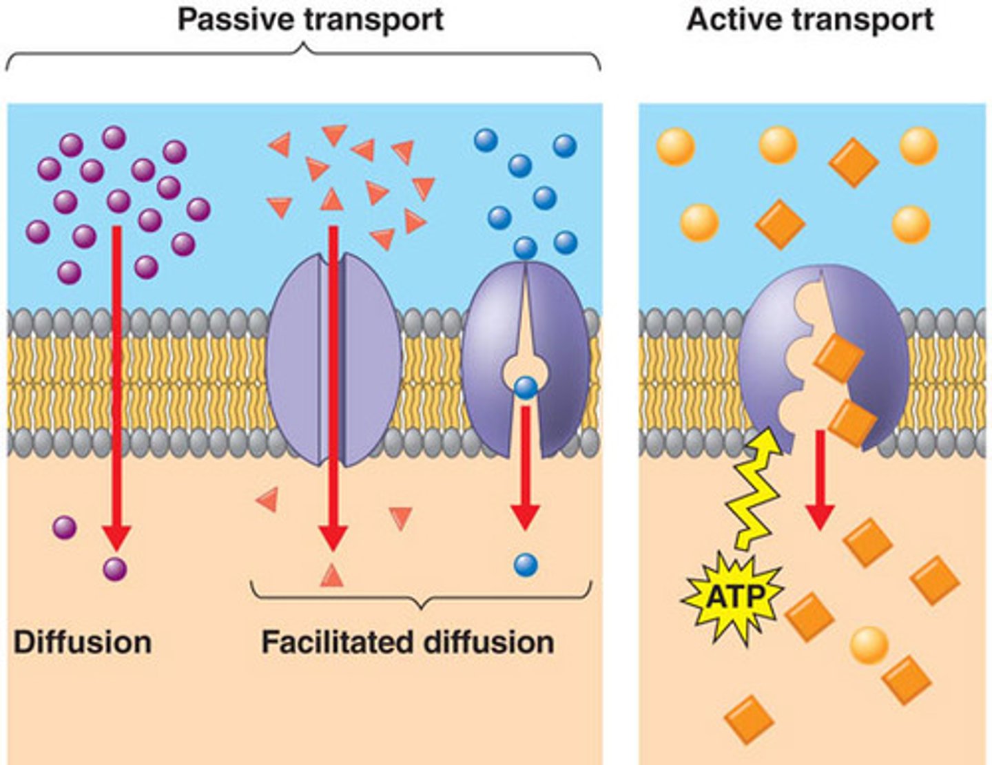
What is the difference between passive and active transport?
Passive Transport doesn't need energy to allow transportation
Active transport does need energy, usually in the form of ATP, can also allow movement against a gradient
What are voltage-gated ion channels?
Ion channels in which they open or close when at a certain voltage (ex. Na+ voltage gated channels open when hit Action Potential threshold)
What are ligand-gated ion channels?
Ion channel that opens when a ligand or molecule binds to channel and changes structure causing it to open or close
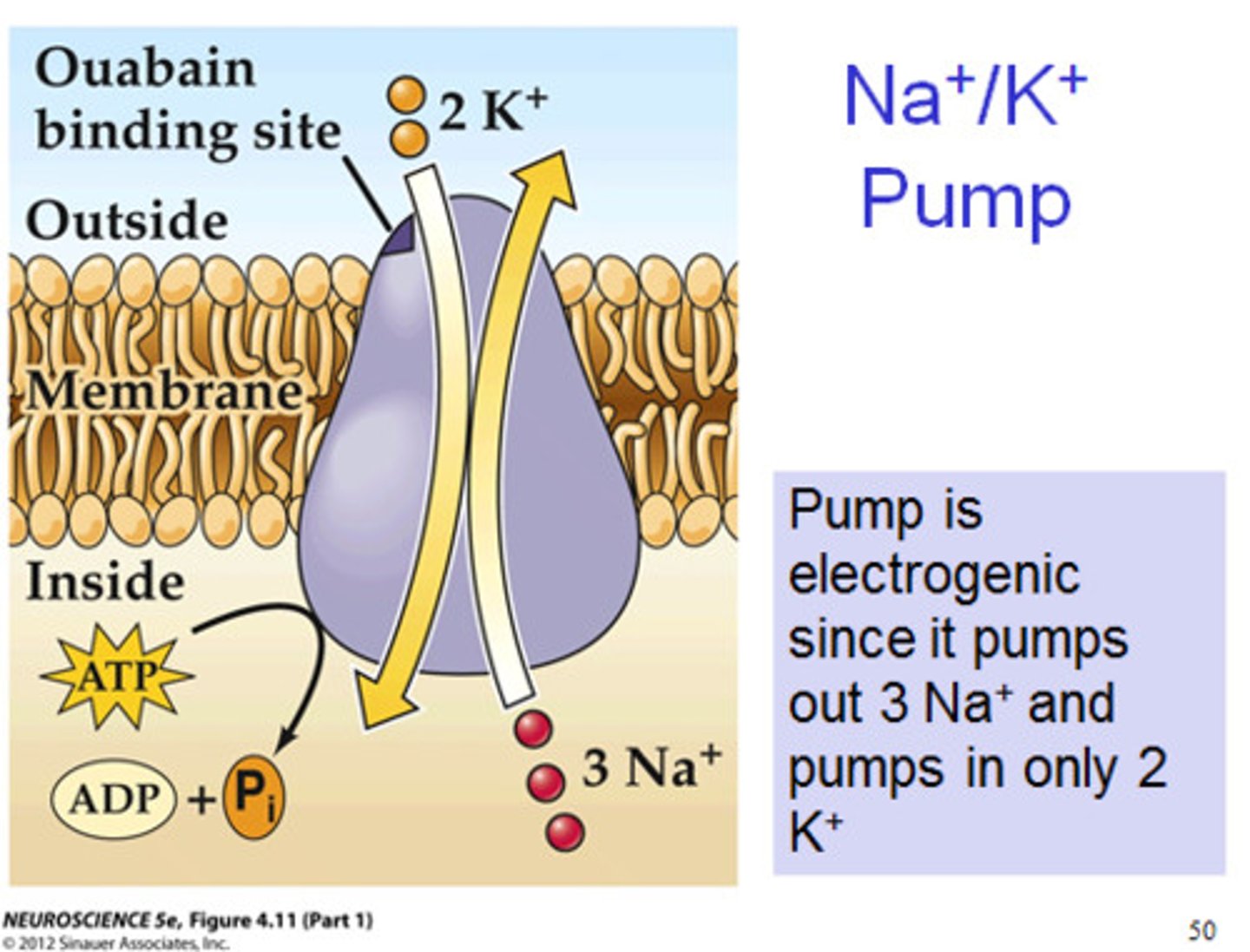
What is the Na+/K+ pump?
The sodium-potassium pump uses energy to push out 3 Na+ ions out of the cell and 2 K+ ions into it. The sodium potassium pump is a good reason as to how the cell stays at resting membrane potential
What is the resting potential?
-70 mV (neurons are more negatively charged intracellularly) this is the voltage of a neuron when it isn't being fired
What is the sequence of voltage changes in an action potential?
Depolarization: Once the neuron reaches the voltage threshold the Na+ voltage channels open and Na+ to flow into the neuron (neuron gets more positive)
Repolarization: Once a neuron hits a certain positive point, K+ voltage channels open and K+ flows out the cell (neuron gets more negative)
Hyperpolarization: While the K+ ion channels do close, it takes a little more time and causes the voltage to dip below the resting membrane potential
Back to Resting: The neuron is then slowing brought back to resting membrane potential because of the Sodium Potassium Pump which brings it back
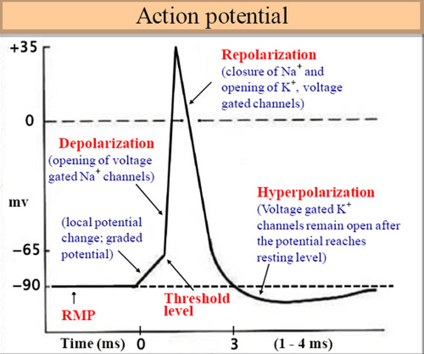
What is the threshold potential?
The threshold potential is the voltage that a neuron must hit to trigger an action potential (~50mV)
What is the all-or-none principle?
The All or none Principle is the idea that a neuron will either fire an action potential (by reaching the threshold) or it won't fire an action potential (AP will always have same size and not lose strength)
What is the refractory period?
A brief period of time in which the neuron is not at resting potential
Absolute Refractory Period: no action potential can be generated
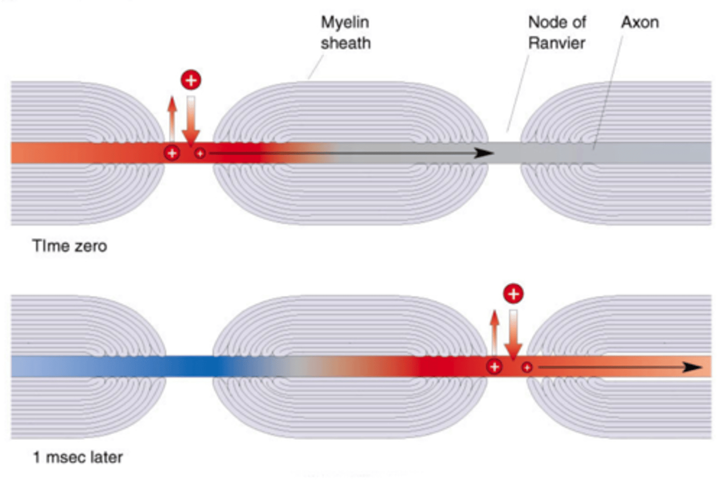
What is saltatory conduction?
As action potentials travel through a myelinated axon, the AP jumps from opening between the myelin called the nodes of ranvier, this makes the propagation of the AP energy efficient and fast.
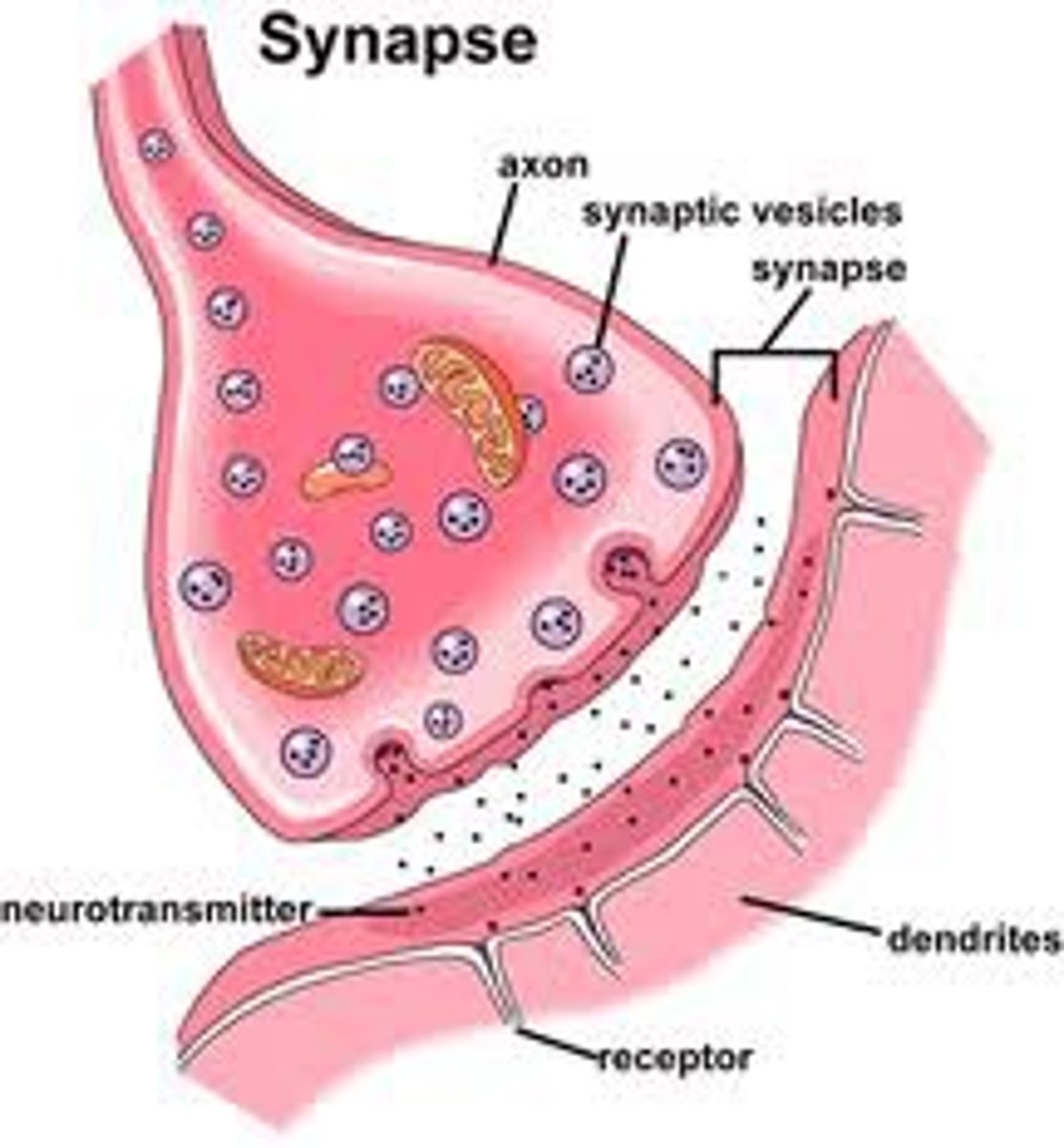
How is the neural signal transmitted at the synapse?
1. Once an AP travels down an axon and to the terminals, it triggers the opening of voltage-gated Ca2+ channels and Ca2+ flows into the cell.
2. Ca2+ then triggers the release of neurotransmitters from the presynaptic terminal to the synaptic cleft
3. They move through synaptic cleft
4. Neurotransmitters bind to receptors which can then lead to a depolarization/hyperpolarization/more
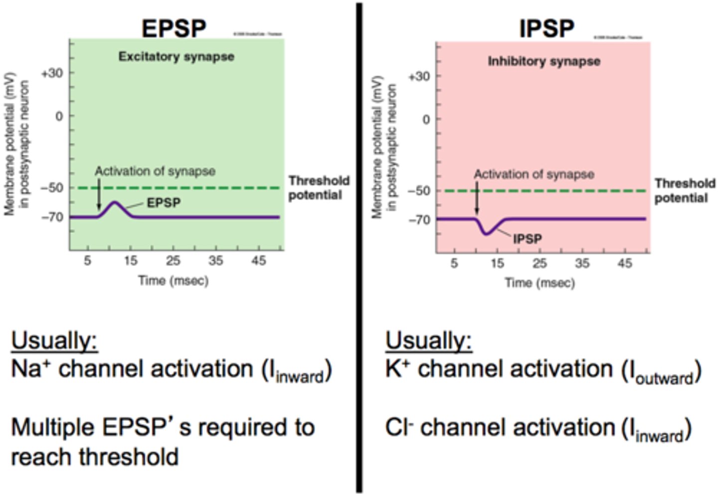
How is an excitatory postsynaptic potential (EPSP) elicited?
Neurotransmitters bind to receptors which can open and positive ions flow into the cell or negative ions flow out causing a positive increase in voltage within the neuron

How is an inhibitory postsynaptic potential (IPSP) elicited?
Neurotransmitters bind to receptors which can open and negative ions flow into the cell or positive ions flow out causing a negative decrease in voltage within the neuron
What is spatial summation of postsynaptic potentials?
On the postsynaptic neuron, in which multiple synapses from different locations on neuron are added up (increased likelihood of AP)
Spatial: Space
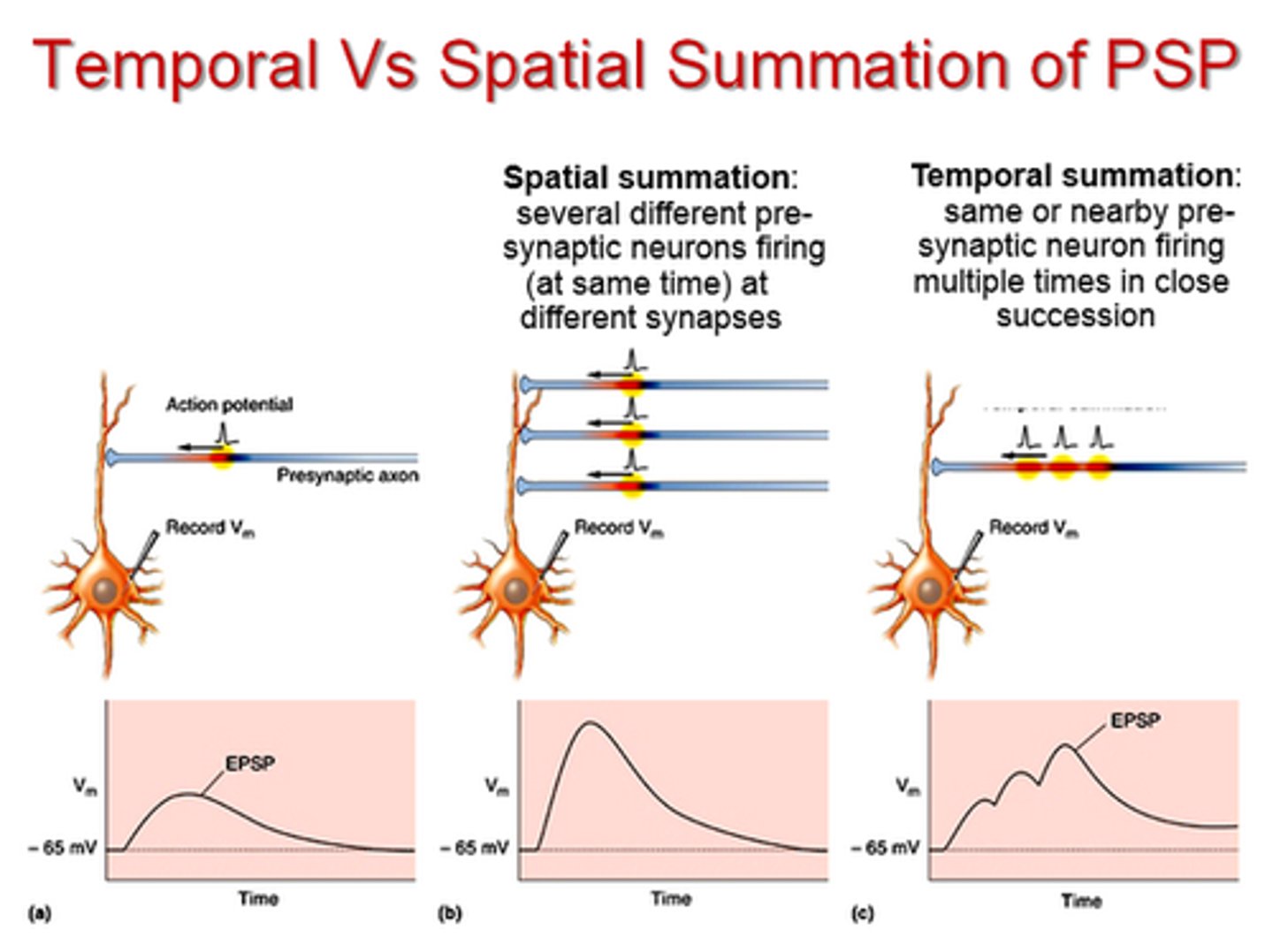
What is temporal summation of postsynaptic potentials?
On the postsynaptic neuron, in which a single synapse fires in rapid succession causing the them to be added over time (increased likelihood of AP)
Temporal: Time

How are neurotransmitters removed from the synaptic cleft?
1. Diffusion (only removes a small amount)
2. Reuptake (NT will be recycled and repacked into vesicles)
3. Enzymatic Breakdown
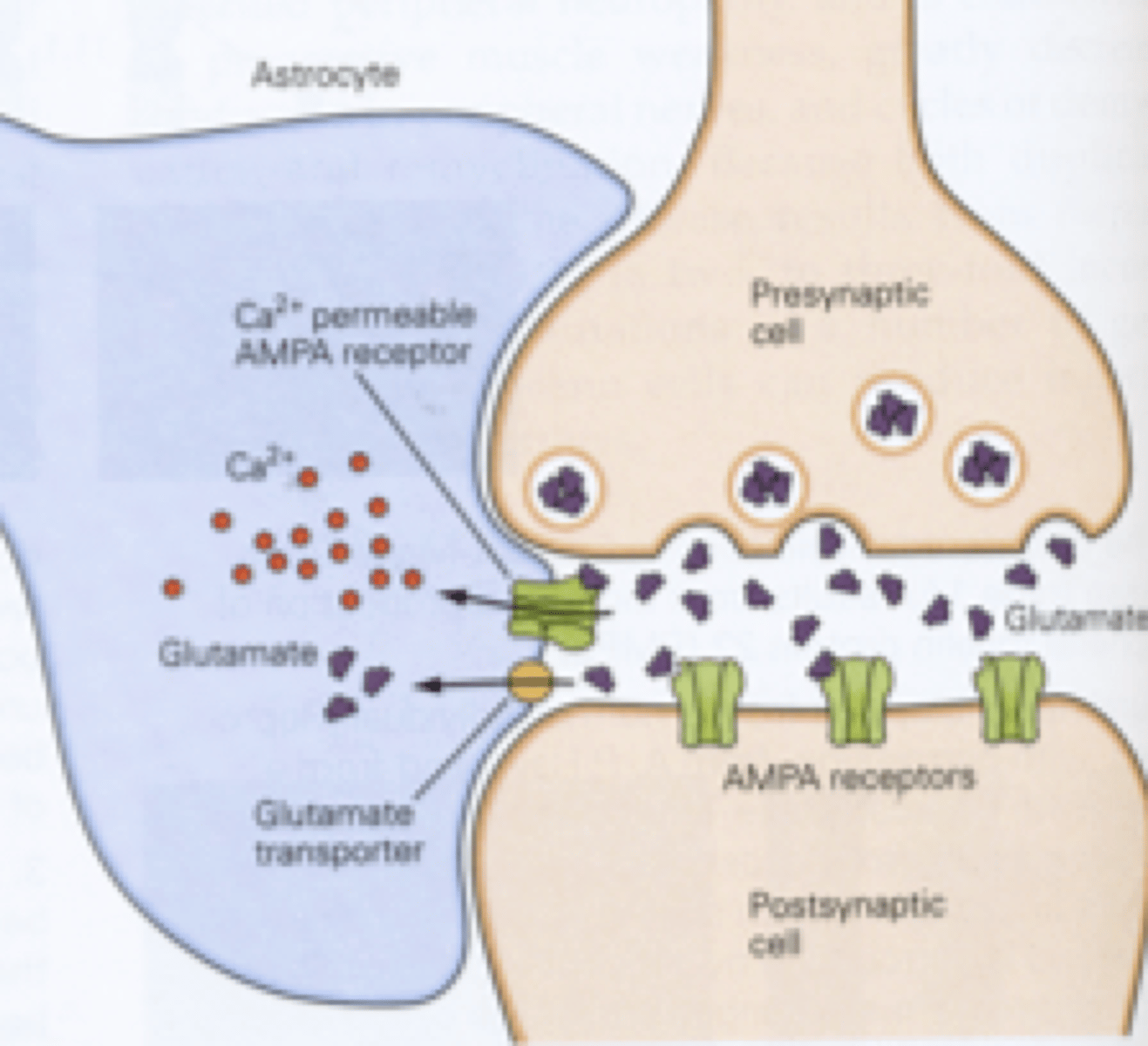
What is the tripartite synapse?
3 parts: Presynaptic neuron, Postsynaptic, and Astrocyte
Astrocytes will stabilize synapses (develop and maintain), provide neurotransmitters for presynaptic cell, remove neurotransmitters from synaptic cleft (reuptake)
Explain the pathway for touch on the head
Sensory info from the head is sent to the brain via the trigeminal nerve
Where is somatotopic representation shown in the cortex? What lobe is it located in
Located in the primary sensory cortex on the postcentral gyrus within the parietal lobe
Explain the pathway for touch below your head
Sensory neurons enter the spinal cord through the dorsal side and is then sent to the brain
basic components of the eye
iris, sclera, pupil(where light enters), lens, retina
main cells in the retina
*photoreceptors (rods and cones)
horizontal cells
amacrine cells
*ganglion cells
*bipolar cells
what are the optic disk, fovea, periphery and optic nerve within the retina?
OPTIC DISK:
-"blind spot" where there are no photoreceptors due to bundles of axons and blood vessels that need to leave the eye to form the optic nerve
FOVEA:
-cone dense area in the middle of the retina
-responds to bright light
-helps with acuity(detail)
PERIPHERY:
-rod dense
-responds to dim light
-does not help with acuity(detail)
OPTIC NERVE:
-bundles of retinal ganglion cell AXONS
-lead to the thalamus (LGN)
Phototransduction
light -> neural activity
-converts light energy from photons into neural activity
rods VS cones
RODS:
-active/useful in low light
-poor detail
-does not process color
-dense in periphery
-1 type
CONES:
-active/useful in bright light
-high detail, processes color
-dense in fovea
-3 different types that process different length wavelengths associated with colors
What is neural convergence of rods and cones with bipolar and retinal ganglion cells?
*photoreceptors -> bipolar cells -> ganglion cell -> firing
-refers to the rate at which information from rods and cones CONVERGE with bipolar cells and ganglion cells. This is the erasion why rods are not useful for acuity and cones are useful for acuity
RODS:
-more convergence
-low acuity
-high sensitivity
-activate ganglion cells in sum (ex: 3:1)
CONES:
-less convergence
-high acuity
-low sensitivity
-activate ganglion cells as ONE (1:1)
NOTE: think about the word 'convergence', how much info of rods vs cones CONVERGES with bipolar and ganglion cells
retina-cortex pathway
retina -> optic disk -> optic nerves -> optic chiasm -> optic tract -> thalamus (LGN) -> optic radiation -> primary visual cortex (V1)
optic chiasm
where the optic nerves "cross"
NOTE: technically only the nasal cross, temporal do not cross, but BOTH types are still considered the optic chiasm
What is the lateral geniculate nucleus (LGN) and what happens there?
-first cortical structure that the retina communicates with
-projects to the primary visual cortex (V1)
-receives information from the cortex
what are receptive fields?
-part of the environment(visual field) that the retina responds to
-response can be inhibitory or excitatory
AKA a region in the visual field that changes the firing of a neuron
CENTER-SURROUND gangion cells: ON-center ganglion cell vs OFFf-center ganglion cell
ON-center ganglion cell = light in the center of the receptive field is excitatory, and light in the surrounding area is inhibitory
OFF-center ganglion cell = light in the center of the receptive field is inhibitory, and light in the surrounding area is excitatory
simple cells VS complex cells
*LGN -> simple cells -> complex cells
SIMPLE cells:
-selectively fire to bars of light at a specific orientation/angle at a specific location in receptive field
-fire more to their preferred orientation and fire less and less the further the light becomes from their preferred orientation and location
-center-surround receptive field
COMPLEX cells:
-respond to bars of light of specific orientation/angle at any point in their receptive field
-convergence of many simple cells
-no center-surround receptive field
how is the primary visual cortex organized?
-retinotopically, where neighboring cortical representation reflect neighboring areas of the visual field
-there is more cortical area of the cortex related to and representing input from the fovea than from the periphery
-columnar architecture with orientation(angle) columns and ocular(eye) dominance columns
what is the primary visual cortex and where is it located?
-where visual information is processed
-located in the posterior-medial area of the brain (occipital lobe)
what is the function of the dorsal stream? What areas does the dorsal stream connect?
-"where"/"how" pathway from occipital lobe to the parietal lobe
-WHERE objects are in space and HOW to act on them
-Spatial navigation(acting on objects)
Ex: how to grab and use the pencil on your desk
What is the ventral steam? What area does the ventral stream connect
-"What" pathway from occipital lobe to temporal cortex
-What something is
-Object recognition
-Involves memory (temporal lobe)
what is area V4 and what happens when it is damaged?
V4 = color perception
damage = cerebral achromotopsia
-color vision in black and white
-NOT the same as color blindness (cone issues)
What is area MT/V5 and what happens when it is damaged?
V5 = visual motion
damage = cerebral akinetopsia
-motion blindness AKA stop motion vision
Which of the two pathways to CNS for pain refer to the "physical sensation of pain"?
Sensory Pathway: sensory neurons synapse in the spinal cord, cross the midline, synapse at the thalamus then projects to the cortex
(ex. Pain in Arm: Neurons in arm bundle to nerve, enter spinal cord on dorsal side and synapse, cross over mid line goes up spinothalamic tract on contralateral side, synapses at thalamus and then projects to cortex)
Which of the two pathways to CNS for pain refer to the "emotional processing of pain"?
Emotional pathway: synapse in the spinal cord, cross midline and project info to hypothalamus and amygdala which contribute to emotional response
Which specialized mechanoreceptors detect pain?
Free Nerve Endings
Which touch receptors are associated with temperature? How are the ion channels activated?
Free Nerve endings
Ion channels are temperature gated, once temperature hits a certain point it can trigger an AP
Name the 4 different type of receptors for temperature
Non-noxious warm: warm Non-noxious cold: cool
Noxious warm: super hot Noxious cold super cold
What are the two types of pain transmissions are what make them different?
A Delta (Aδ) fibers are myelinated, fast conducting, sharp pain and intense heat
C fiber: mom myelinated, slow conducting, dull, throbbing and chronic pain
Where is the vestibular system located?
The inner ear
What is the vestibular sensation?
Sensory input that comes from inner ear that detects head movement, acceleration/deceleration and gravity
What are the receptors for touch called? (Name one that deal with detection of light touch/vibrations and one that focuses on deep touch/vibrations)
Mechanoreceptors
(Meissner's corpuscles: Low frequency vibration, texture, fine touch
Pacinian corpuscles: High frequency vibes, texture)
What are the semicircular canals and what do they detect?
Semicircular canals: senses rotational movement of the head
Also responds to acceleration/deceleration and use hair cells
What are the otolith organs and what do they detect?
2 Otolith organs (saccule and utricle): senses linear acceleration and gravity
Also responds to acceleration/deceleration and use hair cells
Explain the Vestibular Pathway to the Cortex
1. Vestibular information is sent from inner ear (semicircular canals/otoliths) through the vestibular nerve to vestibular nuclei in brainstem
2. Info crosses the midline and ends up in the thalamus
3. Thalamus projects to cortex parieto-insular vestibular cortex (PIVC) for motion perception and spatial orientation
What is the Vestibular-ocular reflex?
The integration of vestibular info with motor signals to eye (oculomotor info) which allows the eyes to maintain a steady gaze on an stimuli while the head is moving
how are frequency and amplitude of sound waves perceived?
frequency = *pitch in Hz
-frequency increases = higher pitch
-frequency decreases = lower pitch
amplitude = *loudness
-large amplitude = louder
-small amplitude = quiet
what range of frequencies is audible to humans?
20-20,000 Hz
cochlea
-fluid filled organ in our inner ear
-snail shaped
-contains organ of corti
organ of corti
-sensory organ within the cochlea
-contains hair cells with stereocilia on top
hair cells
-cells in the cochlear organ of corti
-convert mechanical vibrational energy into an electrical neural signal by "bending"
bone vibration -> cochlear fluid vibration -> hair cell stereocilia bent -> ion channels open
auditory pathway to cortex
cochlear neuron bundles forms cochlear nerve -> ipsilateral cochlear nucleus -> ipsilateral & contralateral superior olive -> ipsilateral and contralateral inferior cochlea colliculus -> ipsilateral MGN (thalamus) -> primary auditory cortex (A1)
tonotopic organization
corresponding cortical areas respond to neighboring frequencies in primary visual cortex
-spatial arrangement of frequencies
-as seen in frequency gradients in:
cochlea, cochlear nucleus, superior olive, inferior colliculus, MGN and A1
What is the primary auditory cortex (A1) and where is it located?
-located in superior temporal lobe
what is the role of the dorsal stream in auditory processing?
-WHERE is the sound coming from and HOW do I reproduce it
A1 -> parietal and frontal cortex
what is the role of the ventral stream in auditory processing?
-WHAT is the sound
-auditory recognition
A1 -> temporal and frontal cortex
What are the papillae, tastebuds, taste receptor cells and tastants?
Papillae = bumps on tongue that contain groups of taste buds
taste bud = contain groups of receptor cells
taste receptor cells = where tastings interact with receptors for processing
tastings = food molecules that produce taste sensation
5 taste qualities
salty
sweet
sour
bitter
umami
taste pathway in the brain
gustatory nerve -> nucleus of the solitary tract (brainstem) -> ventral posterior medial nucleus (thalamus) -> primary gustatory cortex (parietal/insula)
What is flavor?
combo of taste, smell and somatosensory info
olfactory structures
olfactory bulb
odorants
olfactory receptor cells
glomeruli
moral/tufted cells
combinatorial coding
each odor is represented by a combo of activated receptors
ex: 20:1
direct and indirect olfactory projections
direct = limbic structures (amygdala, hippocampus) , primary olfactory cortex (piriform cortex in inferior temporal lobe)
indirect = thalamus
What is the difference between monosynaptic reflex vs polysynaptic reflex?
Monosynaptic: Involves only a single direct synapse between a sensory neuron and a motor neuron (faster, e.g., knee-jerk). Polysynaptic: Involves one or more interneurons between the sensory input and motor output (slower, e.g., withdrawal reflex)
Reciprocal Inhibition
The neural mechanism where the contraction of an agonist muscle automatically triggers the relaxation of its opposing antagonist muscle to allow fluid movement
What neurotransmitter is released at the neuromuscular junction and what is its effects?
Acetylcholine (ACh)is released at the neuromuscular junction and acts exclusively as an excitatory signal to cause skeletal muscle contraction
Central Pattern Generators (CPGs)
Autonomous neural circuits located within the central nervous system (spinal cord/brainstem) that produce rhythmic, repetitive motor outputs without requiring sensory or higher brain inputs
How are Central Pattern Generators modified?
Higher cortical input provides voluntary control to initiate or terminate CPG patterns, while real-time sensory feedback dynamically adjusts the rhythm to adapt to unexpected environmental changes (e.g., tripping)
Direct (GO) Pathway
a) Function
b) Regions involved
Function: Disinhibits the thalamus to facilitate and initiate voluntary movement. Regions involved: Cerebral cortex, Striatum, Globus Pallidus internus (GPi), and Thalamus
Indirect (NO GO) Pathway
a) Function
b) Regions involved
Function: Increases inhibition on the thalamus to suppress unwanted or competing movements. Regions involved: Cerebral cortex, Striatum, Globus Pallidus externus (GPe), Subthalamic Nucleus (STN), GPi, and Thalamus
D1 vs. D2 Receptors in the Striatum
D1 receptors are excitatory and located on striatal neurons of the direct (Go) pathway. D2 receptors are inhibitory and located on striatal neurons of the indirect (No-Go) pathway